


Home / Training / Manuals / Atlas of breast cancer early detection / Cases
Atlas of breast cancer early detection
Go back to the list of case studies
.png) Click on the pictures to magnify and display the legends
Click on the pictures to magnify and display the legends
| Case number: | 055 |
| Age: | 73 |
| Clinical presentation: | Postmenopausal woman with increased risk of developing breast cancer in view of family history of breast cancer presented with a lump in the left breast. Examination revealed a hard irregular lump 2 cm in diameter in the upper quadrant of the left breast. |
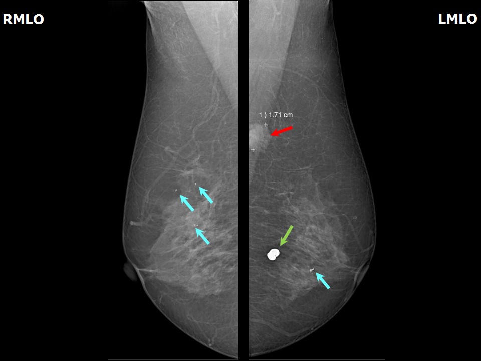
Mammography:
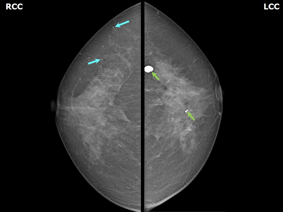 |  |
| Breast composition: | ACR category b (there are scattered areas of fibroglandular density) | Mammography features: |
| ‣ Location of the lesion: | Left breast, upper outer quadrant at 11 oclock, posterior third, at axillary tail |
| ‣ Mass: | |
| • Number: | 1 |
| • Size: | 1.71 cm in greatest dimension |
| • Shape: | Irregular |
| • Margins: | Spiculated |
| • Density: | High |
| ‣ Calcifications: | |
| • Typically benign: | None |
| • Suspicious: | None |
| • Distribution: | None |
| ‣ Architectural distortion: | None |
| ‣ Asymmetry: | None |
| ‣ Intramammary node: | None |
| ‣ Skin lesion: | None |
| ‣ Solitary dilated duct: | None |
| ‣ Associated features: | None |
| Breast composition: | ACR category b (there are scattered areas of fibroglandular density) | Mammography features: |
| ‣ Location of the lesion: | Left breast, lower outer quadrant at 45 oclock, posterior third |
| ‣ Mass: | |
| • Number: | 0 |
| • Size: | None |
| • Shape: | None |
| • Margins: | None |
| • Density: | None |
| ‣ Calcifications: | |
| • Typically benign: | Typically benign round, rod-like, macrocalcifications and vascular calcifications |
| • Suspicious: | None |
| • Distribution: | Diffuse |
| ‣ Architectural distortion: | None |
| ‣ Asymmetry: | None |
| ‣ Intramammary node: | None |
| ‣ Skin lesion: | None |
| ‣ Solitary dilated duct: | None |
| ‣ Associated features: | None |
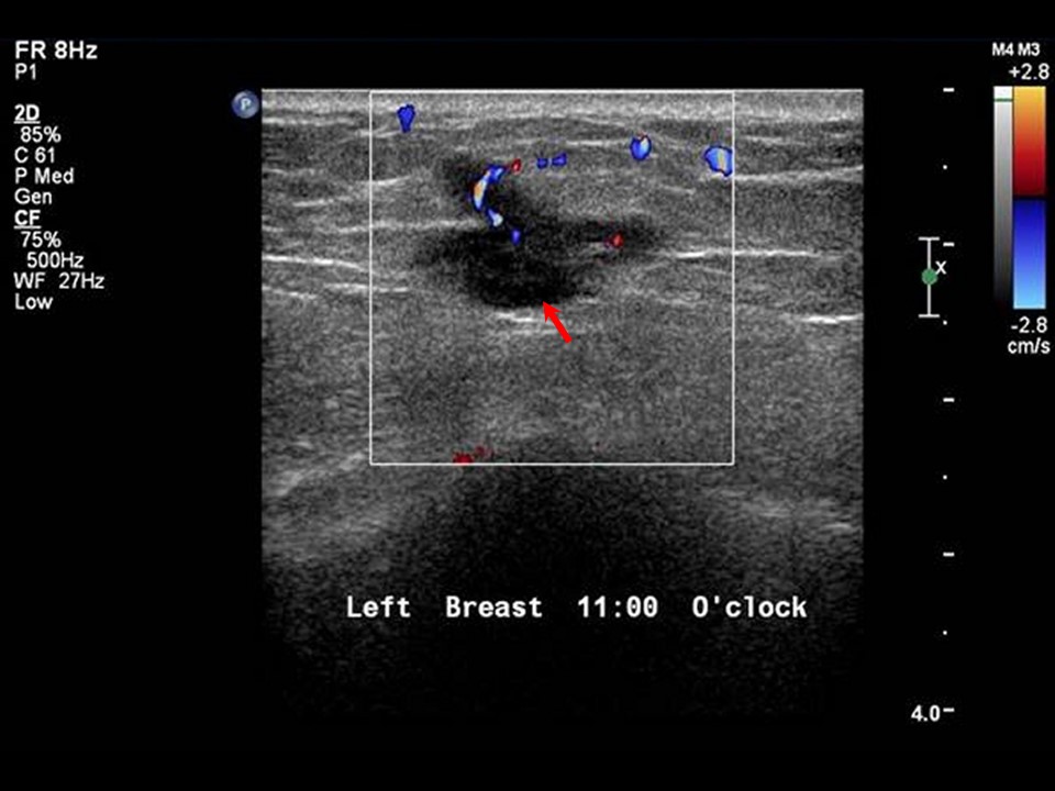
Ultrasound:
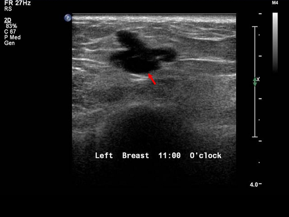 |  |
| Ultrasound features: Left breast, upper inner quadrant at 11 oclock | |
| ‣ Mass | |
| • Location: | Left breast, upper inner quadrant at 11 oclock |
| • Number: | 1 |
| • Size: | 1.4 × 1.1 cm |
| • Shape: | Irregular |
| • Orientation: | Not parallel |
| • Margins: | Angular |
| • Echo pattern: | Hypoechoic |
| • Posterior features: | No posterior features |
| ‣ Calcifications: | None |
| ‣ Associated features: | Internal vascularity |
| ‣ Special cases: | None |
BI-RADS:
BI-RADS Category: 5 (highly suggestive of malignancy)Further assessment:
Further assessment advised: Referral for cytologyCytology:
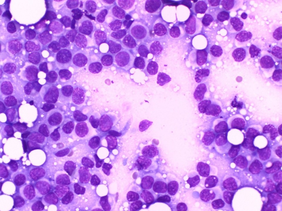 |
| Cytology features: | |
| ‣ Type of sample: | FNAC |
| ‣ Site of biopsy: | |
| • Laterality: | Left |
| • Quadrant: | |
| • Localization technique: | Palpation |
| • Nature of aspirate: | whitish |
| ‣ Cytological description: | Highly cellular smear reveals loosely cohesive sheets of malignant cells, which are large with high N:C ratio and hyperchromatic nuclei with coarse chromatin and occasional prominent nucleoli. Cytoplasm is moderate with occasional vacuolations |
| ‣ Reporting category: | Malignant |
| ‣ Diagnosis: | Carcinoma |
| ‣ Comments: | None |
Histopathology:
Lumpectomy
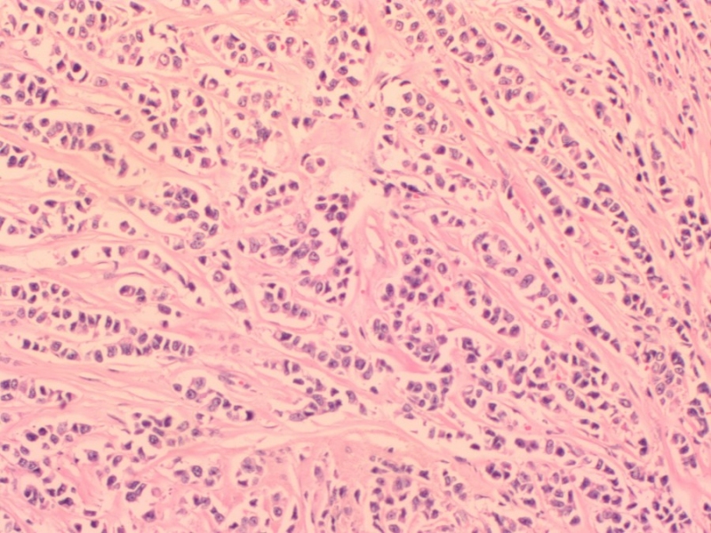 |
| Histopathology features: | |
| ‣ Specimen type: | Lumpectomy |
| ‣ Laterality: | Left |
| ‣ Macroscopy: | Specimen 6.0 × 4.0 × 2.5 cm with a skin flap 4.0 × 1.5 cm. On serial sectioning, firm greyish white tumour (1.8 × 1.0 × 1.7 cm) is identified. It is located 0.8 cm from the skin (anterior margin), 0.8 cm from the posterior margin, 1.5 cm from the superior margin, 1.7 cm from the inferior margin, 1.5 cm from the medial margin, and 2.0 cm from the lateral margin |
| ‣ Histological type: | Infiltrating lobular carcinoma |
| ‣ Histological grade: | Grade 2 (3 + 2 + 1 = 6) |
| ‣ Mitosis: | 5 |
| ‣ Maximum invasive tumour size: | 1.8 cm in greatest dimension |
| ‣ Lymph node status: | 0/20 |
| ‣ Peritumoural lymphovascular invasion: | Present |
| ‣ DCIS/EIC: | Absent; lobular carcinoma in situ not seen |
| ‣ Margins: | Free of tumour, 0.8 cm from the anterior and posterior margins |
| ‣ Pathological stage: | pT1N0 |
| ‣ Biomarkers: | |
| ‣ Comments: |
Case summary:
| Postmenopausal woman presented with left breast lump. Diagnosed as left breast carcinoma, BI-RADS 5 on imaging, as breast carcinoma on cytology, and as invasive lobular carcinoma, pT1N0 on histopathology. |
Learning points:
|