


Home / Training / Manuals / Atlas of breast cancer early detection / Cases
Atlas of breast cancer early detection
Go back to the list of case studies
.png) Click on the pictures to magnify and display the legends
Click on the pictures to magnify and display the legends
| Case number: | 099 |
| Age: | 77 |
| Clinical presentation: | Woman was diagnosed with right breast carcinoma and underwent MRM in 2000. She noticed a left breast lump 68 months ago. On clinical examination, a hard lump was palpable in the upper quadrant of the left breast. |
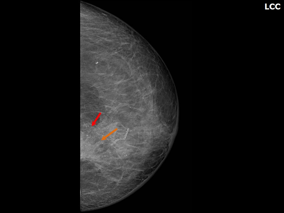
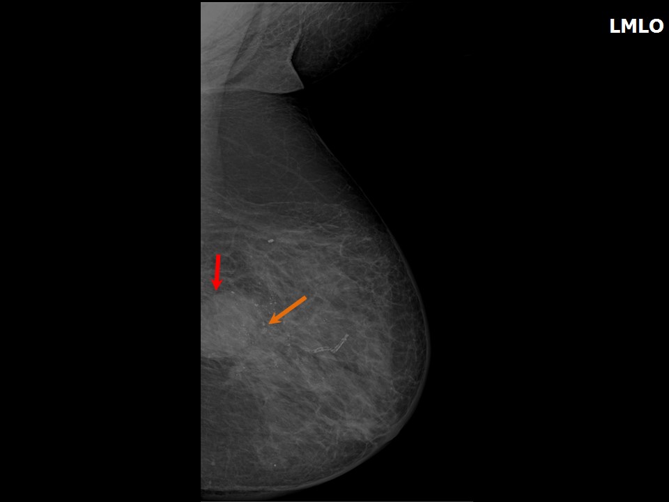
Mammography:
 |  |
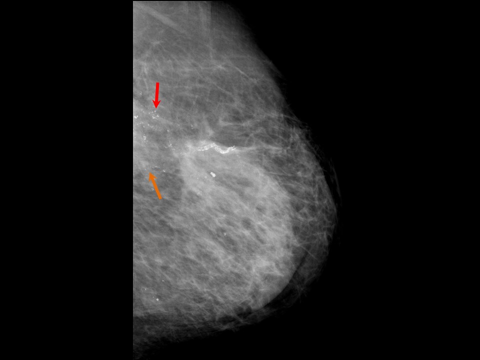 |
| Breast composition: | ACR category b (there are scattered areas of fibroglandular density) | Mammography features: |
| ‣ Location of the lesion: | Left breast, upper inner quadrant at 1011 oclock, posterior third |
| ‣ Mass: | |
| • Number: | 1 |
| • Size: | 4.5 × 3.4 cm |
| • Shape: | Irregular |
| • Margins: | Indistinct |
| • Density: | High |
| ‣ Calcifications: | |
| • Typically benign: | None |
| • Suspicious: | Fine pleomorphic |
| • Distribution: | Regional |
| ‣ Architectural distortion: | Present |
| ‣ Asymmetry: | Focal |
| ‣ Intramammary node: | None |
| ‣ Skin lesion: | None |
| ‣ Solitary dilated duct: | None |
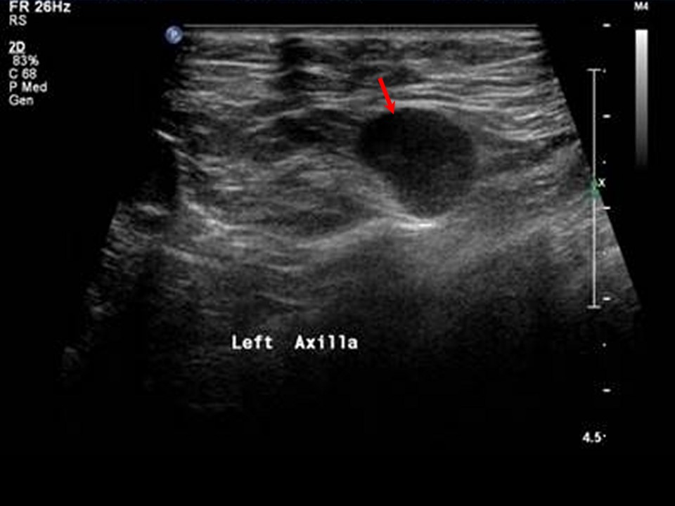
| ‣ Associated features: | Trabecular thickening, architectural distortion, pleomorphic microcalcifications, skin thickening, and axillary adenopathy nodes with thickened cortex and loss of fatty hilum |
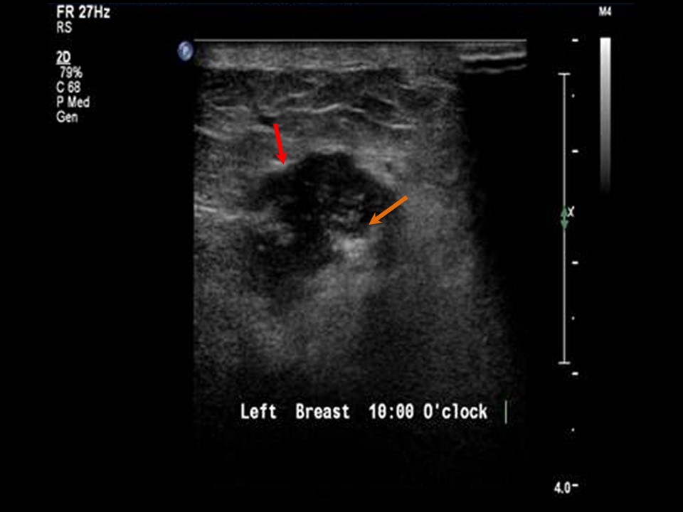
Ultrasound:
 | 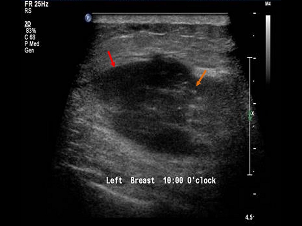 |
 |
| Ultrasound features: Left breast, upper inner quadrant at 1011 oclock position | |
| ‣ Mass | |
| • Location: | Left breast, upper inner quadrant at 1011 oclock position |
| • Number: | 23, grouped together |
| • Size: | Largest 4.5 × 2.5 cm |
| • Shape: | Irregular |
| • Orientation: | Not parallel |
| • Margins: | Angular |
| • Echo pattern: | Hypoechoic |
| • Posterior features: | No posterior features |
| ‣ Calcifications: | Present in mass |
| ‣ Associated features: | Architectural distortion, skin thickening, internal vascularity, oedema, and axillary lymphadenopathy |
| ‣ Special cases: | None |
BI-RADS:
BI-RADS Category (Right MRM): 5 (highly suggestive of malignancy)Further assessment:
Further assessment advised: Referral for cytology and for core biopsyCytology:
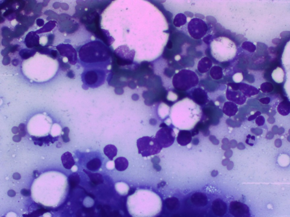 |
| Cytology features: | |
| ‣ Type of sample: | FNAC |
| ‣ Site of biopsy: | |
| • Laterality: | Left |
| • Quadrant: | Upper inner |
| • Localization technique: | Palpation |
| • Nature of aspirate: | Whitish |
| ‣ Cytological description: | Loosely cohesive sheets of large malignant cells with marked nuclear pleomorphism |
| ‣ Reporting category: | Malignant |
| ‣ Diagnosis: | Carcinoma high grade |
| ‣ Comments: | None |
Histopathology:
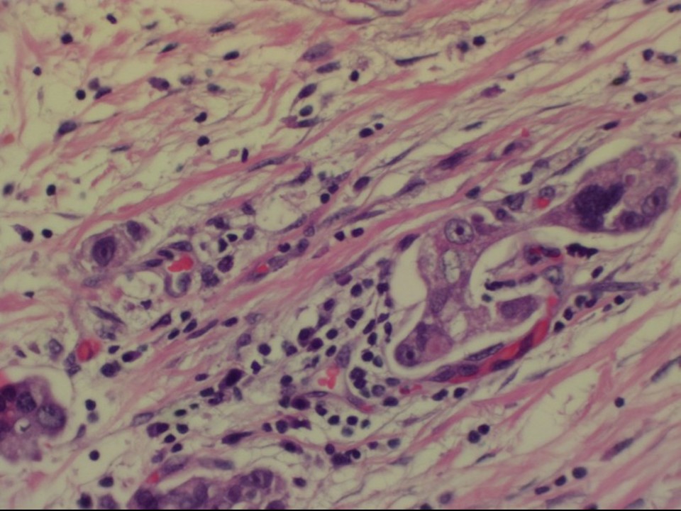
Core needle biopsy
 |
| Histopathology features: | |
| ‣ Specimen type: | Core needle biopsy |
| ‣ Laterality: | Left |
| ‣ Macroscopy: | 5 cores |
| ‣ Histological type: | Invasive carcinoma of no special type |
| ‣ Histological grade: | Grade 3 (3 + 3 + 2 = 8) |
| ‣ Mitosis: | 18 |
| ‣ Maximum invasive tumour size: | |
| ‣ Lymph node status: | |
| ‣ Peritumoural lymphovascular invasion: | |
| ‣ DCIS/EIC: | |
| ‣ Margins: | |
| ‣ Pathological stage: | |
| ‣ Biomarkers: | |
| ‣ Comments: |
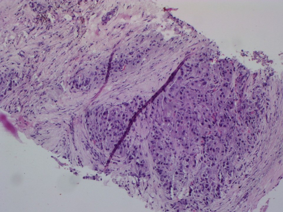
MRM
 | 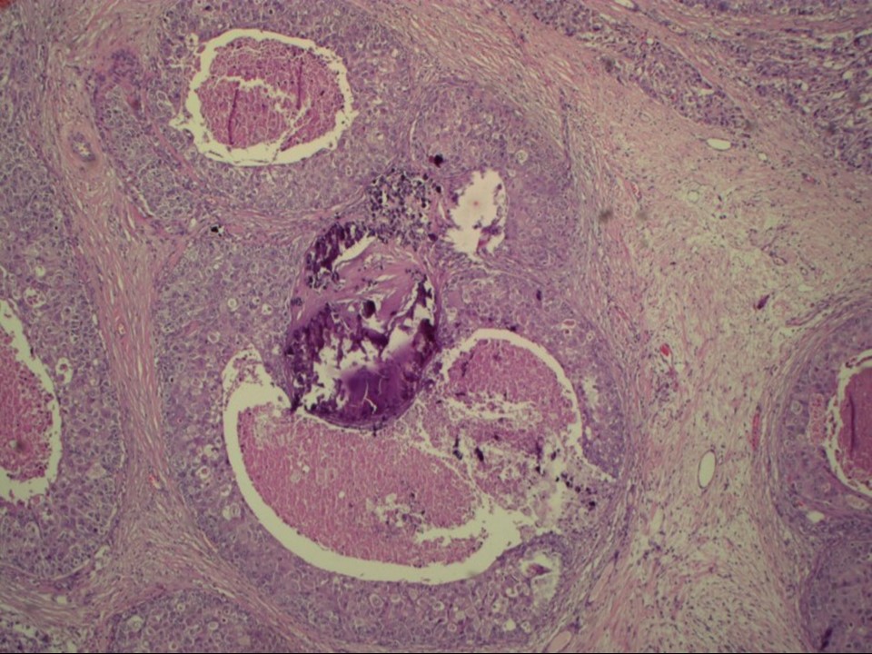 |
| Histopathology features: | |
| ‣ Specimen type: | MRM |
| ‣ Laterality: | Left |
| ‣ Macroscopy: | On serial sectioning, a firm greyish white tumour (6.8 × 4.5 × 2.8 cm) is identified. It is located 1.4 cm below the skin and 0.2 cm from the base |
| ‣ Histological type: | Invasive carcinoma of no special type |
| ‣ Histological grade: | Grade 3 (3 + 3 + 3 = 9) |
| ‣ Mitosis: | 22 |
| ‣ Maximum invasive tumour size: | 6.8 cm |
| ‣ Lymph node status: | 3/19 |
| ‣ Peritumoural lymphovascular invasion: | Present |
| ‣ DCIS/EIC: | Comedo DCIS high grade |
| ‣ Margins: | Free of tumour |
| ‣ Pathological stage: | pT3N3 |
| ‣ Biomarkers: | |
| ‣ Comments: | The remaining breast tissue shows extensive areas of fibrosis |
Case summary:
| Postmenopausal woman, operated 15 years ago for right breast carcinoma, presented with a large, hard left breast lump. Diagnosed as left breast carcinoma with clustered pleomorphic microcalcifications, BI-RADS 5 on imaging, as carcinoma on cytology, and as invasive breast carcinoma of no special type, pT3N3 on histopathology. |
Learning points:
|