


Home / Training / Manuals / Atlas of breast cancer early detection / Cases
Atlas of breast cancer early detection
Go back to the list of case studies
.png) Click on the pictures to magnify and display the legends
Click on the pictures to magnify and display the legends
| Case number: | 004 |
| Age: | 78 |
| Clinical presentation: | Postmenopausal woman with average risk of developing breast cancer presented with a large lump in the right breast. On examination, she had a large firm right breast lump. |
Mammography:
 |  |
| Breast composition: | ACR Category a (the breasts are almost entirely fatty) | Mammography features: |
| ‣ Location of the lesion: | Right breast, upper inner quadrant at 121 oclock, middle third |
| ‣ Mass: | |
| • Number: | 1 |
| • Size: | 3 cm |
| • Shape: | Oval |
| • Margins: | Circumscribed |
| • Density: | Equal |
| ‣ Calcifications: | |
| • Typically benign: | None |
| • Suspicious: | None |
| • Distribution: | None |
| ‣ Architectural distortion: | None |
| ‣ Asymmetry: | None |
| ‣ Intramammary node: | None |
| ‣ Skin lesion: | None |
| ‣ Solitary dilated duct: | None |
| ‣ Associated features: | None |
| Breast composition: | ACR category a (the breasts are almost entirely fatty) | Mammography features: |
| ‣ Location of the lesion: | Left breast, upper outer quadrant at 1 oclock, middle third |
| ‣ Mass: | |
| • Number: | 2 |
| • Size: | Largest 0.5 cm |
| • Shape: | Round |
| • Margins: | Circumscribed |
| • Density: | Equal |
| ‣ Calcifications: | |
| • Typically benign: | None |
| • Suspicious: | None |
| • Distribution: | None |
| ‣ Architectural distortion: | None |
| ‣ Asymmetry: | None |
| ‣ Intramammary node: | None |
| ‣ Skin lesion: | None |
| ‣ Solitary dilated duct: | None |
| ‣ Associated features: | None |
Ultrasound:
 |  |
 |
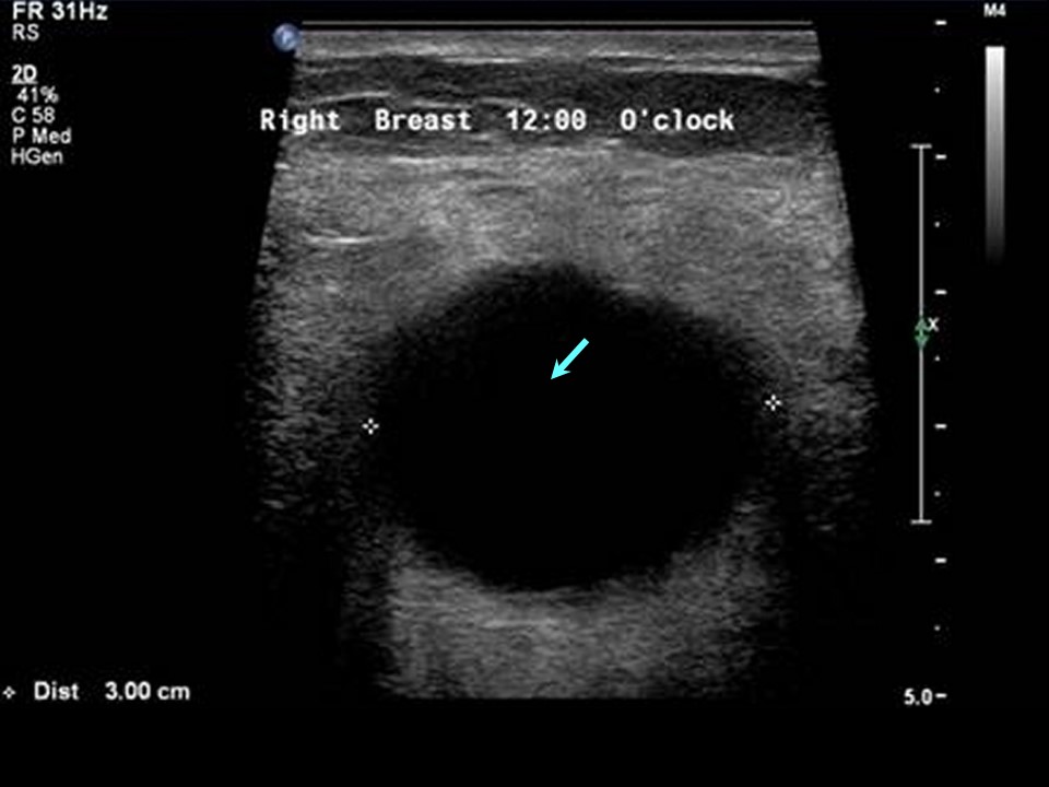
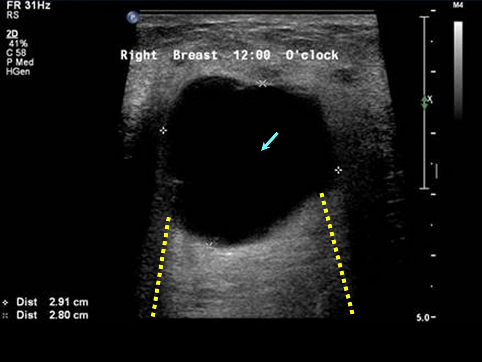
| Ultrasound features: Right breast, central portion of the breast at 12 oclock | |
| ‣ Mass | |
| • Location: | Right breast, central portion of the breast at 12 oclock |
| • Number: | Multiple |
| • Size: | Largest 3.0 cm |
| • Shape: | Round |
| • Orientation: | Parallel |
| • Margins: | Circumscribed |
| • Echo pattern: | Anechoic |
| • Posterior features: | Posterior shadowing |
| ‣ Calcifications: | None |
| ‣ Associated features: | None |
| ‣ Special cases: | Simple cyst |
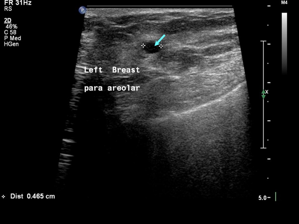
| Ultrasound features: Left breast, central portion of the breast at 1 oclock | |
| ‣ Mass | |
| • Location: | Left breast, central portion of the breast at 1 oclock |
| • Number: | Multiple |
| • Size: | Largest 0.5 cm in greatest dimension |
| • Shape: | Oval |
| • Orientation: | Parallel |
| • Margins: | Circumscribed |
| • Echo pattern: | Anechoic |
| • Posterior features: | Posterior shadowing |
| ‣ Calcifications: | None |
| ‣ Associated features: | None |
| ‣ Special cases: | Simple cyst |
BI-RADS:
BI-RADS Category: 2 (benign)Further assessment:
Further assessment advised: Referral for cytologyCytology:
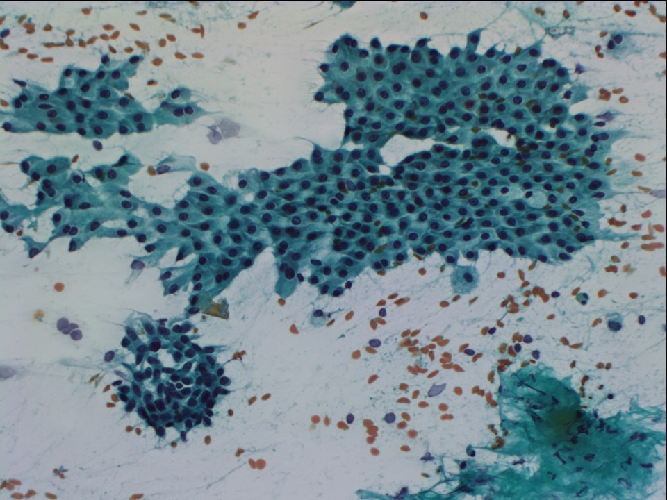 | 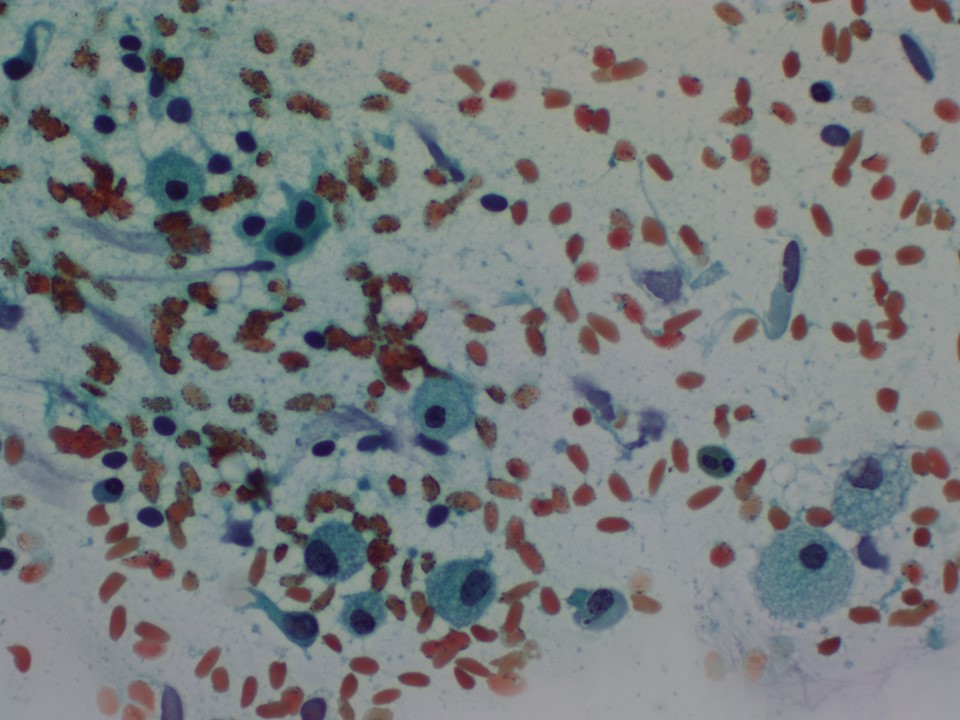 |
| Cytology features: | |
| ‣ Type of sample: | FNAC done on four occasions |
| ‣ Site of biopsy: | |
| • Laterality: | Right |
| • Quadrant: | Upper and central |
| • Localization technique: | Palpation |
| • Nature of aspirate: | 26 mL of yellow fluid aspirated each time |
| ‣ Cytological description: | Smears show tight clusters of benign ductal epithelial cells, some with apocrine metaplasia and many foamy histiocytes on a proteinaceous background |
| ‣ Reporting category: | Benign |
| ‣ Diagnosis: | Proliferative fibrocystic change |
| ‣ Comments: | None |
Histopathology:
Lump excision
 | 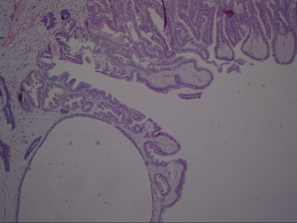 |
| Histopathology features: | |
| ‣ Specimen type: | Lump excision |
| ‣ Laterality: | Right |
| ‣ Macroscopy: | Irregular fibrofatty tissue bit (4.5 × 3.5 × 2.5 cm) with small piece of skin on one side (2.0 × 0.7 cm). Cut surface is yellowish, soft to firm, with a few whitish fibrous areas ranging in size from 0.3 cm to 1.0 cm and small cysts ranging in size from 0.3 cm to 0.5 cm in largest dimension |
| ‣ Histological type: | Many of the ducts are cystically dilated, some lined by flattened epithelium; a few show mild epitheliosis and papillary apocrine hyperplasia |
| ‣ Histological grade: | |
| ‣ Mitosis: | |
| ‣ Maximum invasive tumour size: | |
| ‣ Lymph node status: | |
| ‣ Peritumoural lymphovascular invasion: | |
| ‣ DCIS/EIC: | Not identified |
| ‣ Margins: | |
| ‣ Pathological stage: | |
| ‣ Biomarkers: | |
| ‣ Comments: | Negative for ADH/DCIS/malignancy |
Case summary:
| Postmenopausal woman presented with right breast lump. Diagnosed as multiple simple cysts in both breasts, BI-RADS 2 on imaging and as proliferative fibrocystic change on cytology and histopathology. |
Learning points:
|
Cysts in older women:
|