


Home / Training / Manuals / Atlas of breast cancer early detection / Cases
Atlas of breast cancer early detection
Go back to the list of case studies
.png) Click on the pictures to magnify and display the legends
Click on the pictures to magnify and display the legends
| Case number: | 018 |
| Age: | 56 |
| Clinical presentation: | Postmenopausal woman presented with a lump in the right breast, which she had had for many years. She had an increased risk of developing breast cancer with family history of breast cancer. Examination revealed a firm 2 cm lump in the upper outer quadrant of the right breast. |
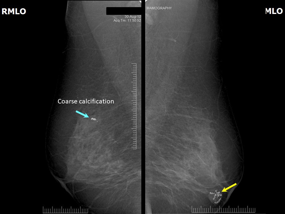
Mammography:
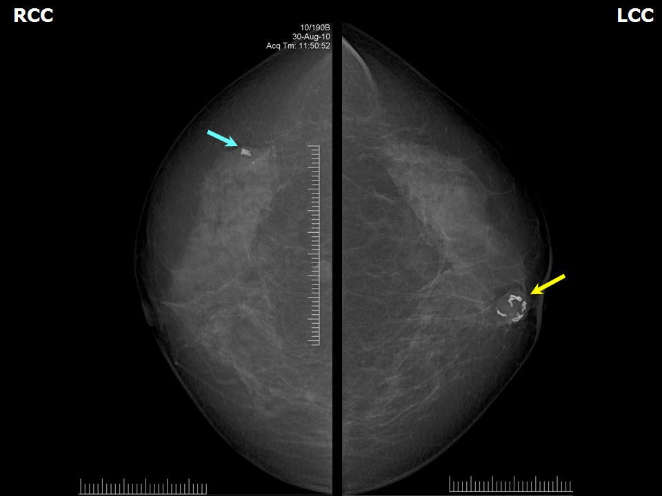 |  |
| Breast composition: | ACR category b (there are scattered areas of fibroglandular density) | Mammography features: |
| ‣ Location of the lesion: | Right breast, upper outer quadrant at 10 oclock, middle third |
| ‣ Mass: | |
| • Number: | 0 |
| • Size: | None |
| • Shape: | None |
| • Margins: | None |
| • Density: | None |
| ‣ Calcifications: | |
| • Typically benign: | Coarse |
| • Suspicious: | None |
| • Distribution: | None |
| ‣ Architectural distortion: | None |
| ‣ Asymmetry: | None |
| ‣ Intramammary node: | None |
| ‣ Skin lesion: | None |
| ‣ Solitary dilated duct: | None |
| ‣ Associated features: | Calcifications |
| Breast composition: | ACR category b (there are scattered areas of fibroglandular density) | Mammography features: |
| ‣ Location of the lesion: | Left breast, central portion of the breast, anterior third |
| ‣ Mass: | |
| • Number: | 1 |
| • Size: | 1.5 × 1.3 cm |
| • Shape: | Oval |
| • Margins: | Circumscribed |
| • Density: | Equal |
| ‣ Calcifications: | |
| • Typically benign: | Popcorn-like |
| • Suspicious: | None |
| • Distribution: | Within mass |
| ‣ Architectural distortion: | None |
| ‣ Asymmetry: | None |
| ‣ Intramammary node: | None |
| ‣ Skin lesion: | None |
| ‣ Solitary dilated duct: | None |
| ‣ Associated features: | Calcifications |
Ultrasound:
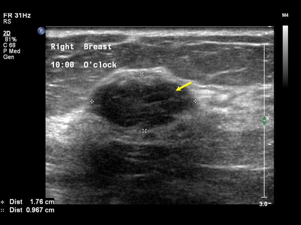 | 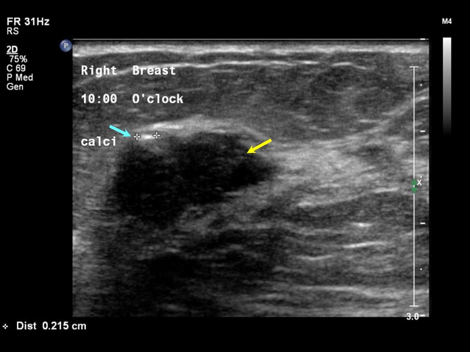 |
| Ultrasound features: Right breast, upper outer quadrant at 10 oclock | |
| ‣ Mass | |
| • Location: | Right breast, upper outer quadrant at 10 oclock |
| • Number: | 1 |
| • Size: | 1.8 × 1.0 cm |
| • Shape: | Oval |
| • Orientation: | Parallel |
| • Margins: | Circumscribed |
| • Echo pattern: | Hypoechoic |
| • Posterior features: | No posterior features |
| ‣ Calcifications: | Present in mass |
| ‣ Associated features: | None |
| ‣ Special cases: | None |
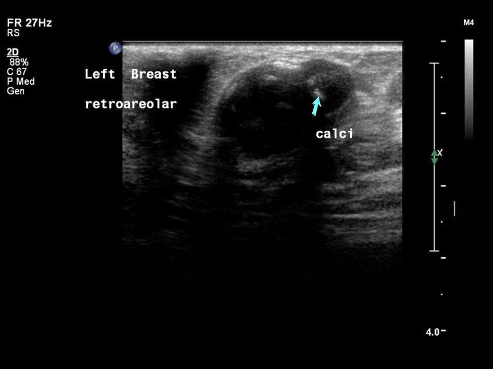 | 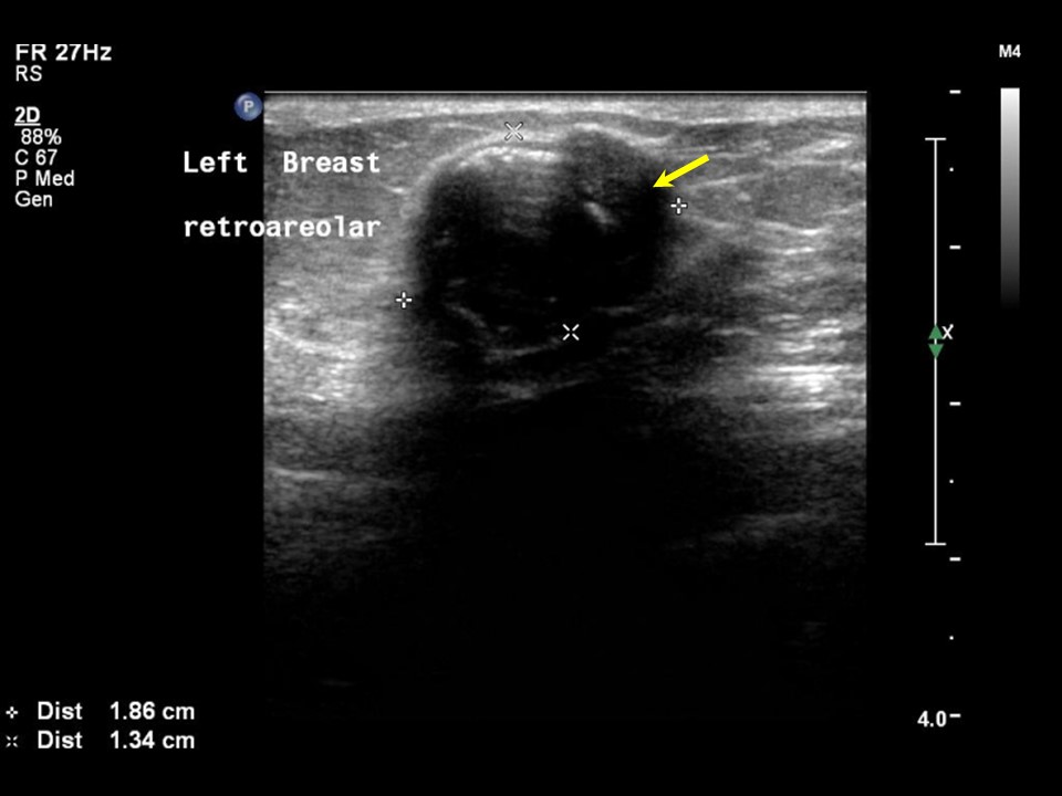 |
| Ultrasound features: Left breast, central portion of the breast | |
| ‣ Mass | |
| • Location: | Left breast, central portion of the breast |
| • Number: | 1 |
| • Size: | 2.0 × 1.3 cm |
| • Shape: | Oval |
| • Orientation: | Parallel |
| • Margins: | Circumscribed |
| • Echo pattern: | Hypoechoic |
| • Posterior features: | Posterior shadowing |
| ‣ Calcifications: | Present in mass |
| ‣ Associated features: | None |
| ‣ Special cases: | None |
BI-RADS:
BI-RADS Category: 2 (benign)Case summary:
| Postmenopausal woman presented with right breast lump. Diagnosed as involuting (partially calcified) fibroadenoma in both breasts, BI-RADS 2 on imaging. |
Learning points:
|