


Home / Training / Manuals / Atlas of breast cancer early detection / Cases
Atlas of breast cancer early detection
Go back to the list of case studies
.png) Click on the pictures to magnify and display the legends
Click on the pictures to magnify and display the legends
| Case number: | 178 |
| Age: | 46 |
| Clinical presentation: | Premenopausal woman with average risk of developing breast cancer presented with an area of firmness in the upper inner quadrant of the left breast first noticed a few days ago. |
Mammography:
 |  |
 |  |
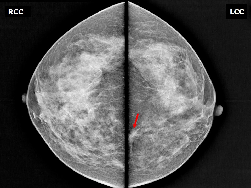
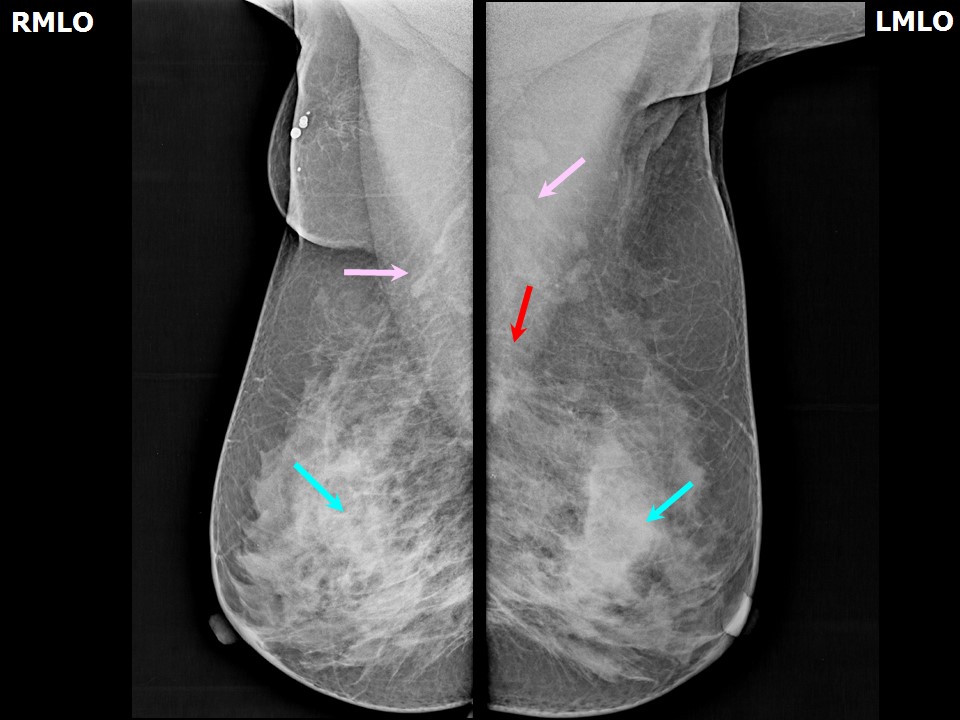
| Breast composition: | ACR category c (the breasts are heterogeneously dense, which may obscure small masses) | Mammography features: |
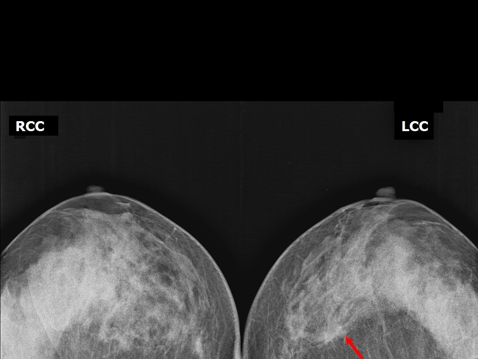
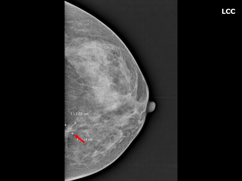
| ‣ Location of the lesion: | Left breast, upper inner quadrant at 11 oclock, posterior third |
| ‣ Mass: | |
| • Number: | 1 |
| • Size: | 2.0 × 1.2 cm |
| • Shape: | Irregular |
| • Margins: | Spiculated |
| • Density: | Equal |
| ‣ Calcifications: | |
| • Typically benign: | None |
| • Suspicious: | None |
| • Distribution: | None |
| ‣ Architectural distortion: | Present |
| ‣ Asymmetry: | None |
| ‣ Intramammary node: | None |
| ‣ Skin lesion: | None |
| ‣ Solitary dilated duct: | None |
| ‣ Associated features: | Architectural distortion with focal spiculations |
Ultrasound:
 |  |
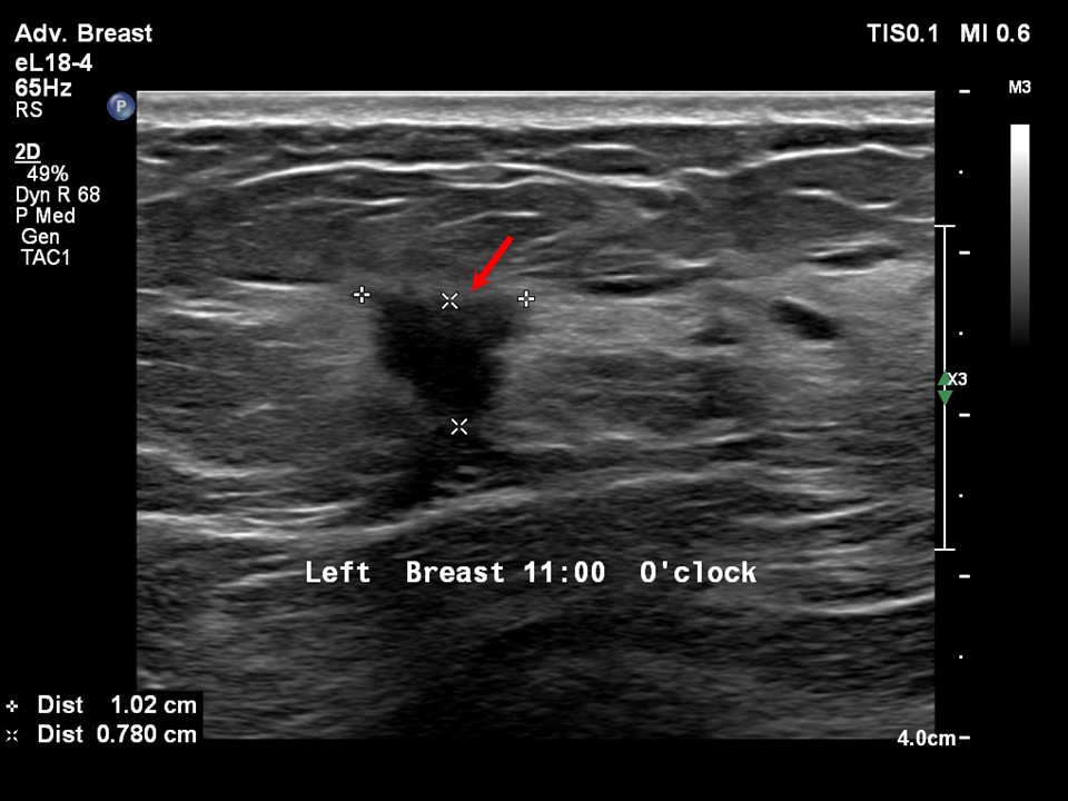 |  |
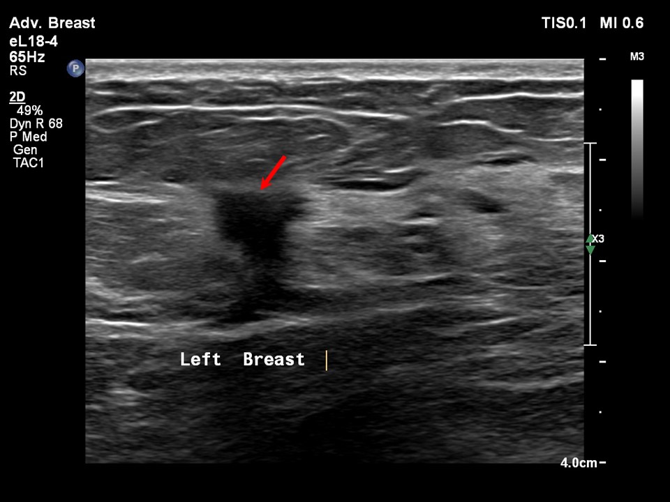
| Ultrasound features: Left breast, upper inner quadrant at 11 oclock, 8.0 cm from the nipple and at 1.8 cm skin depth | |
| ‣ Mass | |
| • Location: | Left breast, upper inner quadrant at 11 oclock, 8.0 cm from the nipple and at 1.8 cm skin depth |
| • Number: | 1 |
| • Size: | 1.0 × 0.8 cm |
| • Shape: | Irregular |
| • Orientation: | Not parallel |
| • Margins: | Spiculated |
| • Echo pattern: | Isoechoic |
| • Posterior features: | No posterior features |
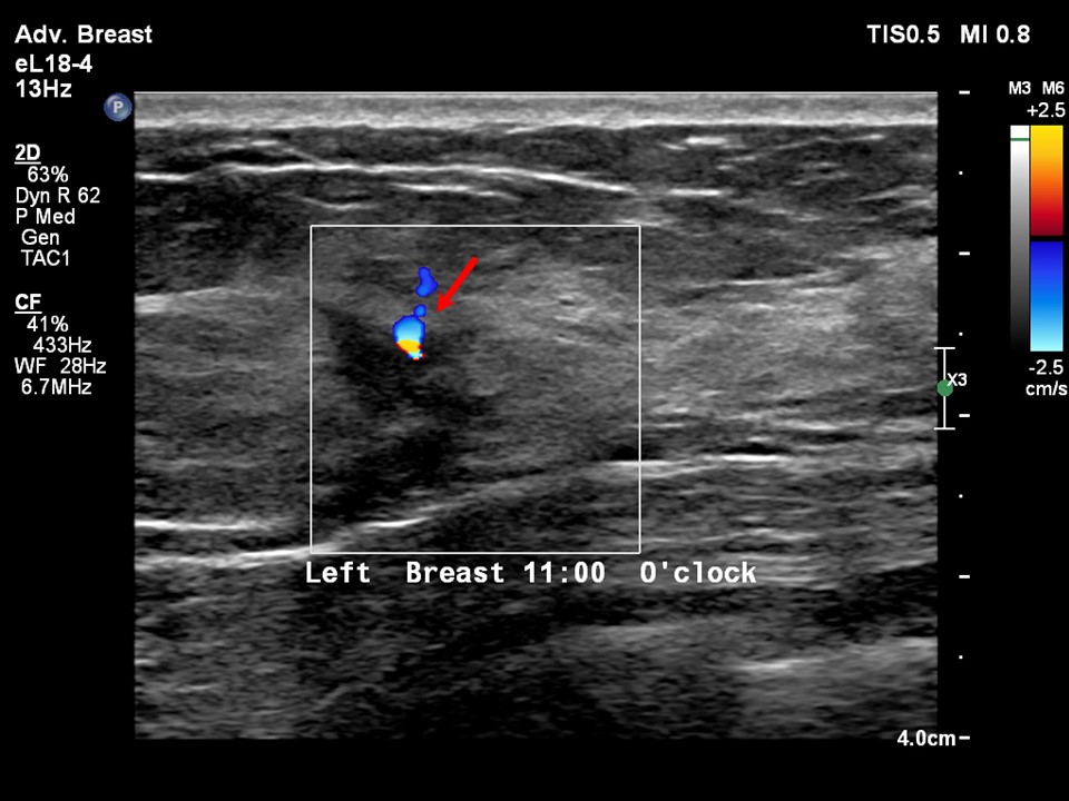
| ‣ Calcifications: | None |
| ‣ Associated features: | Increased central vascularity |
| ‣ Special cases: | None |
BI-RADS:
BI-RADS Category: 4C (high suspicion for malignancy)Further assessment:
Further assessment advised: Referral for cytologyCytology:
 |
| Cytology features: | |
| ‣ Type of sample: | FNAC |
| ‣ Site of biopsy: | |
| • Laterality: | Left |
| • Quadrant: | Upper inner |
| • Localization technique: | Ultrasound-guided FNAC |
| • Nature of aspirate: | Whitish |
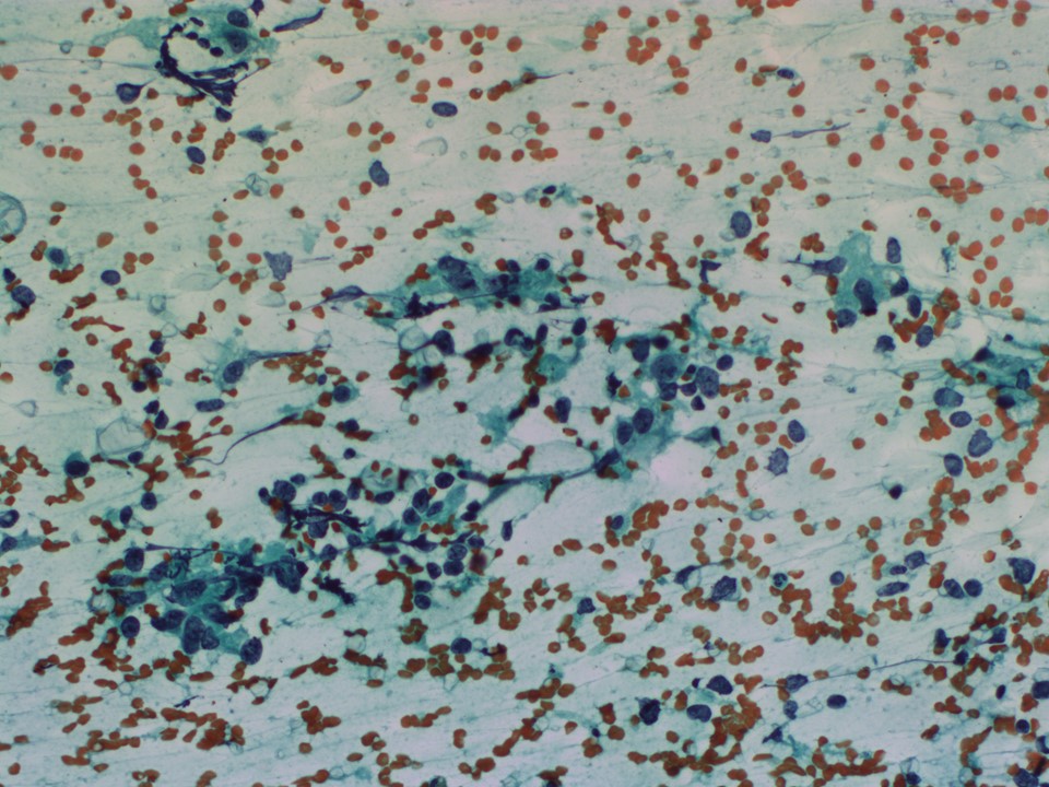
| ‣ Cytological description: | Smears from first pass show proteinaceous fluid, foamy macrophages and benign ductal epithelial cells. Smears from second pass show loosely cohesive sheets of ductal epithelial cells showing high N:C ratio and hyperchromasia in places. Cytoplasm is moderate in amount and shows a few vacuoles |
| ‣ Reporting category: | Suspicious, probably in situ or invasive carcinoma |
| ‣ Diagnosis: | Suspicious, probably in situ or invasive carcinoma |
| ‣ Comments: | None |
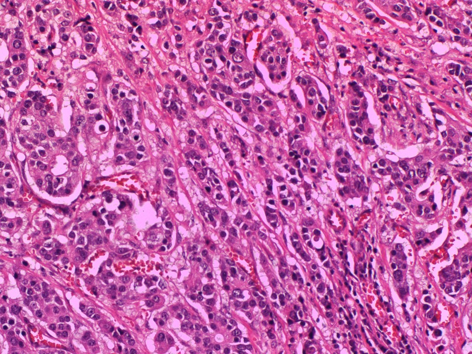
Histopathology:
Lumpectomy
 |  |
| Histopathology features: | |
| ‣ Specimen type: | Lumpectomy |
| ‣ Laterality: | Left |
| ‣ Macroscopy: | Lumpectomy specimen (6.0 × 5.0 × 4.0 cm) oriented with long suture laterally and short suture superiorly. Skin flap (2.7 × 2.8 cm). On serial sectioning, an ill-defined whitish tumour (1.7 × 1.4 × 1.0 cm) with adjacent fibrosed areas was identified. It is located 2.4 cm from the skin (anterior margin), 1.6 cm from the posterior margin, 1.7 cm from the superior margin, 0.8 cm from the inferior margin, 1.1 cm from the medial margin and 1.8 cm from the lateral margin. The remaining breast parenchyma is normal |
| ‣ Histological type: | Invasive breast carcinoma of no special type |
| ‣ Histological grade: | Grade 2 (2 + 3 + 1 = 6) |
| ‣ Mitosis: | 6 |
| ‣ Maximum invasive tumour size: | 1.7 cm |
| ‣ Lymph node status: | 0/20 |
| ‣ Peritumoural lymphovascular invasion: | Absent |
| ‣ DCIS/EIC: | DCIS cribriform type, low grade mainly |
| ‣ Margins: | Free of tumour |
| ‣ Pathological stage: | pT1cNo |
| ‣ Biomarkers: | ER: Positive (100% of cells show nuclear staining with moderate intensity. Allred score 7/8). PR: Positive (100% of cells show nuclear staining with moderate intensity. Allred score 7/8). HER2: Equivocal (score 2). Testing for HER2 by fluorescence in situ hybridization was suggested because of the equivocal score |
| ‣ Comments: | The adjacent breast shows fibrocystic change, apocrine and columnar metaplasia, and areas of ADH and UDH |
Case summary:
| Premenopausal woman presented with a firm area in the left breast. Diagnosed as left breast architectural distortion with spiculations from a point, BI-RADS 4C on imaging, as suspicious for malignancy on cytology, and as invasive breast carcinoma of no special type, pT1cN0 along with fibrocystic disease on histopathology. |
Learning points:
|