


Home / Training / Manuals / Atlas of breast cancer early detection / Cases
Atlas of breast cancer early detection
Go back to the list of case studies
.png) Click on the pictures to magnify and display the legends
Click on the pictures to magnify and display the legends
| Case number: | 118 |
| Age: | 75 |
| Clinical presentation: | Postmenopausal woman who was a known case of bilateral breast cancer and had undergone right breast-conserving surgery, left MRM, and a reconstruction with rectus abdominis muscle presented for a follow-up mammography. |
Mammography:
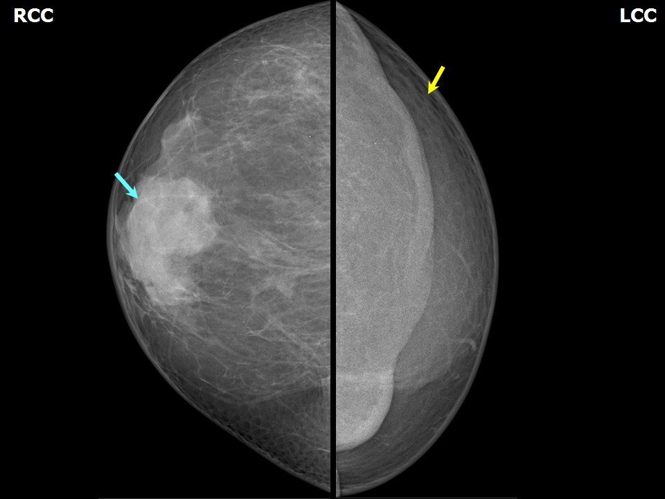 | 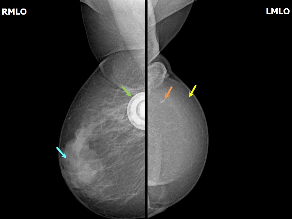 |
| Breast composition: | ACR category b (there are scattered areas of fibroglandular density) | Mammography features: |
| ‣ Location of the lesion: | Right breast, upper quadrant breast-conserving surgery scar tissue with chemoport in situ |
| ‣ Mass: | |
| • Number: | 0 |
| • Size: | No |
| • Shape: | None |
| • Margins: | None |
| • Density: | None |
| ‣ Calcifications: | |
| • Typically benign: | None |
| • Suspicious: | None |
| • Distribution: | None |
| ‣ Architectural distortion: | None |
| ‣ Asymmetry: | None |
| ‣ Intramammary node: | None |
| ‣ Skin lesion: | None |
| ‣ Solitary dilated duct: | None |
| ‣ Associated features: | None |
| Breast composition: | ACR category b (there are scattered areas of fibroglandular density) | Mammography features: |
| ‣ Location of the lesion: | Left breast, MRM status, reconstructed TRAM flap seen |
| ‣ Mass: | |
| • Number: | 0 |
| • Size: | No |
| • Shape: | None |
| • Margins: | None |
| • Density: | None |
| ‣ Calcifications: | |
| • Typically benign: | Solitary round calcification focus in upper quadrant of the flap |
| • Suspicious: | None |
| • Distribution: | None |
| ‣ Architectural distortion: | None |
| ‣ Asymmetry: | None |
| ‣ Intramammary node: | None |
| ‣ Skin lesion: | None |
| ‣ Solitary dilated duct: | None |
| ‣ Associated features: | None |
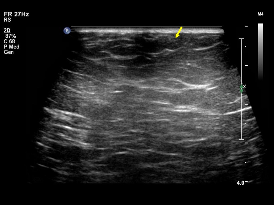
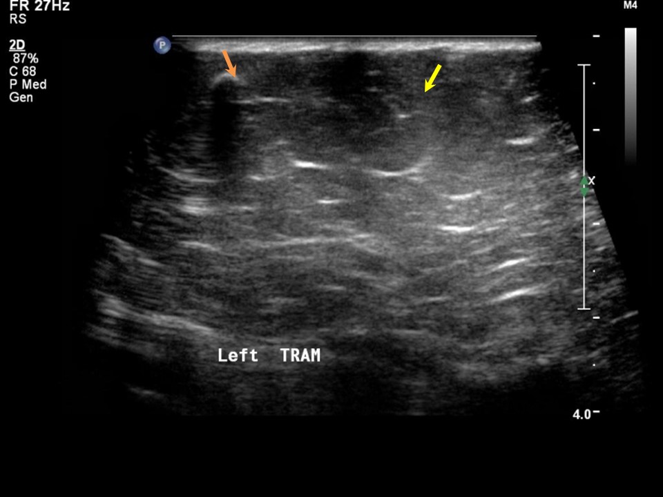
Ultrasound:
 |  |
BI-RADS:
BI-RADS Category: 2 (benign)Case summary:
| Postmenopausal woman, a known case of bilateral breast cancer who had undergone right breast-conserving surgery and left MRM with rectus abdominis muscle reconstruction, is on regular follow-up mammography. On imaging, heterogeneously dense breast parenchyma in the right breast and TRAM flap reconstruction of the left breast are seen. A chemoport is seen in situ in the right breast. |
Learning points:
| <
IARC, 150 Cours Albert Thomas, 69372 Lyon CEDEX 08, France - Tel: +33 (0)4 72 73 84 85 - Fax: +33 (0)4 72 73 85 75
© IARC 2024 - All Rights Reserved.
© IARC 2024 - All Rights Reserved.