


Home / Training / Manuals / Atlas of breast cancer early detection / Cases
Atlas of breast cancer early detection
Go back to the list of case studies
.png) Click on the pictures to magnify and display the legends
Click on the pictures to magnify and display the legends
| Case number: | 179 |
| Age: | 70 |
| Clinical presentation: | Postmenopausal woman with increased risk of developing breast cancer presented with a lump in the left axillary tail. The patient had ovarian carcinoma detected 1 year ago and had undergone a hysterectomy. |
Mammography:
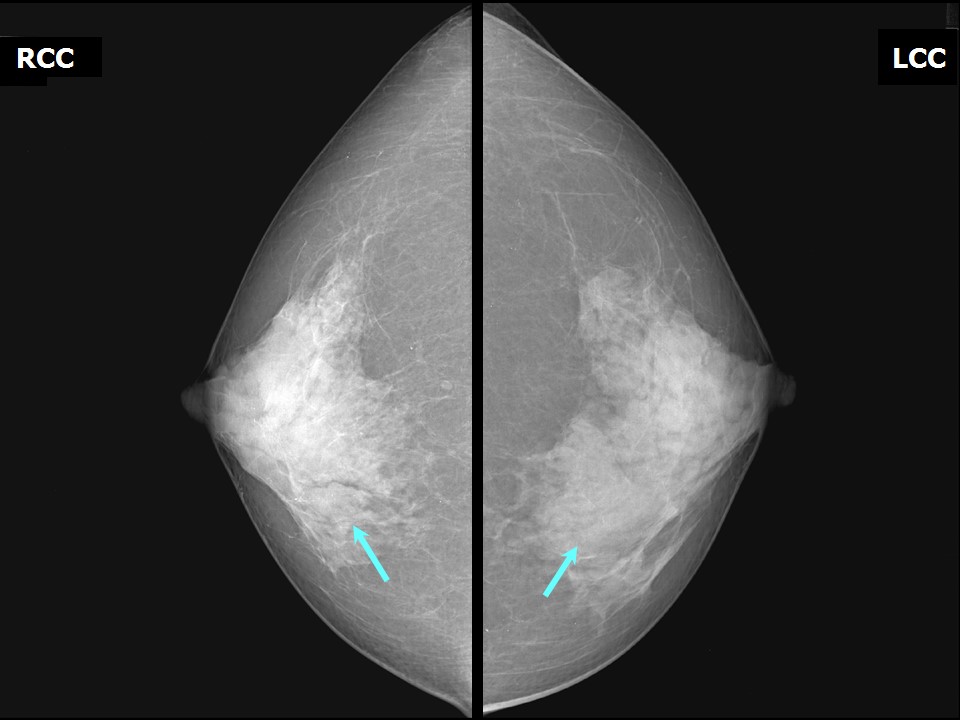 | 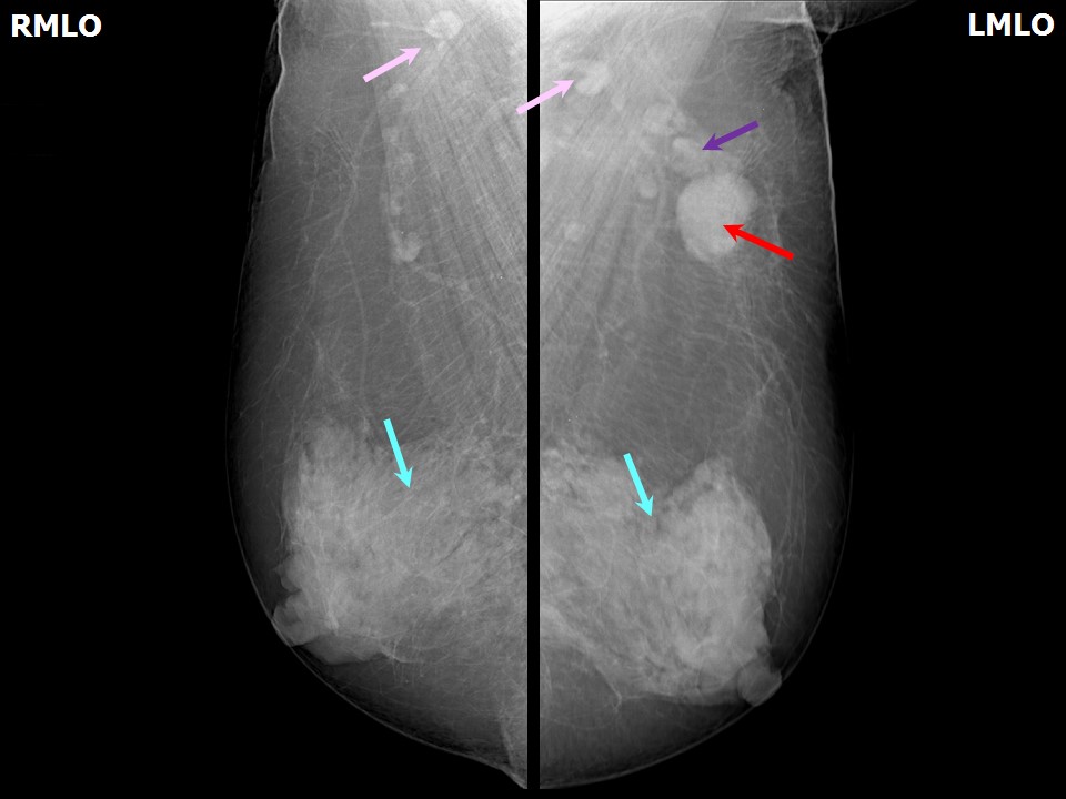 |
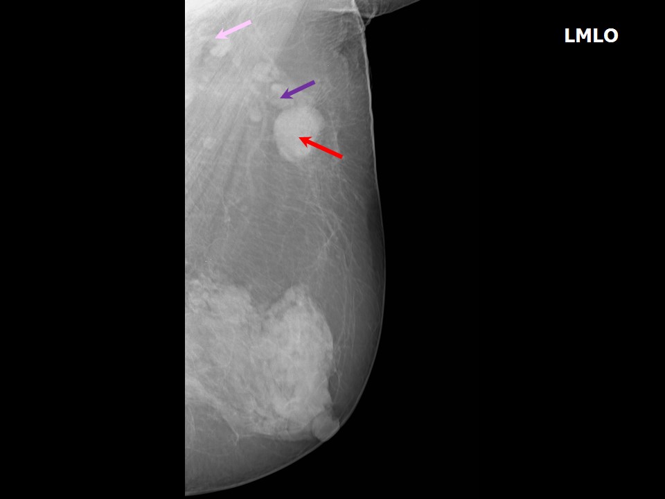 |
| Breast composition: | ACR category d (the breasts are extremely dense, which lowers the sensitivity of mammography) | Mammography features: |
| ‣ Location of the lesion: | Left axilla, enlarged axillary lymph node (2.4 × 1.4 cm) of altered morphology with loss of fatty hilum |
| ‣ Mass: | |
| • Number: | 1 |
| • Size: | None |
| • Shape: | None |
| • Margins: | None |
| • Density: | None |
| ‣ Calcifications: | |
| • Typically benign: | None |
| • Suspicious: | None |
| • Distribution: | None |
| ‣ Architectural distortion: | Present |
| ‣ Asymmetry: | None |
| ‣ Intramammary node: | None |
| ‣ Skin lesion: | None |
| ‣ Solitary dilated duct: | None |
| ‣ Associated features: | None |
Ultrasound:
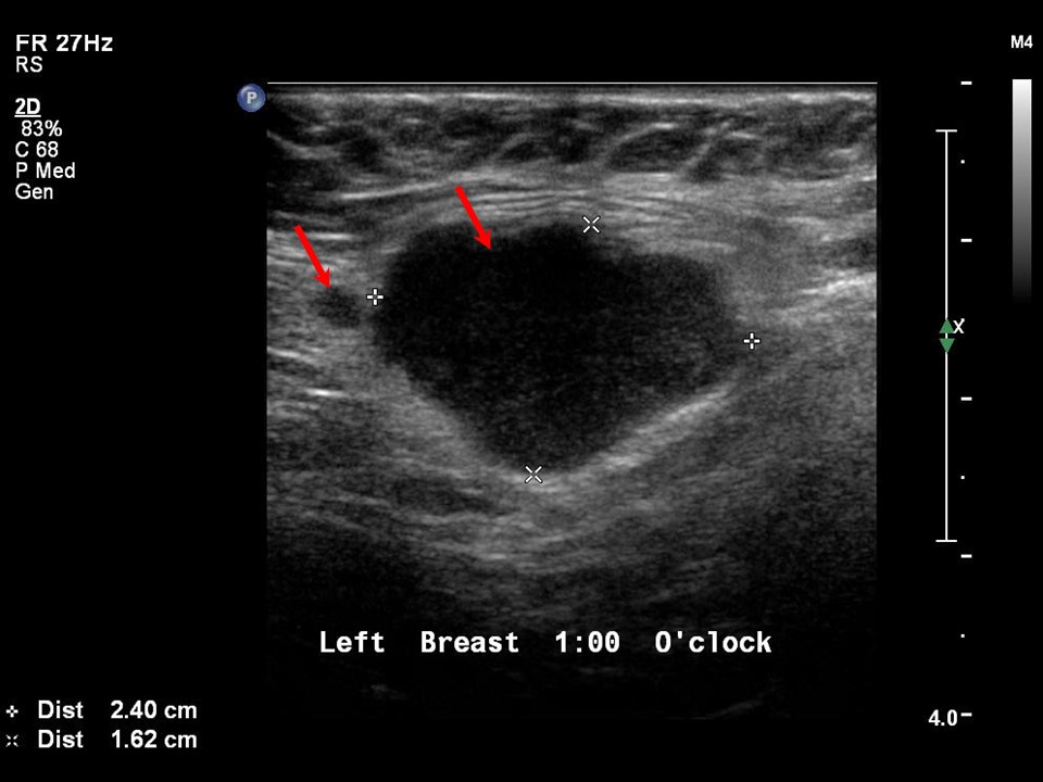 | 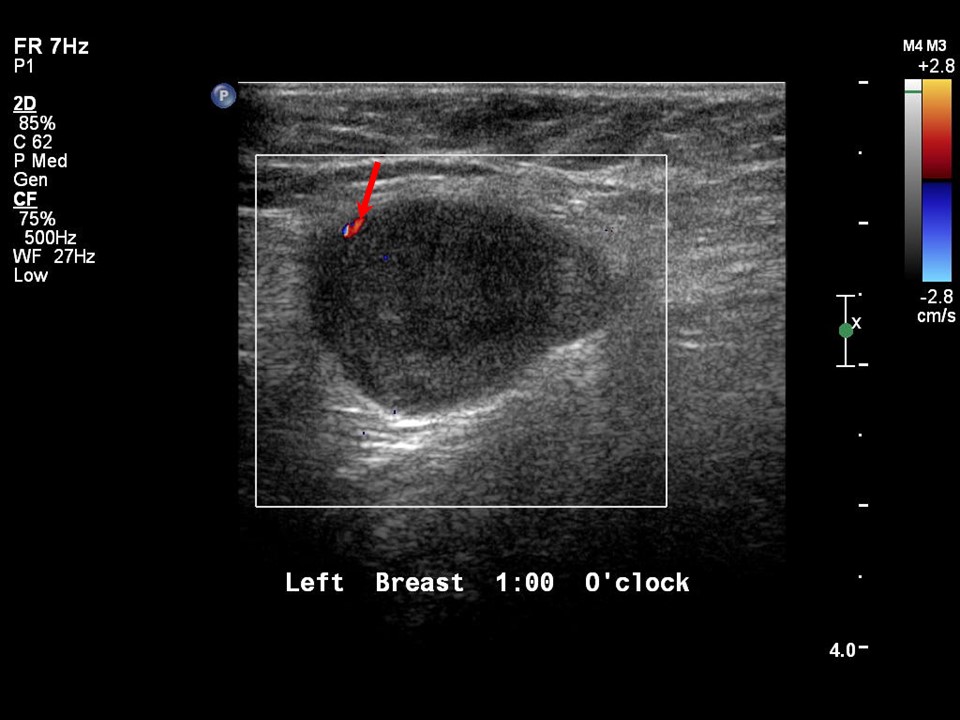 |
| Ultrasound features: None | |
| ‣ Mass | |
| • Location: | None |
| • Number: | 1 |
| • Size: | None |
| • Shape: | None |
| • Orientation: | None |
| • Margins: | None |
| • Echo pattern: | None |
| • Posterior features: | No posterior features |
| ‣ Calcifications: | None |
| ‣ Associated features: | None |
| ‣ Special cases: | Lymph nodes, axillary |
BI-RADS:
BI-RADS Category: 4C (high suspicion for malignancy)Further assessment:
Further assessment advised: Referral for cytologyCytology:
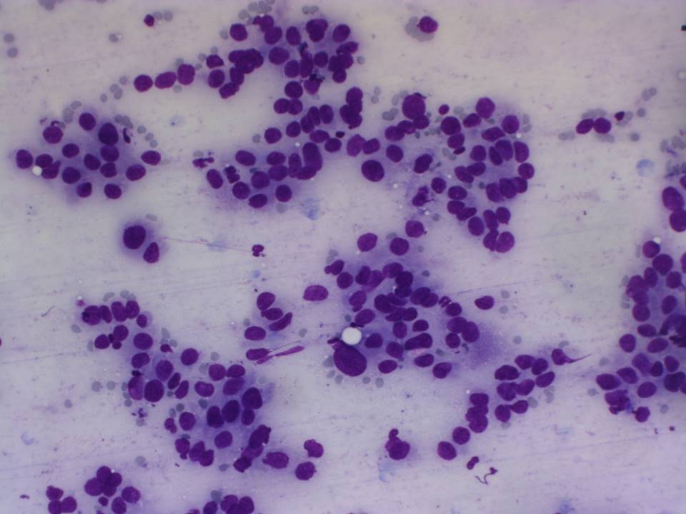 |
| Cytology features: | |
| ‣ Type of sample: | FNAC |
| ‣ Site of biopsy: | |
| • Laterality: | Left axillary node |
| • Quadrant: | No lump in breast |
| • Localization technique: | Palpation |
| • Nature of aspirate: | Whitish |
| ‣ Cytological description: | Smears are very cellular and show dyscohesive clusters of malignant epithelial cells with marked pleomorphism and nuclear hyperchromasia. A glandular arrangement of the epithelial cells is seen in some cell clusters |
| ‣ Reporting category: | Malignant |
| ‣ Diagnosis: | Distant metastases, correlating with the clinical history of ovarian carcinoma |
| ‣ Comments: | None |
Case summary:
| Postmenopausal woman, who had undergone surgery for ovarian carcinoma a year ago, presented with a lump in the left axillary tail. Diagnosed as left axillary tail enlarged dysmorphic lymph nodes, BI-RADS 4C on imaging. No suspicious finding was seen in bilateral breasts on imaging other than the left axillary node findings. On FNAC of the node, malignant cells were seen. |
Learning points:
|