


Home / Training / Manuals / Atlas of breast cancer early detection / Cases
Atlas of breast cancer early detection
Go back to the list of case studies
.png) Click on the pictures to magnify and display the legends
Click on the pictures to magnify and display the legends
| Case number: | 140 |
| Age: | 43 |
| Clinical presentation: | Premenopausal woman with average risk of breast cancer presented with a lump in the left breast of recent onset. Examination revealed a left upper quadrant lump. She has had a stable right breast lump for many years, which is proven fibroadenoma on tissue diagnosis. |
Mammography:
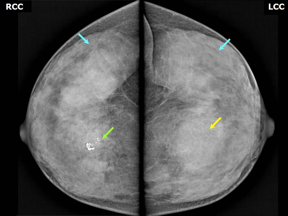 | 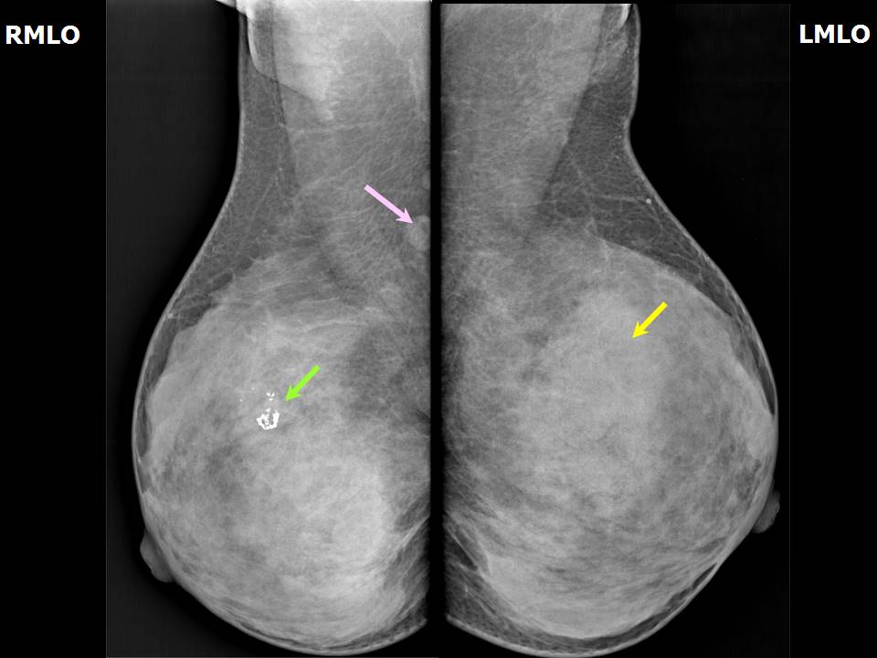 |
| Breast composition: | ACR category d (the breasts are extremely dense, which lowers the sensitivity of mammography) | Mammography features: |
| ‣ Location of the lesion: | Left breast, upper inner quadrant at 1011 oclock, middle third |
| ‣ Mass: | |
| • Number: | 1 |
| • Size: | 4.2 × 4.0 cm |
| • Shape: | Oval |
| • Margins: | Circumscribed |
| • Density: | Equal |
| ‣ Calcifications: | |
| • Typically benign: | None |
| • Suspicious: | None |
| • Distribution: | None |
| ‣ Architectural distortion: | None |
| ‣ Asymmetry: | None |
| ‣ Intramammary node: | None |
| ‣ Skin lesion: | None |
| ‣ Solitary dilated duct: | None |
| ‣ Associated features: | None |
| Breast composition: | ACR category d (the breasts are extremely dense, which lowers the sensitivity of mammography) | Mammography features: |
| ‣ Location of the lesion: | Right breast, upper inner quadrant at 1 oclock, middle third |
| ‣ Mass: | |
| • Number: | 1 |
| • Size: | No |
| • Shape: | None |
| • Margins: | None |
| • Density: | None |
| ‣ Calcifications: | |
| • Typically benign: | Coarse, popcorn-like |
| • Suspicious: | None |
| • Distribution: | None |
| ‣ Architectural distortion: | None |
| ‣ Asymmetry: | None |
| ‣ Intramammary node: | None |
| ‣ Skin lesion: | None |
| ‣ Solitary dilated duct: | None |
| ‣ Associated features: | None |
Ultrasound:
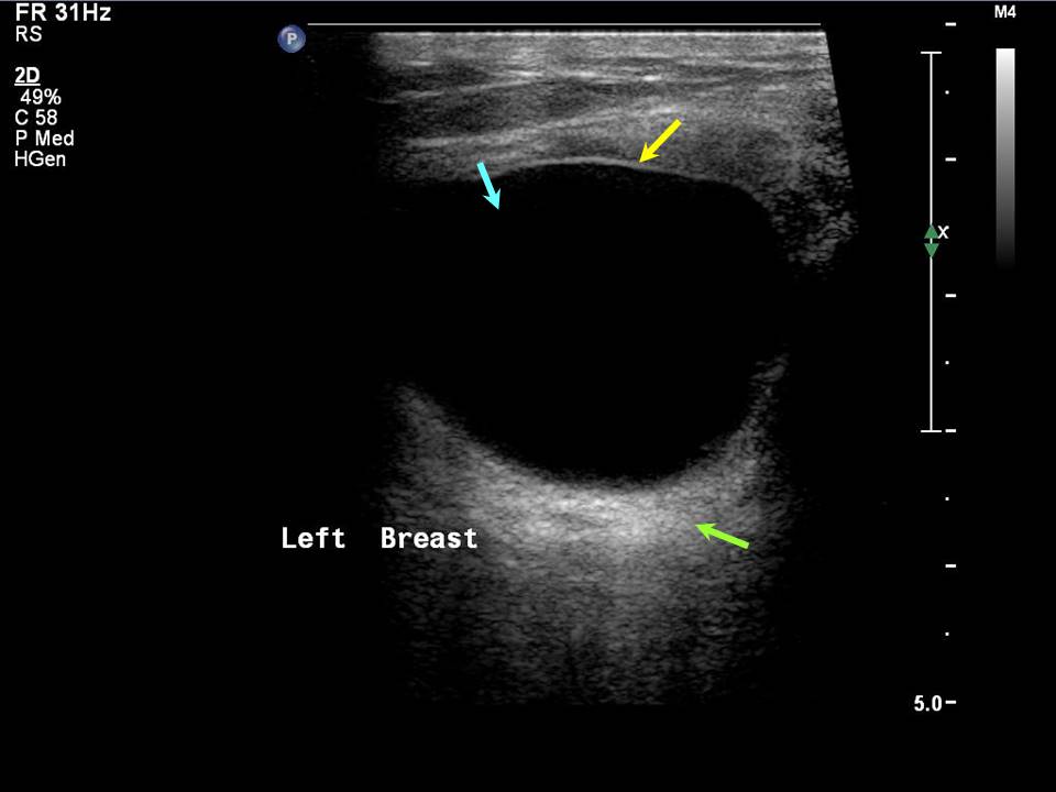 |  |
| Ultrasound features: Left breast, upper inner quadrant at 11 oclock | |
| ‣ Mass | |
| • Location: | Left breast, upper inner quadrant at 11 oclock |
| • Number: | 1 |
| • Size: | 3.5 × 2.5 cm |
| • Shape: | Oval |
| • Orientation: | Parallel |
| • Margins: | Circumscribed |
| • Echo pattern: | Anechoic |
| • Posterior features: | Posterior shadowing |
| ‣ Calcifications: | None |
| ‣ Associated features: | None |
| ‣ Special cases: | Simple cyst |
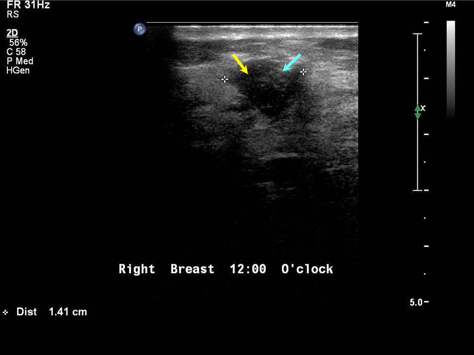
| Ultrasound features: Right breast, upper inner quadrant at 1 oclock | |
| ‣ Mass | |
| • Location: | Right breast, upper inner quadrant at 1 oclock |
| • Number: | 1 |
| • Size: | 1.4 × 1.2 cm |
| • Shape: | Oval |
| • Orientation: | Parallel |
| • Margins: | Circumscribed |
| • Echo pattern: | Hypoechoic |
| • Posterior features: | No posterior features |
| ‣ Calcifications: | Macrocalcifications in mass |
| ‣ Associated features: | Poor vascularity |
| ‣ Special cases: | None |
BI-RADS:
BI-RADS Category: 2 (benign)Further assessment:
Further assessment advised: Referral for cytologyCytology:
 |
| Cytology features: | |
| ‣ Type of sample: | FNAC |
| ‣ Site of biopsy: | |
| • Laterality: | Right |
| • Quadrant: | Lower outer |
| • Localization technique: | Palpation |
| • Nature of aspirate: | Whitish |
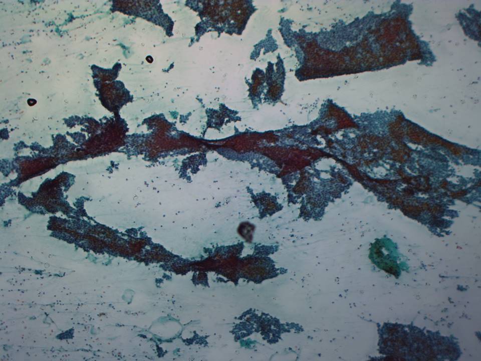
| ‣ Cytological description: | Highly cellular aspirate, showing many cohesive groups of ductal cells with sharply demarcated borders with branching glove-like or antler-horn shapes. Many stripped or bare nuclei are seen in the background |
| ‣ Reporting category: | Benign |
| ‣ Diagnosis: | Fibroadenoma |
| ‣ Comments: | None |
 |
| Cytology features: | |
| ‣ Type of sample: | FNAC |
| ‣ Site of biopsy: | |
| • Laterality: | Left |
| • Quadrant: | Upper inner |
| • Localization technique: | Palpation |
| • Nature of aspirate: | 0.5 mL of brownish fluid |
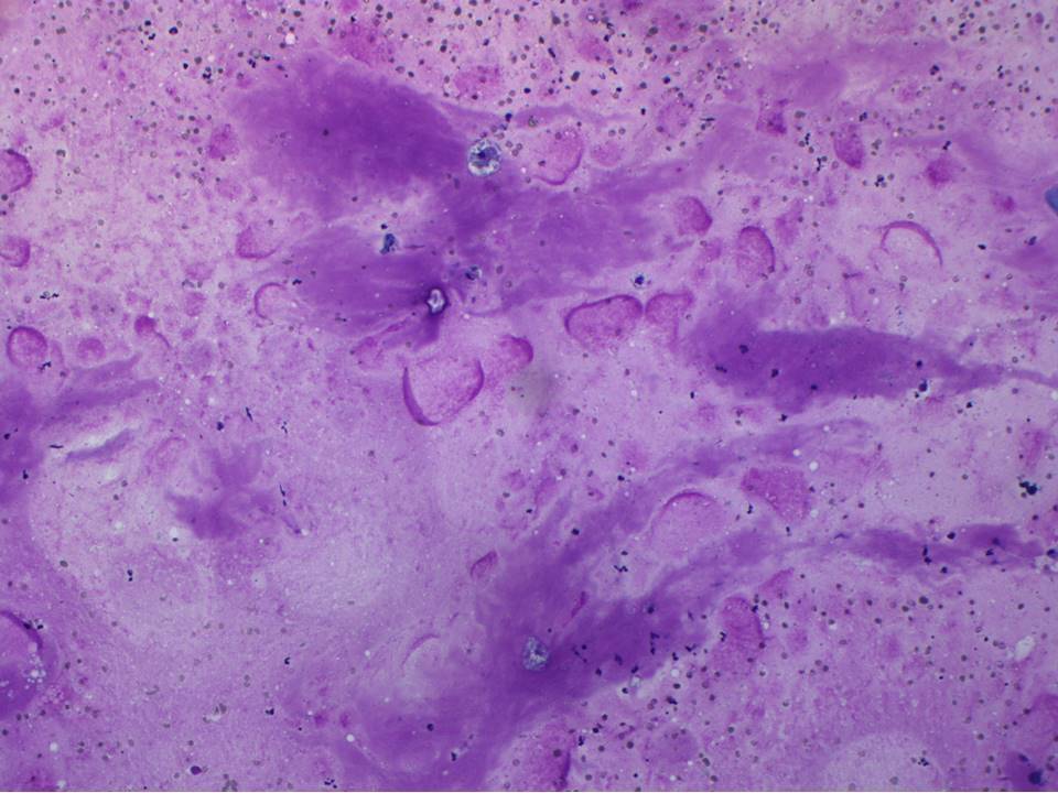
| ‣ Cytological description: | Smears show foamy histiocytes on a thick proteinaceous background. Very a few ductal cells are seen |
| ‣ Reporting category: | Benign |
| ‣ Diagnosis: | Fibrocystic change, non-proliferative |
| ‣ Comments: | None |
Case summary:
| Premenopausal woman presented with left breast lump. Diagnosed as simple cyst, BI-RADS 2 on imaging and as non-proliferative fibrocystic change on cytology. Incidentally noted right breast involuting fibroadenoma, BI-RADS 2 on imaging. |
Learning points:
|