


Home / Training / Manuals / Atlas of breast cancer early detection / Cases
Atlas of breast cancer early detection
Go back to the list of case studies
.png) Click on the pictures to magnify and display the legends
Click on the pictures to magnify and display the legends
| Case number: | 155 |
| Age: | 52 |
| Clinical presentation: | Postmenopausal woPostmenopausal woman with average risk of developing breast cancer attended for mammography screening. On examination no abnormality was detected. man at average risk of developing breast cancer came for mammography screening. On examination no abnormality detected. |
Mammography:
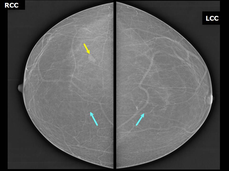 |  |
| Breast composition: | ACR category a (the breasts are almost entirely fatty) | Mammography features: |
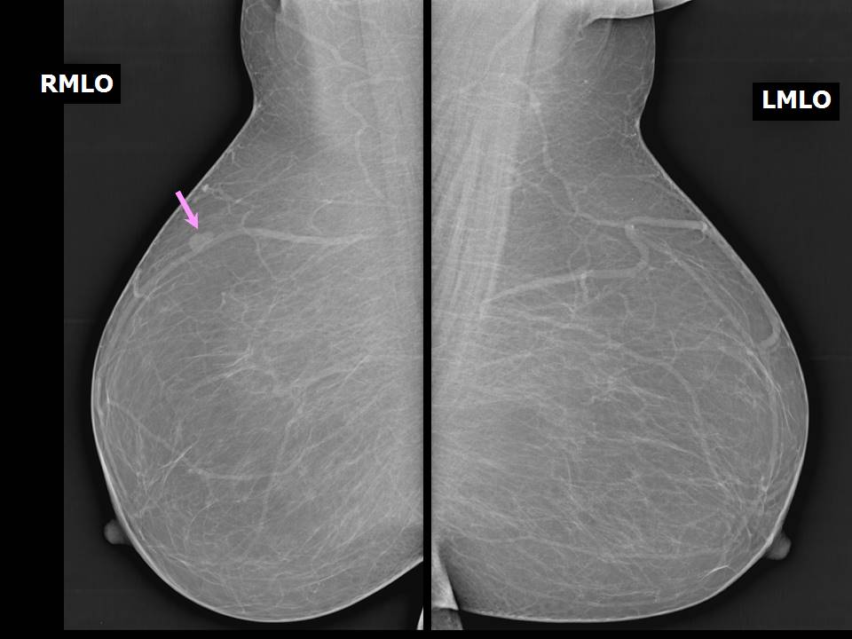
| ‣ Location of the lesion: | Right breast, upper outer quadrant at 11 oclock, posterior third |
| ‣ Mass: | |
| • Number: | 1 |
| • Size: | 1.0 cm in greatest dimension |
| • Shape: | Oval |
| • Margins: | Circumscribed |
| • Density: | Equal |
| ‣ Calcifications: | |
| • Typically benign: | None |
| • Suspicious: | None |
| • Distribution: | None |
| ‣ Architectural distortion: | None |
| ‣ Asymmetry: | None |
| ‣ Intramammary node: | Present |
| ‣ Skin lesion: | None |
| ‣ Solitary dilated duct: | None |
| ‣ Associated features: | None |
Ultrasound:
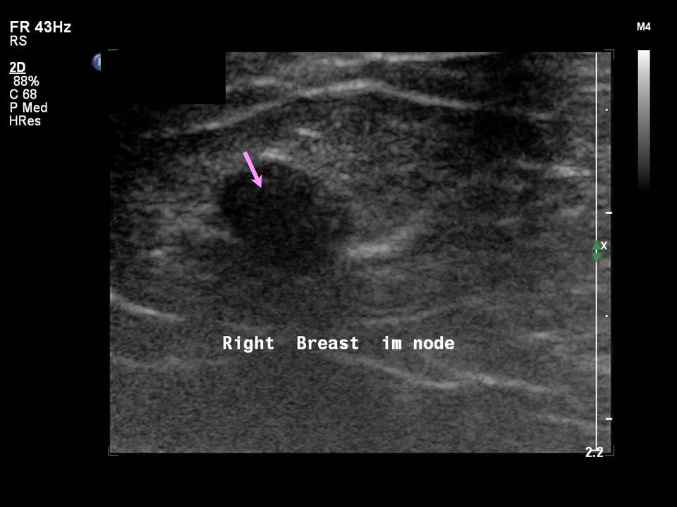 |
| Ultrasound features: Right breast, upper outer quadrant at 10 oclock | |
| ‣ Mass | |
| • Location: | Right breast, upper outer quadrant at 10 oclock |
| • Number: | 1 |
| • Size: | 1.1 cm in greatest dimension |
| • Shape: | Oval |
| • Orientation: | Not parallel |
| • Margins: | Circumscribed |
| • Echo pattern: | Hypoechoic with central sinus |
| • Posterior features: | No posterior features |
| ‣ Calcifications: | None |
| ‣ Associated features: | Hilar vascularity |
| ‣ Special cases: | Lymph nodes, intramammary |
BI-RADS:
BI-RADS Category: 1 (negative)Case summary:
| Postmenopausal woman with average risk of developing breast cancer came for mammography screening. Diagnosed as intramammary node in right breast. BI-RADS 2 on imaging. |
Learning points:
|