


Home / Training / Manuals / Atlas of breast cancer early detection / Cases
Atlas of breast cancer early detection
Filter by language: English / Русский
Go back to the list of case studies
.png) Click on the pictures to magnify and display the legends
Click on the pictures to magnify and display the legends
BI-RADS Category (2015): 4C (high suspicion for malignancy)
| Case number: | 103 |
| Age: | 82 |
| Clinical presentation: | Postmenopausal woman had undergone microdochectomy for right nipple discharge. Histopathology of the excised duct showed benign papilloma. On follow-up a year later, she presented with a left breast lump. Mammography and breast ultrasound dated 2015 reveal a new lesion in the left breast. Compared with a previous mammogram dated 2014, follow-up reveals developing asymmetry in the left breast. Interval change is seen. Diagnosed as highly suspicious for malignancy, BI-RADS 4C on imaging, as mucinous carcinoma on cytology, and as mucinous carcinoma, pT2N0 on histopathology. |
Mammography:
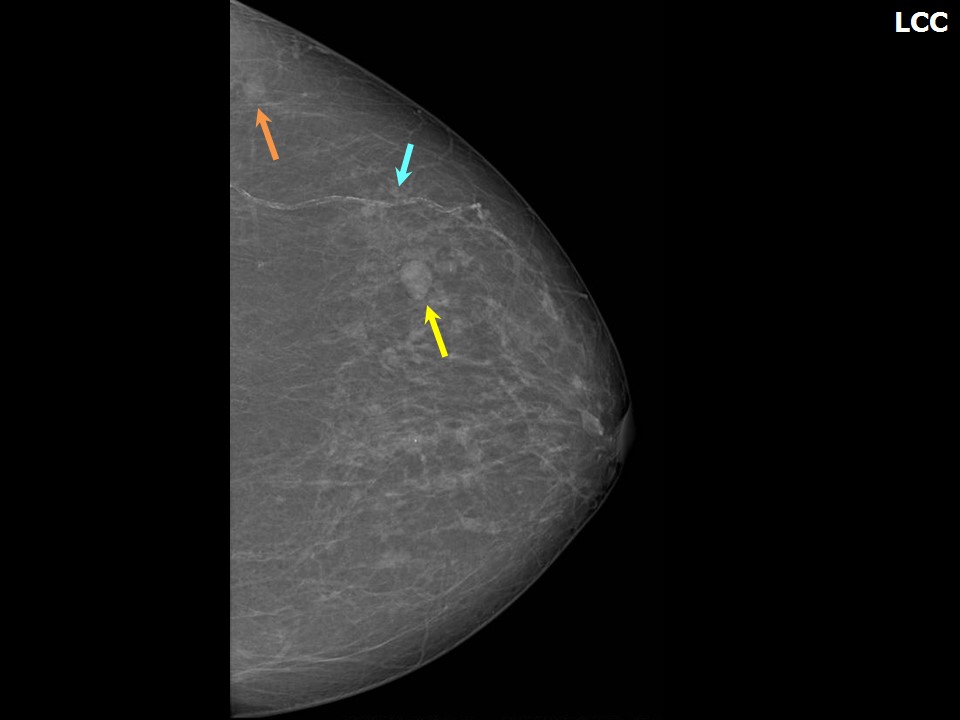 | 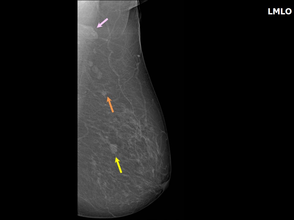 |
| Breast composition: | ACR category b (there are scattered areas of fibroglandular density) | Mammography features: |
| ‣ Location of the lesion: | September 2014: Left breast, upper outer quadrant at 13 oclock, middle third |
| ‣ Mass: | |
| • Number: | Multiple |
| • Size: | Largest 0.4 cm |
| • Shape: | Round |
| • Margins: | Circumscribed |
| • Density: | Equal |
| ‣ Calcifications: | |
| • Typically benign: | None |
| • Suspicious: | None |
| • Distribution: | None |
| ‣ Architectural distortion: | None |
| ‣ Asymmetry: | None |
| ‣ Intramammary node: | None |
| ‣ Skin lesion: | None |
| ‣ Solitary dilated duct: | None |
| ‣ Associated features: | Multiple simple cysts |
 |  |
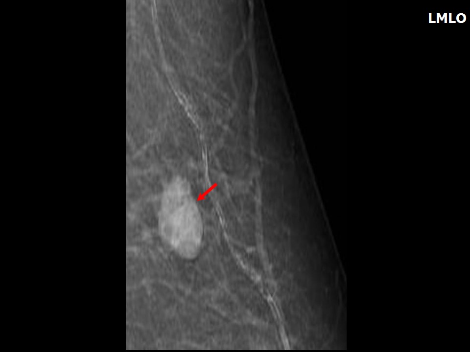 |
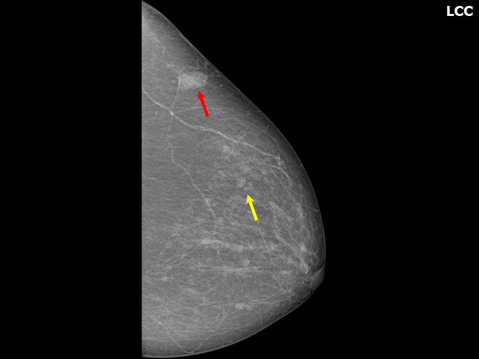
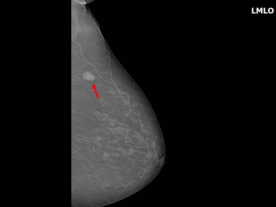
| Breast composition: | ACR category b (there are scattered areas of fibroglandular density) | Mammography features: |
| ‣ Location of the lesion: | July 2015: Left breast, upper outer quadrant at 2 oclock, posterior third |
| ‣ Mass: | |
| • Number: | Multiple |
| • Size: | Largest 1.7 × 1.0 cm (this is the developing asymmetry) |
| • Shape: | Oval |
| • Margins: | Indistinct posterior margin |
| • Density: | High |
| ‣ Calcifications: | |
| • Typically benign: | None |
| • Suspicious: | None |
| • Distribution: | None |
| ‣ Architectural distortion: | None |
| ‣ Asymmetry: | None |
| ‣ Intramammary node: | None |
| ‣ Skin lesion: | None |
| ‣ Solitary dilated duct: | None |
| ‣ Associated features: | None |
Ultrasound:
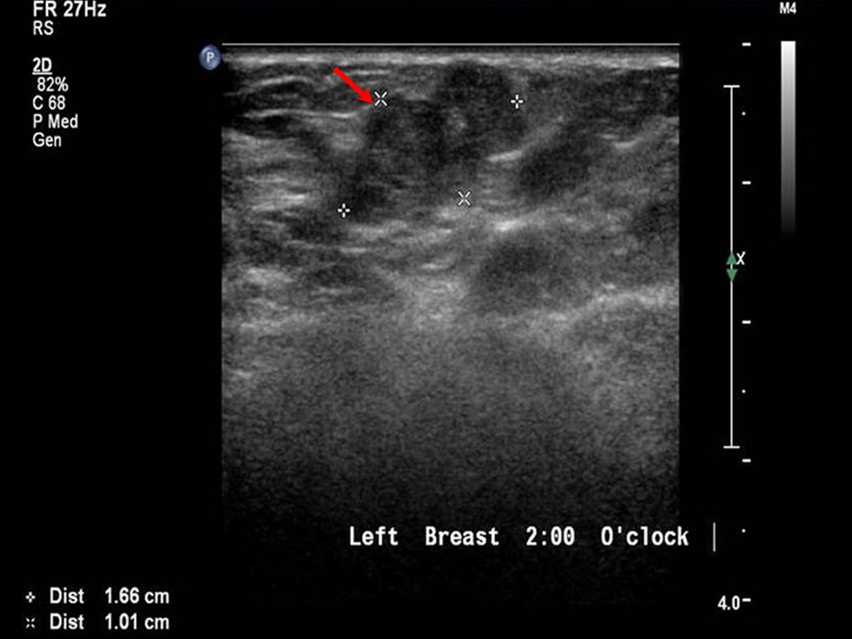 | 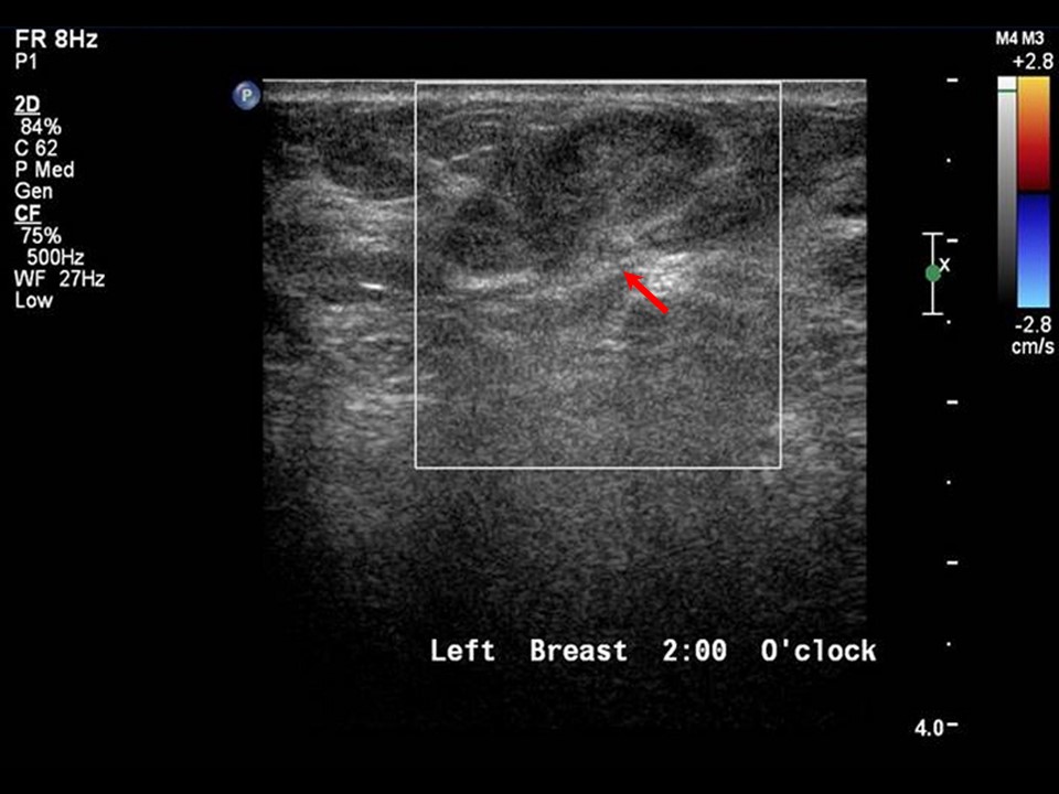 |
| Ultrasound features: July 2015: Left breast, upper outer quadrant at 2 oclock | |
| ‣ Mass | |
| • Location: | July 2015: Left breast, upper outer quadrant at 2 oclock |
| • Number: | 1 |
| • Size: | 2.0 × 1.0 cm (this is the developing asymmetry) |
| • Shape: | Irregular |
| • Orientation: | Not parallel |
| • Margins: | Partly circumscribed and partly indistinct margins |
| • Echo pattern: | Heteroechoic |
| • Posterior features: | No posterior features |
| ‣ Calcifications: | None |
| ‣ Associated features: | None |
| ‣ Special cases: | None |
BI-RADS:
BI-RADS Category (2014): 2 (benign)BI-RADS Category (2015): 4C (high suspicion for malignancy)
Further assessment:
Further assessment advised: Referral for cytologyCytology:
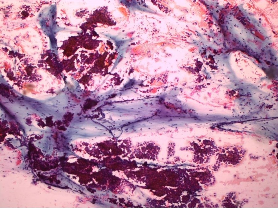 |
| Cytology features: | |
| ‣ Type of sample: | FNAC |
| ‣ Site of biopsy: | |
| • Laterality: | Left |
| • Quadrant: | Upper outer |
| • Localization technique: | Palpation |
| • Nature of aspirate: | 0.2 mL of mucoid material |
| ‣ Cytological description: | Numerous moderately cohesive clusters and balls of malignant cells dispersed in a mucinous background |
| ‣ Reporting category: | Malignant |
| ‣ Diagnosis: | Mucinous carcinoma |
| ‣ Comments: | None |
Histopathology:
MRM
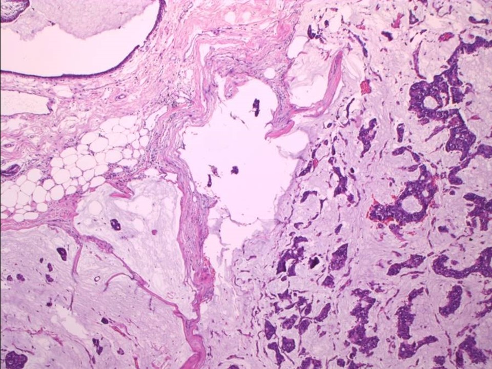 |
| Histopathology features: | |
| ‣ Specimen type: | MRM |
| ‣ Laterality: | Left |
| ‣ Macroscopy: | Specimen (28.0 × 13.0 × 6.0 cm) with an overlying skin flap (11.0 × 5.0 cm). The nipple and areola are unremarkable. On serial sectioning, a small well-circumscribed area (2.1 × 1.3 × 1.0 cm) is identified in the upper outer quadrant, 5.0 cm from the skin and 1.5 cm from the base. Cut surface shows mucoid areas. The remaining breast tissue is unremarkable |
| ‣ Histological type: | Mucinous carcinoma |
| ‣ Histological grade: | Grade 2 (2 + 2 + 1 = 5) |
| ‣ Mitosis: | 4 |
| ‣ Maximum invasive tumour size: | 2.1 cm in greatest dimension |
| ‣ Lymph node status: | 0/22 |
| ‣ Peritumoural lymphovascular invasion: | Absent |
| ‣ DCIS/EIC: | Absent |
| ‣ Margins: | Free of tumour |
| ‣ Pathological stage: | pT2N0 |
| ‣ Biomarkers: | |
| ‣ Comments: |
Case summary:
| Postmenopausal woman had undergone microdochectomy for right nipple discharge, histopathology of the excised duct showed benign papilloma. On follow up a year later, she presented with left breast lump. Mammography and breast ultrasound dated 2015 reveal new lesion in left breast. Compared to previous mammogram dated 2014, follow-up reveals developing asymmetry in left breast. Interval change is seen, diagnosed as highly suspicious of malignancy, BI-RADS category 4C on imaging, mucinous carcinoma on cytology and Mucinous carcinoma, pT2N0 on histopathology. |
Learning points:
|