


Home / Training / Manuals / Atlas of breast cancer early detection / Cases
Atlas of breast cancer early detection
Filter by language: English / Русский
Go back to the list of case studies
.png) Click on the pictures to magnify and display the legends
Click on the pictures to magnify and display the legends
| Case number: | 132 |
| Age: | 50 |
| Clinical presentation: | Perimenopausal woman with average risk of developing breast cancer presented with left breast pain and swelling. |
Mammography:
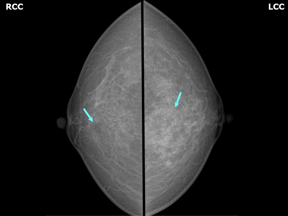 | 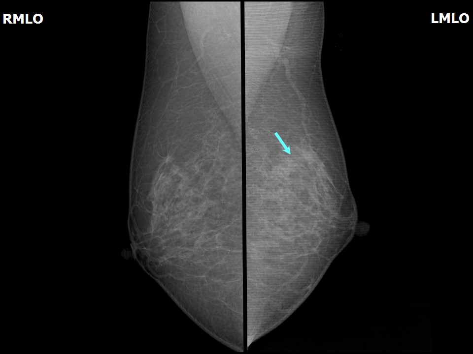 |
| Breast composition: | ACR category b (there are scattered areas of fibroglandular density) | Mammography features: |
| ‣ Location of the lesion: | Left breast, upper quadrants, central zone at 12 oclock, middle third |
| ‣ Mass: | |
| • Number: | 0 |
| • Size: | None |
| • Shape: | None |
| • Margins: | None |
| • Density: | None |
| ‣ Calcifications: | |
| • Typically benign: | None |
| • Suspicious: | None |
| • Distribution: | None |
| ‣ Architectural distortion: | Present |
| ‣ Asymmetry: | Focal |
| ‣ Intramammary node: | None |
| ‣ Skin lesion: | None |
| ‣ Solitary dilated duct: | None |
| ‣ Associated features: | Architectural distortion |
Ultrasound:
 | 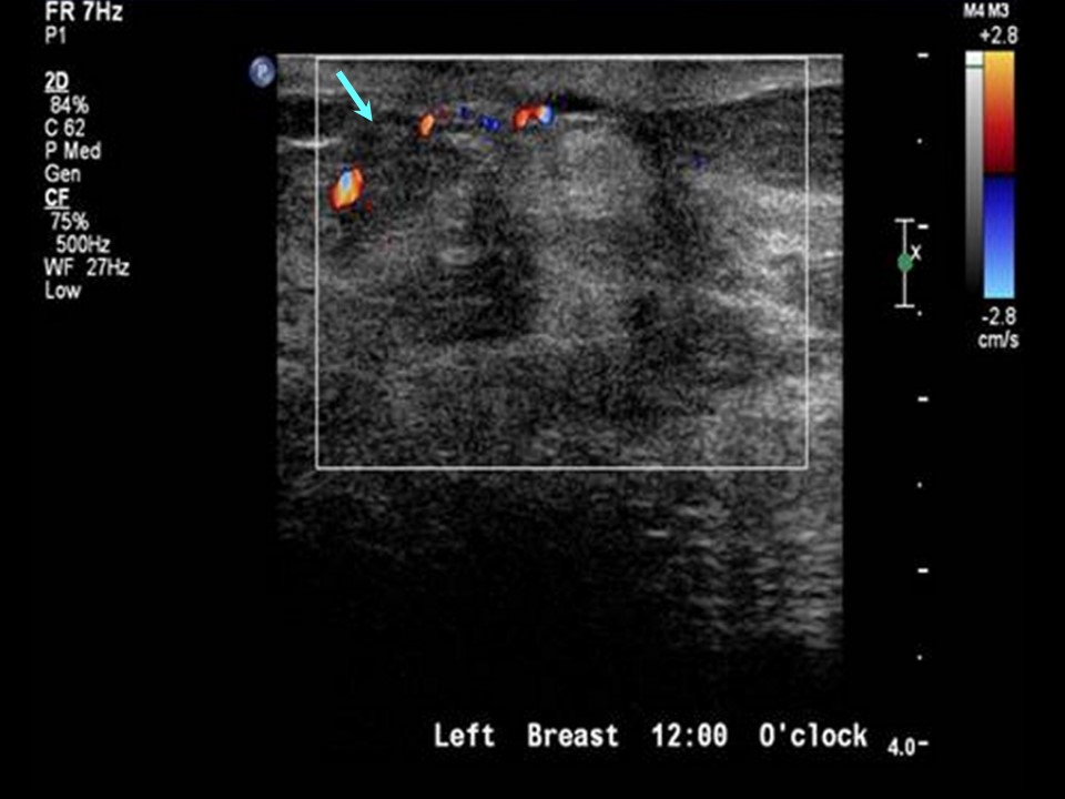 |
| Ultrasound features: Left breast, upper quadrants at 12 oclock, superior areolar margin | |
| ‣ Mass | |
| • Location: | Left breast, upper quadrants at 12 oclock, superior areolar margin |
| • Number: | 1 |
| • Size: | 2.5 × 1.6 cm |
| • Shape: | Irregular |
| • Orientation: | Parallel |
| • Margins: | Indistinct |
| • Echo pattern: | Heterogeneous with areas of breakdown |
| • Posterior features: | No posterior features |
| ‣ Calcifications: | None |
| ‣ Associated features: | Vascularity (vessels in rim) |
| ‣ Special cases: | None |
BI-RADS:
BI-RADS Category: 2 (benign)Further assessment:
Further assessment advised: Referral for cytologyCytology:
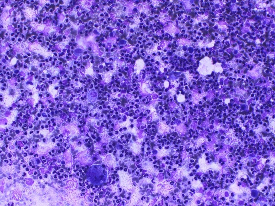 |
| Cytology features: | |
| ‣ Type of sample: | FNAC |
| ‣ Site of biopsy: | |
| • Laterality: | Left |
| • Quadrant: | Upper |
| • Localization technique: | Palpation |
| • Nature of aspirate: | 0.2 mL of purulent material |
| ‣ Cytological description: | Smears show many neutrophils on a necrotic background |
| ‣ Reporting category: | Benign |
| ‣ Diagnosis: | Acute inflammation |
| ‣ Comments: | None |
Case summary:
| Perimenopausal woman presented with left breast pain and swelling. Diagnosed as inflammatory changes in left breast, BI-RADS 2 on imaging and as acute inflammation on cytology. |
Learning points:
|