


Home / Training / Manuals / Atlas of breast cancer early detection / Cases
Atlas of breast cancer early detection
Filter by language: English / Русский
Go back to the list of case studies
.png) Click on the pictures to magnify and display the legends
Click on the pictures to magnify and display the legends
| Case number: | 074 |
| Age: | 59 |
| Clinical presentation: | Postmenopausal woman with average risk of developing breast cancer presented with a left breast lump. Examination revealed a lump 3 cm in diameter in the lower inner quadrant of the left breast. |
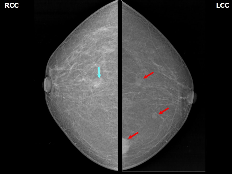
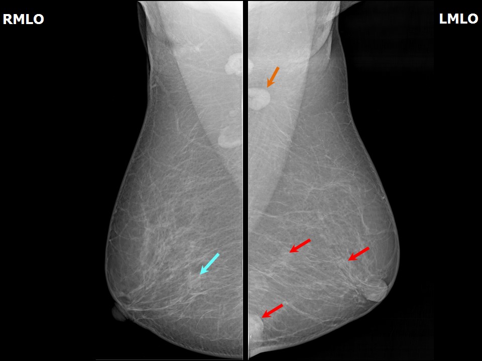
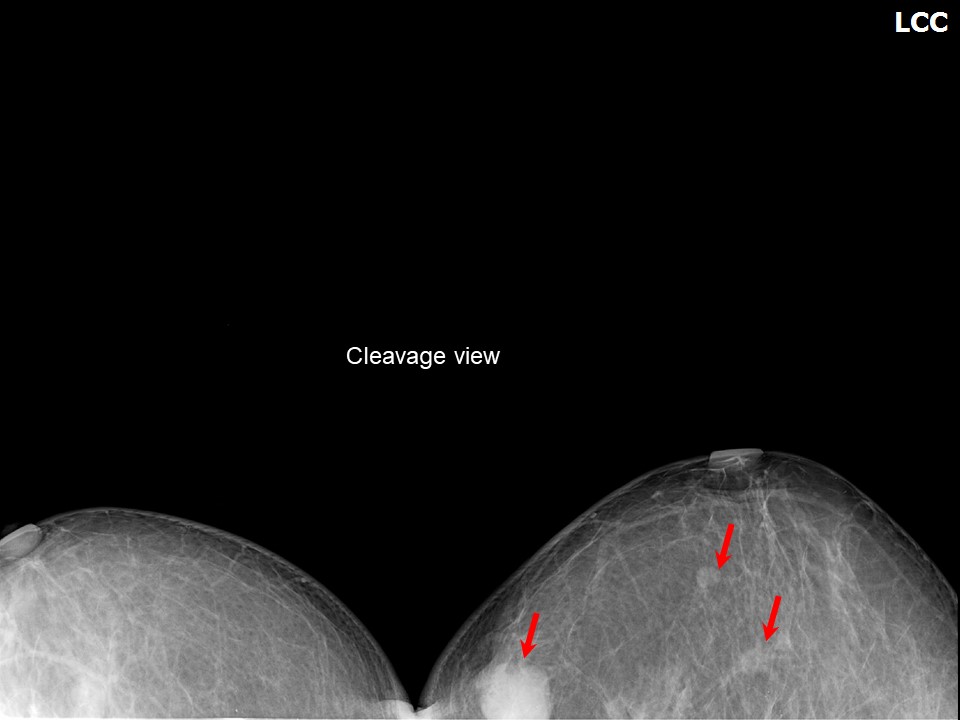
Mammography:
 |  |
 |
| Breast composition: | ACR category a (the breasts are almost entirely fatty) | Mammography features: |
| ‣ Location of the lesion: | Lesion 1: Left breast, lower inner quadrant at 8 oclock, posterior third, at 6.4 cm from the nipple just beneath the dermis |
| ‣ Mass: | |
| • Number: | 1 |
| • Size: | 2.6 × 1.7 cm |
| • Shape: | Irregular |
| • Margins: | Indistinct |
| • Density: | High |
| ‣ Calcifications: | |
| • Typically benign: | None |
| • Suspicious: | None |
| • Distribution: | None |
| ‣ Architectural distortion: | None |
| ‣ Asymmetry: | None |
| ‣ Intramammary node: | None |
| ‣ Skin lesion: | None |
| ‣ Solitary dilated duct: | None |
| ‣ Associated features: | Left enlarged lymph node (1.2 × 1.0 cm) with thickened cortex 4.2 mm |
| Breast composition: | ACR category a (the breasts are almost entirely fatty) | Mammography features: |
| ‣ Location of the lesion: | Lesion 2: Left breast, central portion of the breast, central zone at 6 oclock, posterior third, approximately 7.0 cm superolateral to the primary mass lesion |
| ‣ Mass: | |
| • Number: | 1 |
| • Size: | 1.1 × 0.6 cm |
| • Shape: | Irregular |
| • Margins: | Indistinct |
| • Density: | High |
| ‣ Calcifications: | |
| • Typically benign: | None |
| • Suspicious: | None |
| • Distribution: | None |
| ‣ Architectural distortion: | None |
| ‣ Asymmetry: | None |
| ‣ Intramammary node: | None |
| ‣ Skin lesion: | None |
| ‣ Solitary dilated duct: | None |
| ‣ Associated features: | Left enlarged lymph node (1.2 × 1.0 cm) with thickened cortex 4.2 mm |
| Breast composition: | ACR category a (the breasts are almost entirely fatty) | Mammography features: |
| ‣ Location of the lesion: | Lesion 3: Left breast, upper inner quadrant at 910 oclock, middle third |
| ‣ Mass: | |
| • Number: | 1 |
| • Size: | 0.9 cm in greatest dimension |
| • Shape: | Oval |
| • Margins: | Circumscribed |
| • Density: | Equal |
| ‣ Calcifications: | |
| • Typically benign: | None |
| • Suspicious: | None |
| • Distribution: | None |
| ‣ Architectural distortion: | None |
| ‣ Asymmetry: | None |
| ‣ Intramammary node: | None |
| ‣ Skin lesion: | None |
| ‣ Solitary dilated duct: | None |
| ‣ Associated features: | Left enlarged lymph node (1.2 × 1.0 cm) with thickened cortex 4.2 mm |
| Breast composition: | ACR category a (the breasts are almost entirely fatty) | Mammography features: |
| ‣ Location of the lesion: | Right breast, central portion of the breast at 12 oclock, posterior third |
| ‣ Mass: | |
| • Number: | 1 |
| • Size: | 1.5 × 1.2 cm |
| • Shape: | Oval |
| • Margins: | Circumscribed |
| • Density: | Equal |
| ‣ Calcifications: | |
| • Typically benign: | None |
| • Suspicious: | None |
| • Distribution: | None |
| ‣ Architectural distortion: | None |
| ‣ Asymmetry: | None |
| ‣ Intramammary node: | None |
| ‣ Skin lesion: | None |
| ‣ Solitary dilated duct: | None |
| ‣ Associated features: | None |
Ultrasound:
 |  |
 | 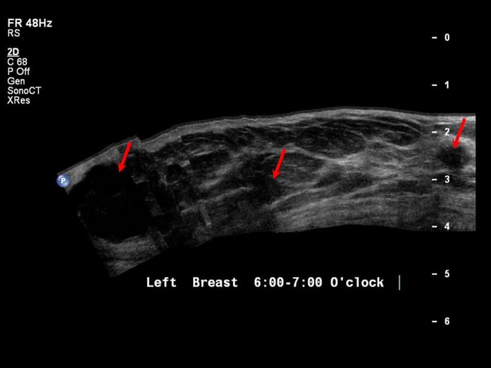 |
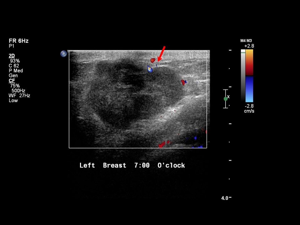
| Ultrasound features: Left breast, lower inner quadrant at 78 oclock | |
| ‣ Mass | |
| • Location: | Left breast, lower inner quadrant at 78 oclock |
| • Number: | 1 |
| • Size: | 2.7 × 1.7 cm |
| • Shape: | Irregular |
| • Orientation: | Not parallel |
| • Margins: | Indistinct |
| • Echo pattern: | Hypoechoic |
| • Posterior features: | No posterior features |
| ‣ Calcifications: | None |
| ‣ Associated features: | Internal vascularity |
| ‣ Special cases: | None |
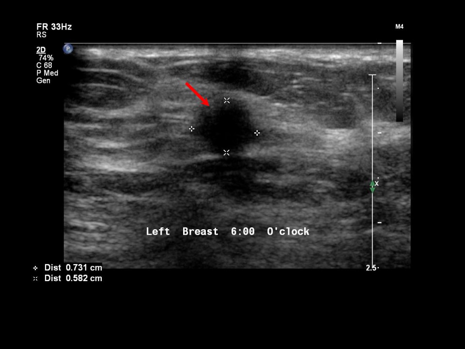
| Ultrasound features: Left breast, lower quadrants at 6 oclock | |
| ‣ Mass | |
| • Location: | Left breast, lower quadrants at 6 oclock |
| • Number: | 1 |
| • Size: | 0.8 × 0.6 cm |
| • Shape: | Irregular |
| • Orientation: | Not parallel |
| • Margins: | Spiculated |
| • Echo pattern: | Hypoechoic |
| • Posterior features: | No posterior features |
| ‣ Calcifications: | None |
| ‣ Associated features: | None |
| ‣ Special cases: | None |
| Ultrasound features: Left breast, outer quadrants at 9 oclock | |
| ‣ Mass | |
| • Location: | Left breast, outer quadrants at 9 oclock |
| • Number: | 1 |
| • Size: | 0.9 cm in greatest dimension |
| • Shape: | Oval |
| • Orientation: | Parallel |
| • Margins: | Circumscribed |
| • Echo pattern: | Hypoechoic |
| • Posterior features: | No posterior features |
| ‣ Calcifications: | None |
| ‣ Associated features: | None |
| ‣ Special cases: | None |
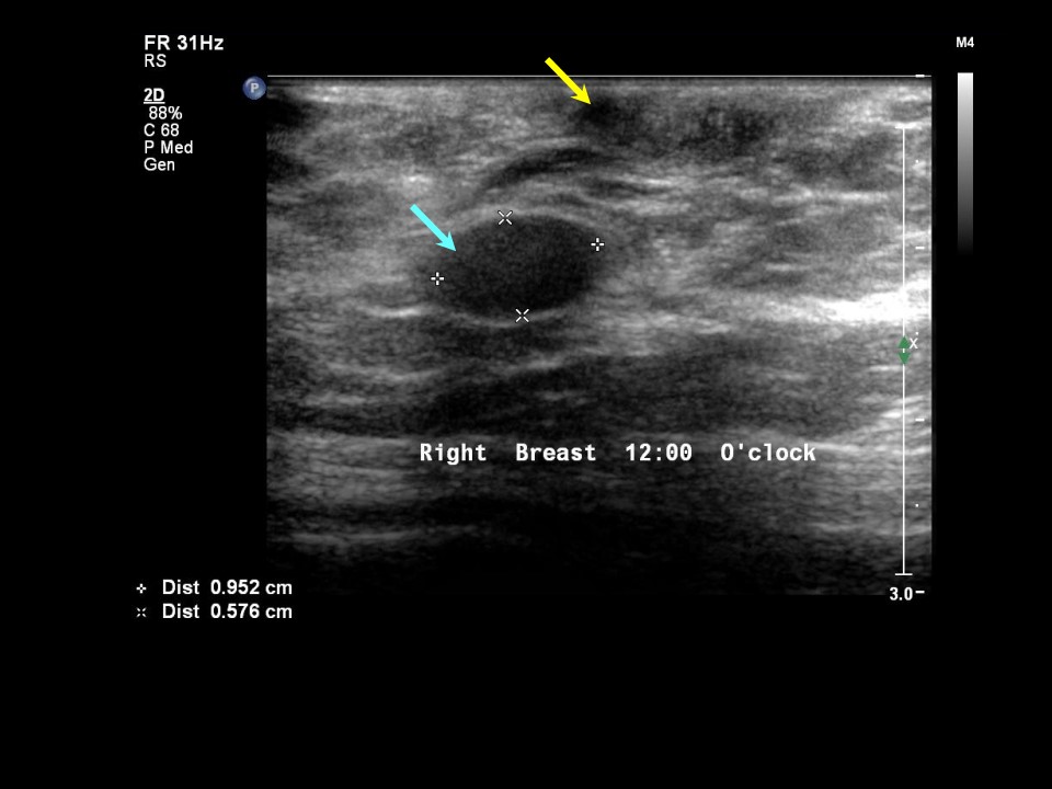 |
| Ultrasound features: Right breast, upper quadrants at 12 oclock | |
| ‣ Mass | |
| • Location: | Right breast, upper quadrants at 12 oclock |
| • Number: | 1 |
| • Size: | 1.0 × 0.6 cm |
| • Shape: | Oval |
| • Orientation: | Parallel |
| • Margins: | Circumscribed |
| • Echo pattern: | Hypoechoic |
| • Posterior features: | No posterior features |
| ‣ Calcifications: | None |
| ‣ Associated features: | None |
| ‣ Special cases: | None |
BI-RADS:
BI-RADS Category: 5 (highly suggestive of malignancy)Further assessment:
Further assessment advised: Referral for cytologyCytology:
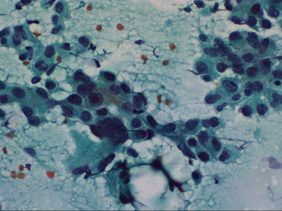 |
| Cytology features: | |
| ‣ Type of sample: | FNAC (solid lesion) |
| ‣ Site of biopsy: | |
| • Laterality: | Left |
| • Quadrant: | Lower inner |
| • Localization technique: | Palpation |
| • Nature of aspirate: | Whitish |
| ‣ Cytological description: | Smears are very cellular with loose clusters of highly pleomorphic malignant cells |
| ‣ Reporting category: | Malignant |
| ‣ Diagnosis: | Carcinoma high grade |
| ‣ Comments: | None |
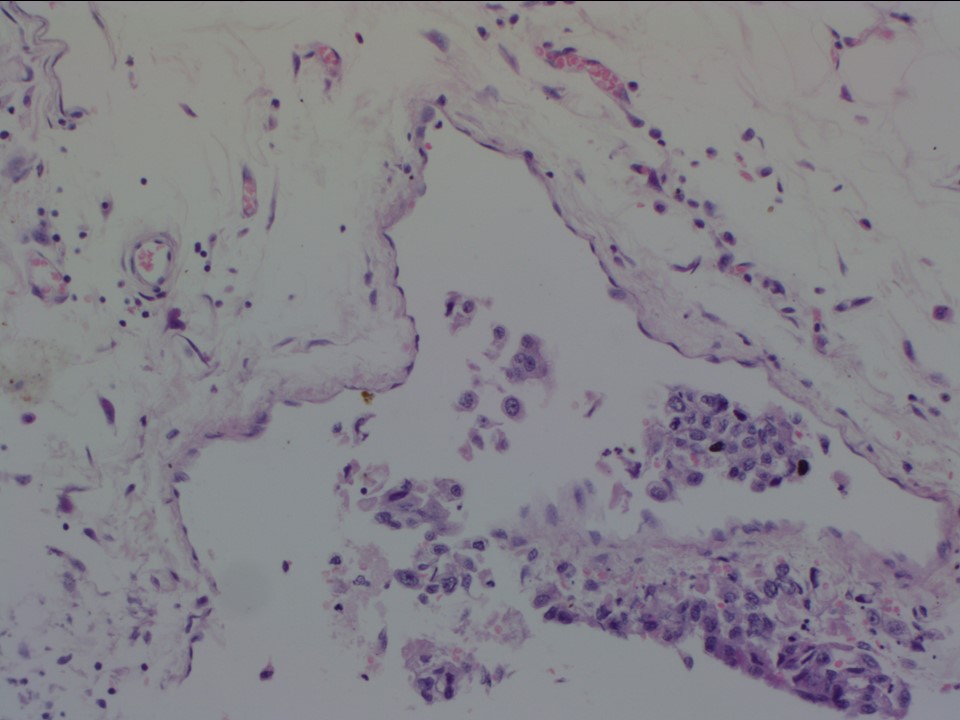
Histopathology:
MRM
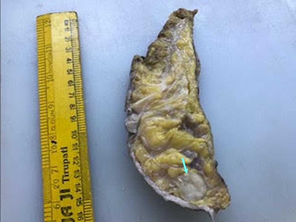 | 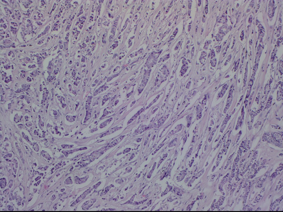 |
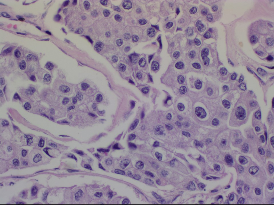 |  |
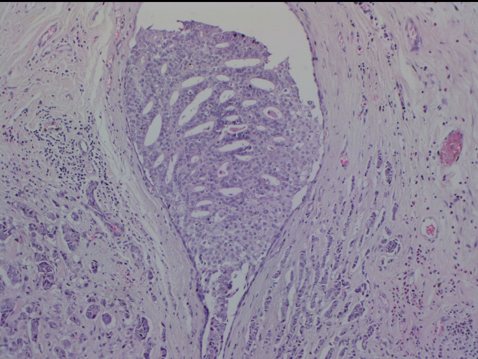 | 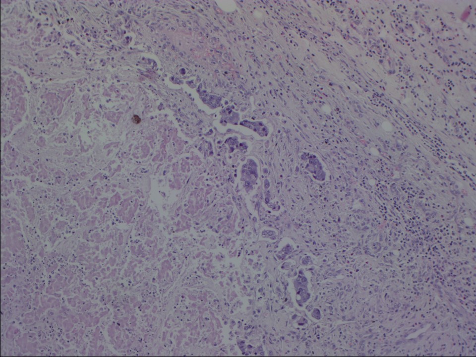 |
| Histopathology features: | |
| ‣ Specimen type: | MRM |
| ‣ Laterality: | Left |
| ‣ Macroscopy: | On serial sectioning three tumours are identified:
|
| ‣ Histological type: | Invasive carcinoma of no special type |
| ‣ Histological grade: | Grade 2 (3 + 2 + 2 = 7) |
| ‣ Mitosis: | 12 |
| ‣ Maximum invasive tumour size: | Largest 3.0 cm (others 2.0 cm and 1.1 cm) |
| ‣ Lymph node status: | 2/21 |
| ‣ Peritumoural lymphovascular invasion: | Present |
| ‣ DCIS/EIC: | Solid, cribriform, and comedo type intermediate nuclear grade |
| ‣ Margins: | Free of tumour |
| ‣ Pathological stage: | pT2(3)N1 |
| ‣ Biomarkers: | |
| ‣ Comments: |
Case summary:
| Postmenopausal woman presented with lump in the left breast, diagnosed as left breast multicentric carcinoma, BI-RADS category 5 on imaging, as breast carcinoma on cytology, and as invasive breast carcinoma of no special type, pT2(3)N1 on histopathology. |
Learning points:
|