


Home / Training / Manuals / Atlas of breast cancer early detection / Cases
Atlas of breast cancer early detection
Filter by language: English / Русский
Go back to the list of case studies
.png) Click on the pictures to magnify and display the legends
Click on the pictures to magnify and display the legends
| Case number: | 035 |
| Age: | 68 |
| Clinical presentation: | Postmenopausal woman with family history of colon cancer presented with a lump in the right breast. Examination revealed a hard, mobile, 3 cm lump in the upper outer quadrant of the right breast. |
Mammography:
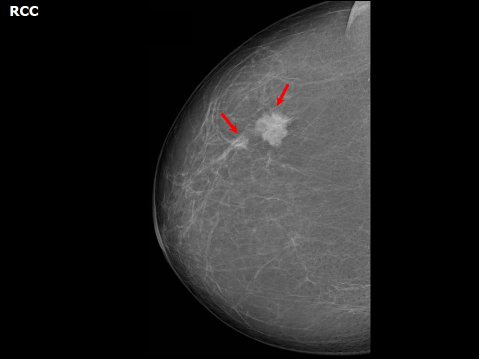 | 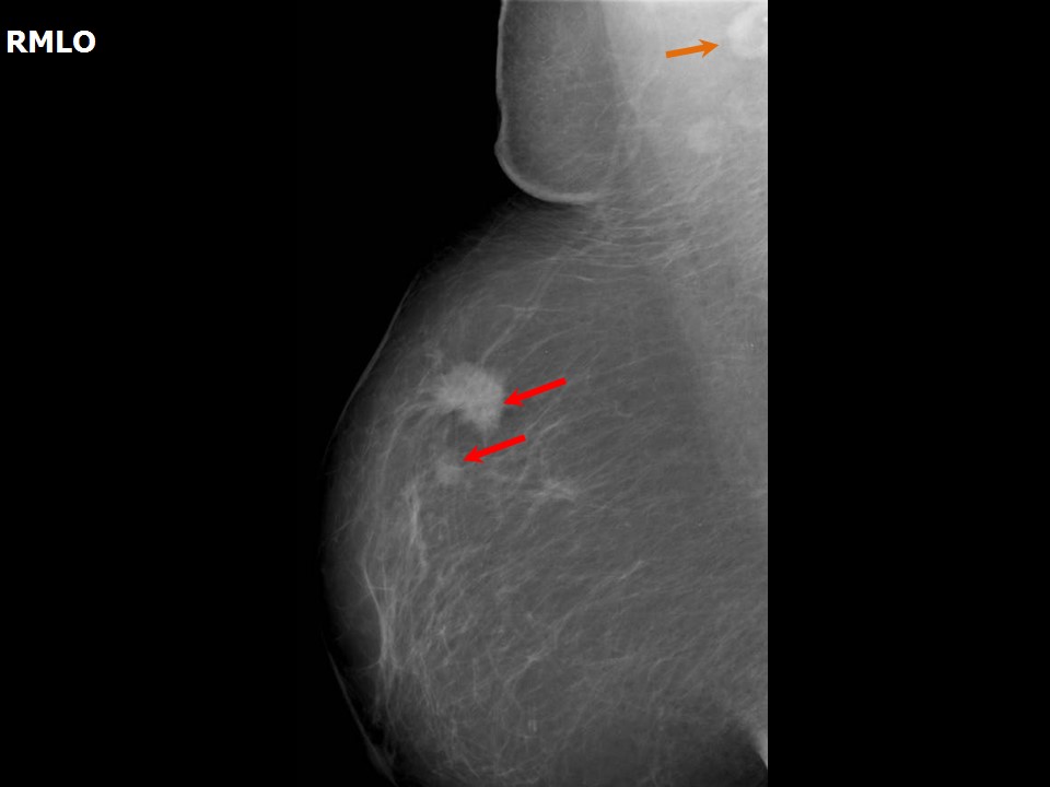 |
| Breast composition: | ACR category a (the breasts are almost entirely fatty) | Mammography features: |
| ‣ Location of the lesion: | Right breast, upper outer quadrant at 11 oclock, middle third |
| ‣ Mass: | |
| • Number: | 2 |
| • Size: | 2.5 × 2.0 cm and 0.7 cm in greatest dimension |
| • Shape: | Irregular |
| • Margins: | Spiculated |
| • Density: | High |
| ‣ Calcifications: | |
| • Typically benign: | None |
| • Suspicious: | None |
| • Distribution: | None |
| ‣ Architectural distortion: | None |
| ‣ Asymmetry: | None |
| ‣ Intramammary node: | None |
| ‣ Skin lesion: | None |
| ‣ Solitary dilated duct: | None |
| ‣ Associated features: | Axillary lymph node |
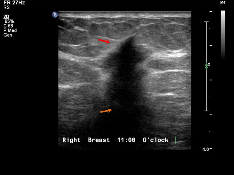
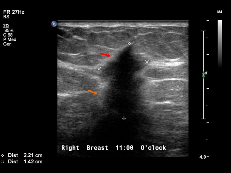
Ultrasound:
 |  |
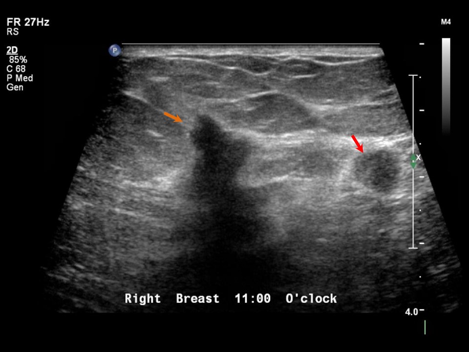 |  |
| Ultrasound features: Right breast, upper outer quadrant at 11 oclock | |
| ‣ Mass | |
| • Location: | Right breast, upper outer quadrant at 11 oclock |
| • Number: | 2 |
| • Size: | 2.2 × 1.4 cm and 0.8 × 0.6 cm |
| • Shape: | Irregular |
| • Orientation: | Not parallel |
| • Margins: | Angular |
| • Echo pattern: | Hypoechoic |
| • Posterior features: | Posterior shadowing |
| ‣ Calcifications: | None |
| ‣ Associated features: | Internal vascularity |
| ‣ Special cases: | None |
BI-RADS:
BI-RADS Category: 5 (highly suggestive of malignancy)Further assessment:
Further assessment advised: Referral for cytologyCytology:
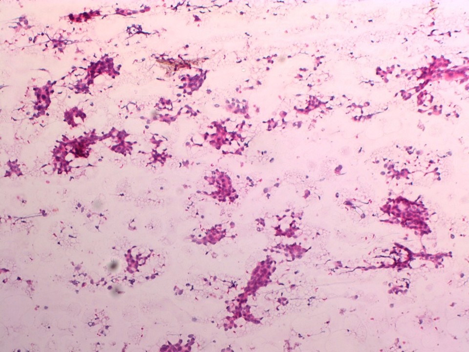 |
| Cytology features: | |
| ‣ Type of sample: | FNAC |
| ‣ Site of biopsy: | |
| • Laterality: | Right |
| • Quadrant: | Upper outer |
| • Localization technique: | Palpation |
| • Nature of aspirate: | Whitish |
| ‣ Cytological description: | Smears are cellular and reveal many loosely cohesive clusters and sheets of large atypical cells with large, pleomorphic and hyperchromatic nuclei and moderate amount of cytoplasm. Nucleoli are seen in a few cells. There are many stromal fragments |
| ‣ Reporting category: | Malignant |
| ‣ Diagnosis: | Carcinoma breast |
| ‣ Comments: | None |
Histopathology:
MRM
 |  |
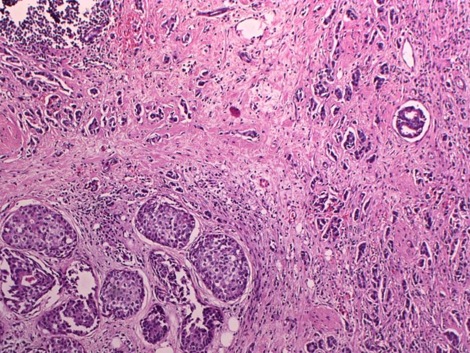 |
| Histopathology features: | |
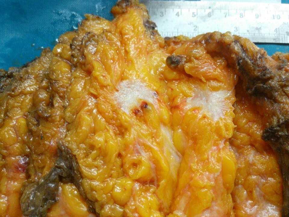
| ‣ Specimen type: | MRM |
| ‣ Laterality: | Right |
| ‣ Macroscopy: | Specimen (24.0 × 18.0 × 7.0 cm), with overlying skin flap (19.0 × 8.0 cm). The nipple and areola are unremarkable. On serial sectioning, a firm, gritty, greyish white tumour (2.5 × 2.0 × 2.0 cm) is identified located in the upper outer quadrant. It is located 2.5 cm from the skin and 1.5 cm from the base. Another firm, gritty, greyish white, nodular tumour (1.0 × 0.8 × 0.6 cm) is located 1.5 cm from the primary tumour and lower and lateral to it. It is 4.0 cm from the base and 1.0 cm below the overlying skin |
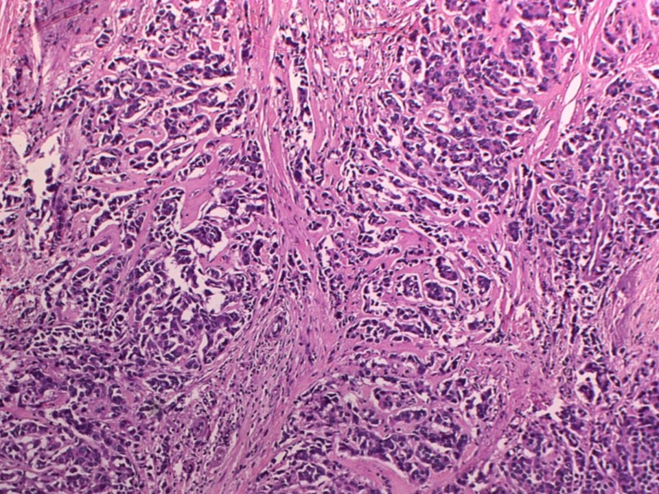
| ‣ Histological type: | Invasive breast carcinoma |
| ‣ Histological grade: | Grade 2 (3 + 2 + 2 = 7) |
| ‣ Mitosis: | 16 |
| ‣ Maximum invasive tumour size: | 2.5 cm |
| ‣ Lymph node status: | 1/22 |
| ‣ Peritumoural lymphovascular invasion: | Absent |
| ‣ DCIS/EIC: | DCIS of solid and comedo type high grade |
| ‣ Margins: | Free of tumour |
| ‣ Pathological stage: | pT2(2)N1 |
| ‣ Biomarkers: | |
| ‣ Comments: |
Case summary:
| Postmenopausal woman presented with a lump in the right breast. Diagnosed as right breast carcinoma (multifocal) with right axillary metastatic node, BI-RADS 5 on imaging, as breast carcinoma on cytology, and as invasive breast carcinoma of no special type, pT2(2)N1 on histopathology. |
Learning points:
|