


Home / Training / Manuals / Atlas of breast cancer early detection / Cases
Atlas of breast cancer early detection
Filter by language: English / Русский
Go back to the list of case studies
.png) Click on the pictures to magnify and display the legends
Click on the pictures to magnify and display the legends
| Case number: | 071 |
| Age: | 74 |
| Clinical presentation: | Postmenopausal woman with a family history of breast cancer in her mother, presented with a left breast lump. On examination, she had two lumps palpable in her left breast. The larger lump was in the lower inner quadrant, 3 cm in diameter, hard, and fixed to the muscle. The other lump was 2 cm in diameter and in the upper outer quadrant. |
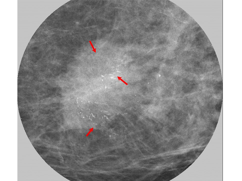
Mammography:
 |  |
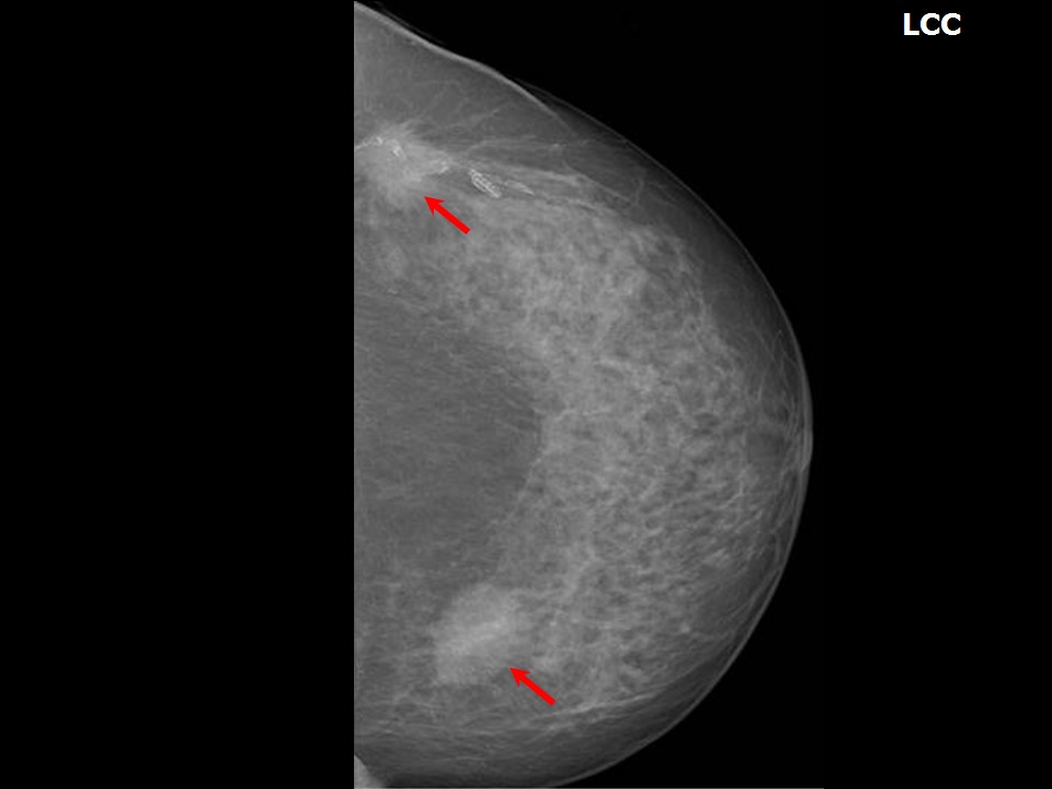 |
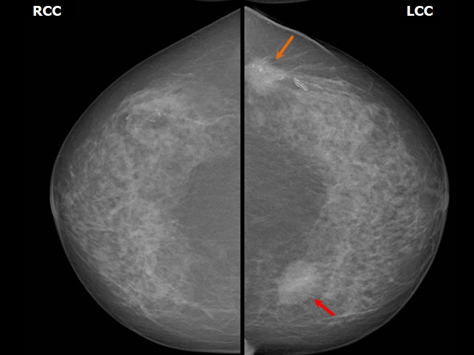
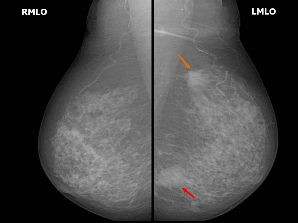
| Breast composition: | ACR category b (there are scattered areas of fibroglandular density) | Mammography features: |
| ‣ Location of the lesion: | Left breast, upper outer quadrant at 2 oclock, posterior third |
| ‣ Mass: | |
| • Number: | 1 |
| • Size: | 2.8 × 2.3 cm |
| • Shape: | Irregular |
| • Margins: | Spiculated |
| • Density: | High |
| ‣ Calcifications: | |
| • Typically benign: | None |
| • Suspicious: | Suspicious calcifications |
| • Distribution: | Groups of pleomorphic calcifications within the mass |
| ‣ Architectural distortion: | None |
| ‣ Asymmetry: | None |
| ‣ Intramammary node: | None |
| ‣ Skin lesion: | None |
| ‣ Solitary dilated duct: | None |
| ‣ Associated features: | None |
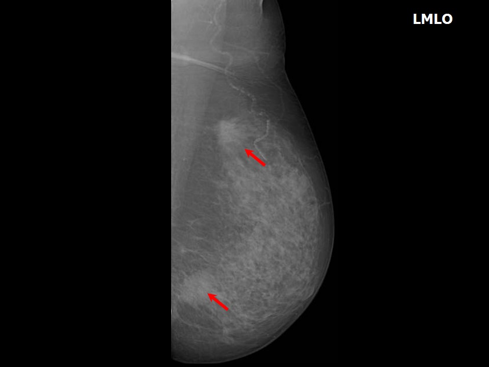 |  |
| Breast composition: | ACR category b (there are scattered areas of fibroglandular density) | Mammography features: |
| ‣ Location of the lesion: | Left breast, lower inner quadrant at 89 oclock, posterior third |
| ‣ Mass: | |
| • Number: | 1 |
| • Size: | 3.0 × 2.5 cm |
| • Shape: | Irregular |
| • Margins: | Spiculated |
| • Density: | High |
| ‣ Calcifications: | |
| • Typically benign: | None |
| • Suspicious: | Fine linear and pleomorphic microcalcifications |
| • Distribution: | Groups of pleomorphic calcifications within the mass |
| ‣ Architectural distortion: | None |
| ‣ Asymmetry: | Focal |
| ‣ Intramammary node: | None |
| ‣ Skin lesion: | None |
| ‣ Solitary dilated duct: | None |
| ‣ Associated features: | Fine linear pleomorphic microcalcifications within the mass |
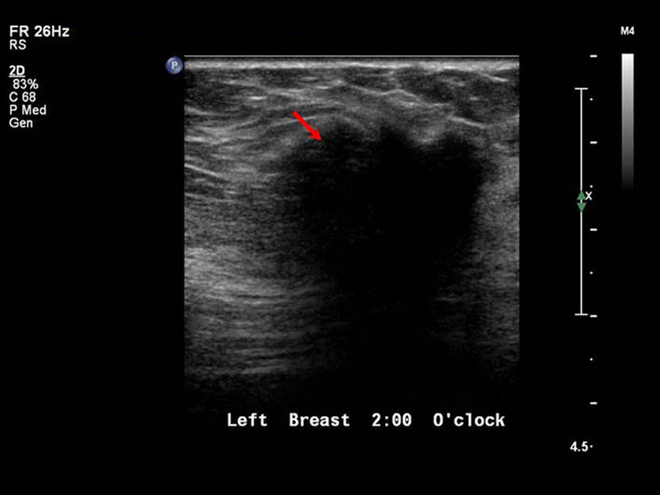
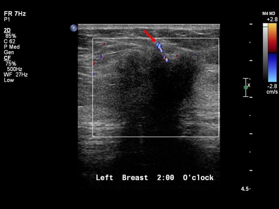
Ultrasound:
 |  |
 | 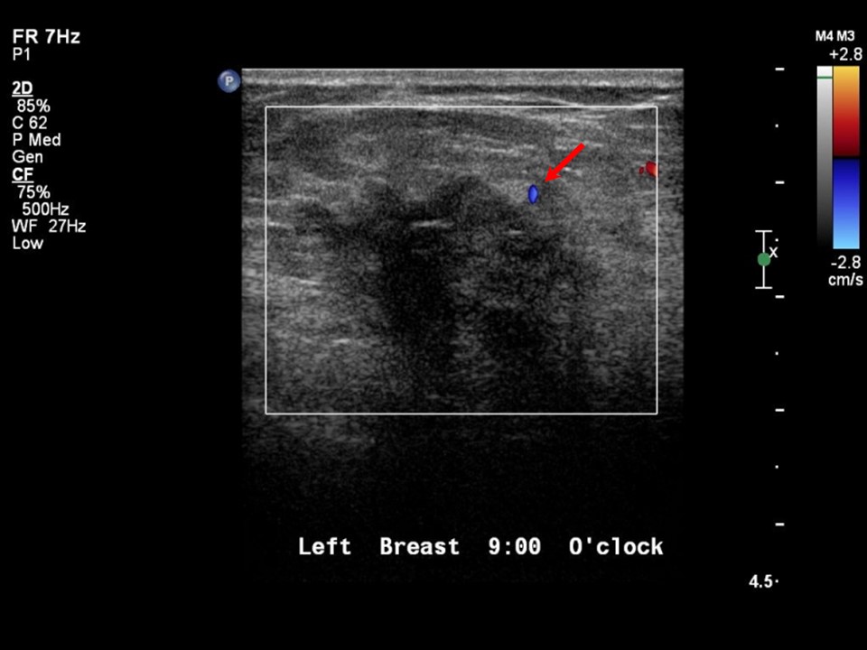 |
| Ultrasound features: Left breast, upper outer quadrant at 2 oclock and inner quadrant at 89 oclock | |
| ‣ Mass | |
| • Location: | Left breast, upper outer quadrant at 2 oclock and inner quadrant at 89 oclock |
| • Number: | 2 |
| • Size: |
|
| • Shape: | Irregular |
| • Orientation: | Not parallel |
| • Margins: | Spiculated |
| • Echo pattern: | Hypoechoic |
| • Posterior features: | Posterior shadowing |
| ‣ Calcifications: | Calcifications in mass at 9 oclock |
| ‣ Associated features: | Internal vascularity |
| ‣ Special cases: | None |
BI-RADS:
BI-RADS Category: 5 (highly suggestive of malignancy)Further assessment:
Further assessment advised: Referral for cytologyCytology:
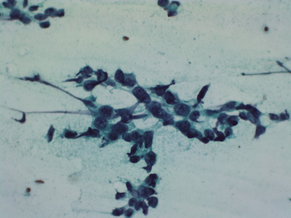 |
| Cytology features: | |
| ‣ Type of sample: | FNAC (solid lesion) |
| ‣ Site of biopsy: | |
| • Laterality: | Left |
| • Quadrant: | Upper outer |
| • Localization technique: | Palpation |
| • Nature of aspirate: | Whitish |
| ‣ Cytological description: | Paucicellular, a few scattered single isolated malignant seen in the smears. Many fibrous tissue fragments seen |
| ‣ Reporting category: | Malignant |
| ‣ Diagnosis: | Carcinoma |
| ‣ Comments: | None |
Histopathology:
MRM
 |  |
| Histopathology features: | |
| ‣ Specimen type: | MRM |
| ‣ Laterality: | Left |
| ‣ Macroscopy: | On serial sectioning, a greyish white tumour (3.5 × 2.5 × 2.5 cm) was identified. Two centimetres lateral to this tumour is another tumour (2.0 × 2.0 × 1.5 cm) |
| ‣ Histological type: | Invasive carcinoma of no special type |
| ‣ Histological grade: | Grade 2 (3 + 2 + 2 = 7) |
| ‣ Mitosis: | 12 |
| ‣ Maximum invasive tumour size: | Largest 4.0 cm in greatest dimension (smaller one 2.0 cm) |
| ‣ Lymph node status: | 0/15 |
| ‣ Peritumoural lymphovascular invasion: | Not identified |
| ‣ DCIS/EIC: | Comedo, cribriform high grade |
| ‣ Margins: | Free of tumour |
| ‣ Pathological stage: | pT2(2)N0 |
| ‣ Biomarkers: | |
| ‣ Comments: | Stroma shows dense fibrosis and desmoplasia. Focal area of calcification present |
Case summary:
| Postmenopausal woman presented with two palpable lumps in the upper inner and lower outer quadrants of left breast. Diagnosed as left breast multicentric carcinoma, BI-RADS 5 on imaging, as carcinoma breast on cytology, and as invasive breast carcinoma of no special type, pT2(2)N0 on histopathology. |
Learning points:
|