


Home / Training / Manuals / Atlas of breast cancer early detection / Cases
Atlas of breast cancer early detection
Filter by language: English / Русский
Go back to the list of case studies
.png) Click on the pictures to magnify and display the legends
Click on the pictures to magnify and display the legends
| Case number: | 062 |
| Age: | 63 |
| Clinical presentation: | Postmenopausal woman with average risk of developing breast cancer presented with a lump in the left breast. On examination, she was found to have a hard 3 cm lump in her left breast. Axillary nodes were not palpable. |
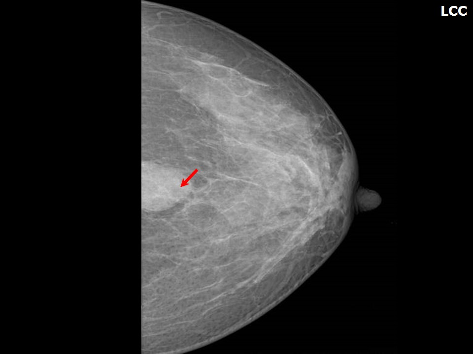
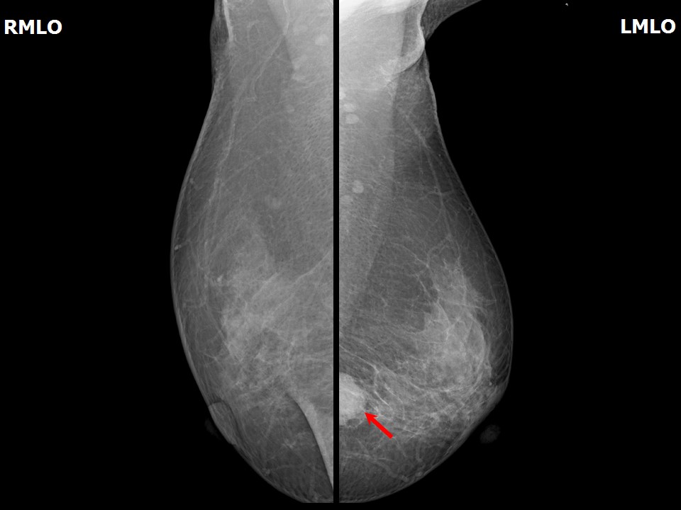
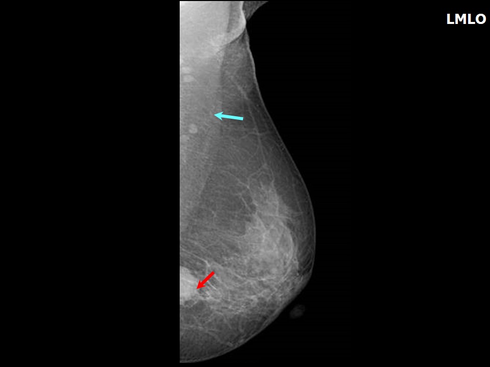
Mammography:
 |  |
 |  |
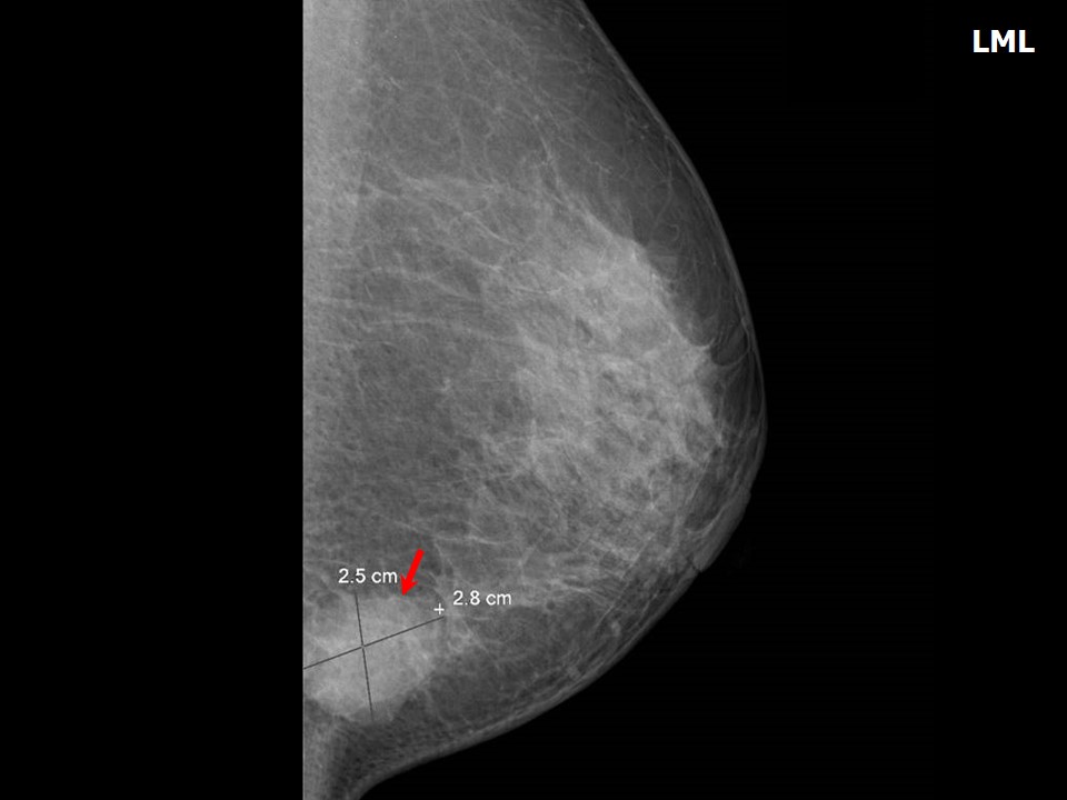 |
| Breast composition: | ACR category b (there are scattered areas of fibroglandular density) | Mammography features: |
| ‣ Location of the lesion: | Left breast, lower quadrants at 6 oclock, posterior third |
| ‣ Mass: | |
| • Number: | 1 |
| • Size: | 2.8 × 2.5 cm |
| • Shape: | Irregular |
| • Margins: | Indistinct |
| • Density: | High |
| ‣ Calcifications: | |
| • Typically benign: | None |
| • Suspicious: | None |
| • Distribution: | None |
| ‣ Architectural distortion: | None |
| ‣ Asymmetry: | None |
| ‣ Intramammary node: | None |
| ‣ Skin lesion: | None |
| ‣ Solitary dilated duct: | None |
| ‣ Associated features: | None |
Ultrasound:
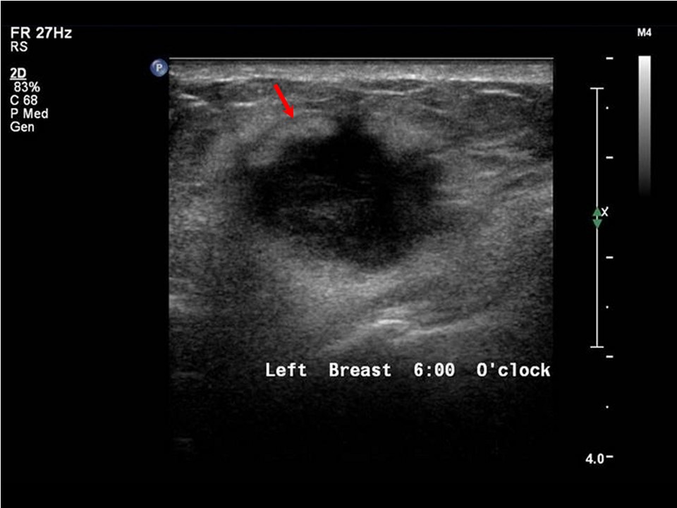 | 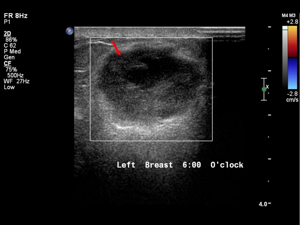 |
| Ultrasound features: Left breast, lower quadrants at 6 oclock | |
| ‣ Mass | |
| • Location: | Left breast, lower quadrants at 6 oclock |
| • Number: | 1 |
| • Size: | 2.5 × 1.6 cm |
| • Shape: | Oval |
| • Orientation: | Parallel |
| • Margins: | Spiculated |
| • Echo pattern: | Heteroechoic |
| • Posterior features: | No posterior features |
| ‣ Calcifications: | None |
| ‣ Associated features: | Minimal internal vascularity |
| ‣ Special cases: | None |
BI-RADS:
BI-RADS Category: 5 (highly suggestive of malignancy)Further assessment:
Further assessment advised: Referral for cytologyCytology:
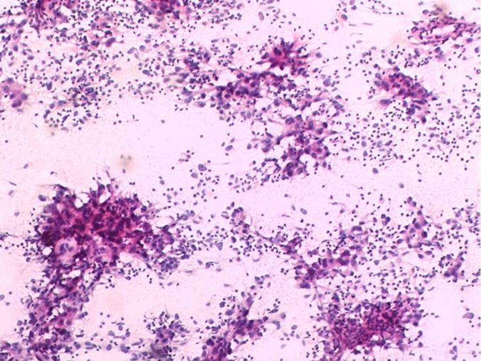 |
| Cytology features: | |
| ‣ Type of sample: | FNAC |
| ‣ Site of biopsy: | |
| • Laterality: | Left |
| • Quadrant: | Lower at 6 oclock |
| • Localization technique: | Palpation, soft feel to the needling |
| • Nature of aspirate: | Whitish |
| ‣ Cytological description: | Highly cellular smears show poorly cohesive clusters and dispersed malignant cells. These cells are large and pleomorphic with prominent nucleoli. Many lymphocytes are seen in the background |
| ‣ Reporting category: | Malignant |
| ‣ Diagnosis: | Carcinoma high grade. Medullary carcinoma to be considered in view of the lymphocytes |
| ‣ Comments: | None |
Histopathology:
Breast-conserving surgery
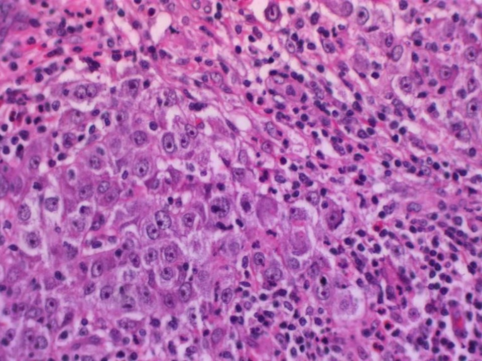 |
| Histopathology features: | |
| ‣ Specimen type: | Breast-conserving surgery |
| ‣ Laterality: | Left |
| ‣ Macroscopy: | Lumpectomy specimen (7.5 × 6.0 × 3.5 cm) oriented with long suture laterally and short suture superiorly. Skin flap is 7.0 × 2.5 cm. On serial sectioning a firm greyish white tumour (2.5 × 2.1 × 2.5 cm) is identified with an intact cystic cavity (1.6 × 1.2 cm) filled with blood |
| ‣ Histological type: | Invasive breast carcinoma of no special type with medullary pattern |
| ‣ Histological grade: | Grade 3 (3 + 2 + 3 = 8) |
| ‣ Mitosis: | 28 |
| ‣ Maximum invasive tumour size: | 2.5 cm in greatest dimension |
| ‣ Lymph node status: | 0/29 |
| ‣ Peritumoural lymphovascular invasion: | Present |
| ‣ DCIS/EIC: | Absent |
| ‣ Margins: | Free of tumour |
| ‣ Pathological stage: | pT2N0 |
| ‣ Biomarkers: | |
| ‣ Comments: |
Case summary:
| Postmenopausal woman presented with lump in the left breast. Diagnosed as left breast carcinoma, BI-RADS 5 on imaging, as breast carcinoma on cytology, and as invasive breast carcinoma of no specific type with medullary pattern, pT2N0 on histopathology. |
Learning points:
|