


Home / Training / Manuals / Atlas of breast cancer early detection / Cases
Atlas of breast cancer early detection
Filter by language: English / Русский
Go back to the list of case studies
.png) Click on the pictures to magnify and display the legends
Click on the pictures to magnify and display the legends
| Case number: | 070 |
| Age: | 45 |
| Clinical presentation: | Premenopausal woman with average risk of developing breast cancer presented with a lump in the left breast. On examination, she had a freely mobile lump in the upper inner quadrant of the left breast. |
Mammography:
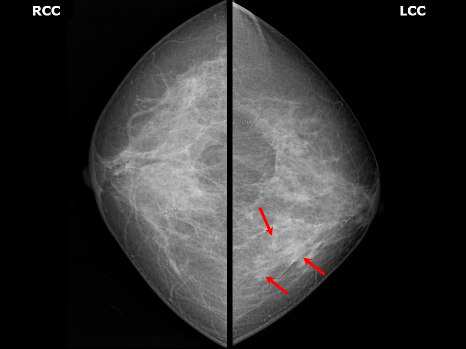 | 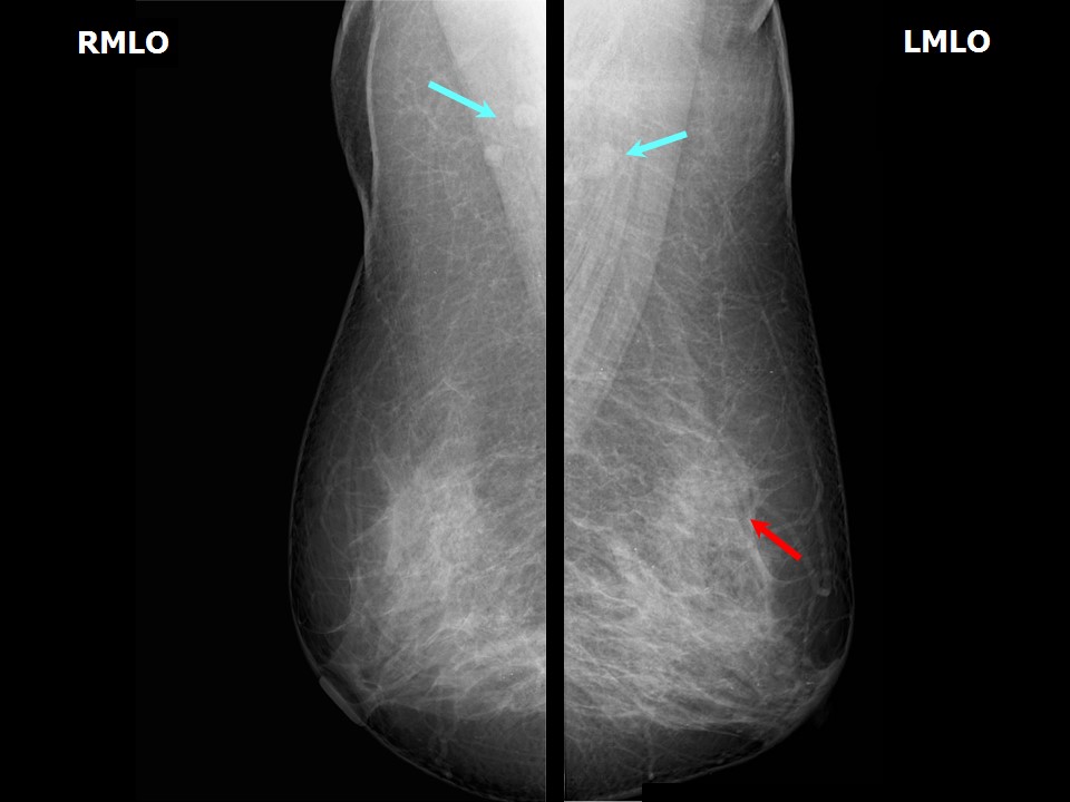 |
| Breast composition: | ACR category c (the breasts are heterogeneously dense, which may obscure small masses) | Mammography features: |
| ‣ Location of the lesion: | Left breast, upper inner quadrant at 11 oclock, middle third |
| ‣ Mass: | |
| • Number: | 1 |
| • Size: | Not measurable, obscured by the glandular parenchyma |
| • Shape: | Irregular |
| • Margins: | Indistinct |
| • Density: | Equal |
| ‣ Calcifications: | |
| • Typically benign: | None |
| • Suspicious: | None |
| • Distribution: | None |
| ‣ Architectural distortion: | Present |
| ‣ Asymmetry: | Focal |
| ‣ Intramammary node: | None |
| ‣ Skin lesion: | None |
| ‣ Solitary dilated duct: | None |
| ‣ Associated features: | Architectural distortion |
Ultrasound:
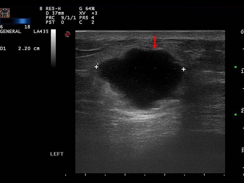 | 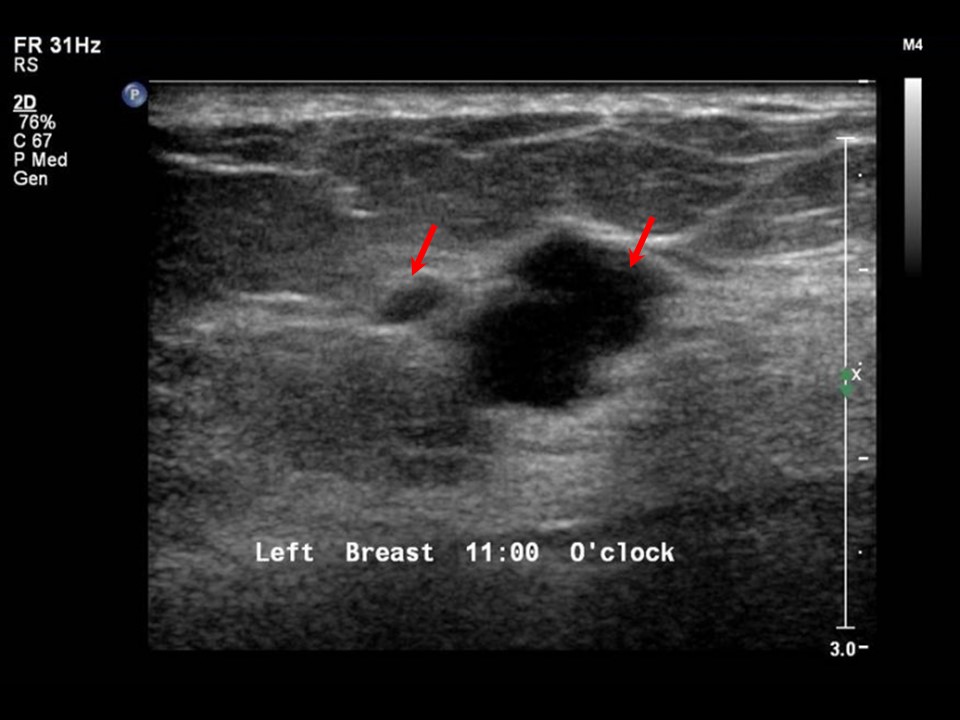 |
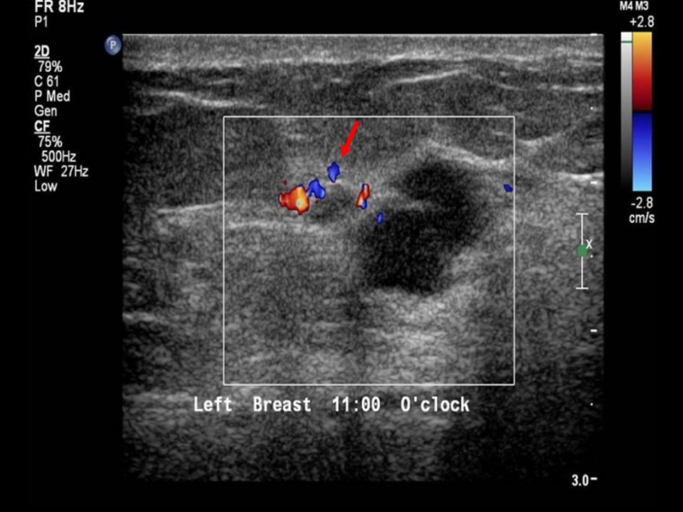 | 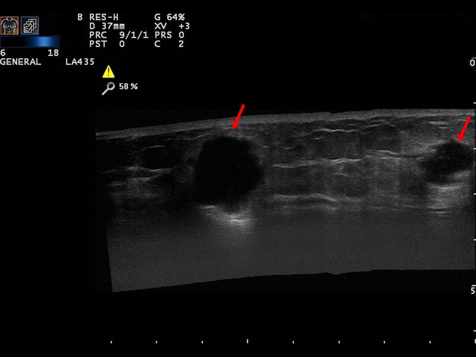 |
| Ultrasound features: Left breast, upper inner quadrant at 1011 oclock | |
| ‣ Mass | |
| • Location: | Left breast, upper inner quadrant at 1011 oclock |
| • Number: | 3 |
| • Size: | Three hypoechoic lesions, taller than wider, with irregular margins in upper inner quadrant:
|
| • Shape: | Irregular |
| • Orientation: | Not parallel |
| • Margins: | Angular |
| • Echo pattern: | Hypoechoic |
| • Posterior features: | No posterior features |
| ‣ Calcifications: | None |
| ‣ Associated features: | Duct changes and internal vascularity |
| ‣ Special cases: | None |
BI-RADS:
BI-RADS Category: 5 (highly suggestive of malignancy)Further assessment:
Further assessment advised: Referral for cytologyCytology:
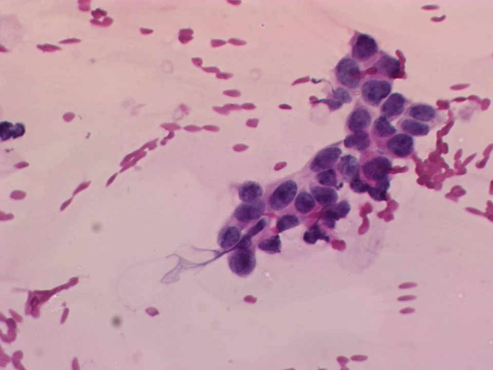 |
| Cytology features: | |
| ‣ Type of sample: | FNAC (solid lesion) |
| ‣ Site of biopsy: | |
| • Laterality: | Left |
| • Quadrant: | Upper inner |
| • Localization technique: | Palpation |
| • Nature of aspirate: | Whitish |
| ‣ Cytological description: | Smear shows loosely cohesive clusters of malignant cells. The cells have marked nuclear pleomorphism, prominent nucleoli, and irregular nuclear membranes. Many lymphocytes are seen in the background |
| ‣ Reporting category: | Malignant |
| ‣ Diagnosis: | Carcinoma high grade |
| ‣ Comments: | None |
Histopathology:
MRM
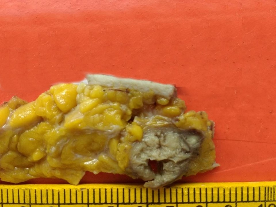 | 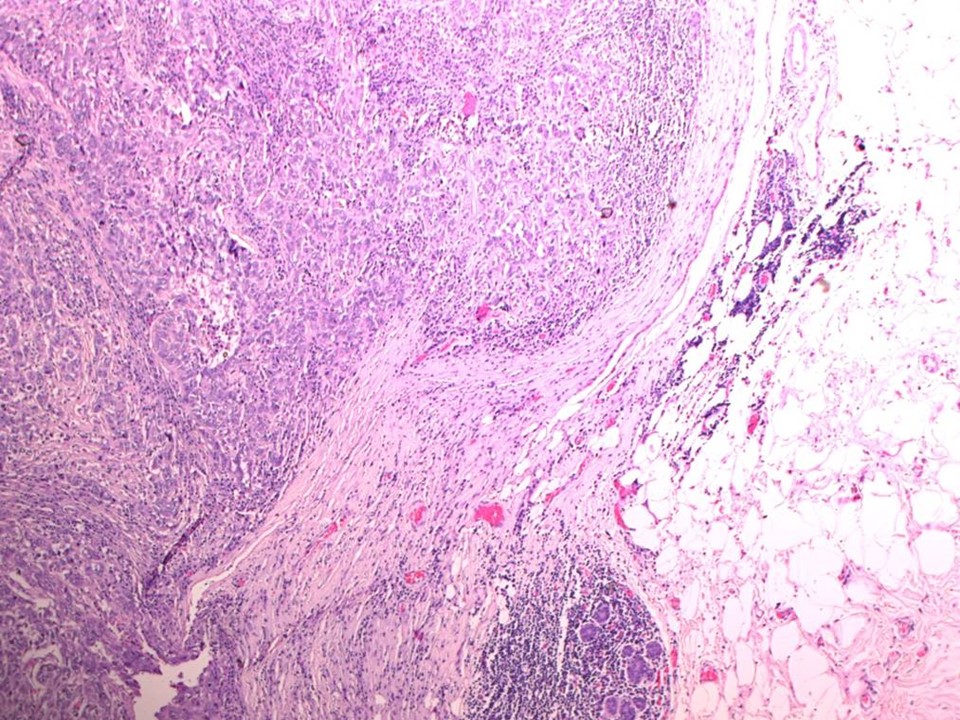 |
| Histopathology features: | |
| ‣ Specimen type: | MRM |
| ‣ Laterality: | Left |
| ‣ Macroscopy: | On serial sectioning, a greyish white, friable, soft necrotic tumour (2.8 × 2.1 × 2.0 cm) is identified located in the upper inner quadrant, 2.0 cm from the skin, which is flush with the base. The tumour was 1.0 cm from the medial surgical resection margin. Another firm greyish white area (1.4 × 1.0 × 1.0 cm) is seen 3.0 cm inferior and lateral to the first tumour. It is 2.8 cm from the skin and 1.0 cm from the base. |
| ‣ Histological type: | Invasive carcinoma of no special type |
| ‣ Histological grade: | Grade 3 (3 + 3 + 2 = 8) |
| ‣ Mitosis: | 14 |
| ‣ Maximum invasive tumour size: | Largest 2.8 cm in greatest dimension (another smaller of 1.4 cm) |
| ‣ Lymph node status: | 0/24 |
| ‣ Peritumoural lymphovascular invasion: | Not identified |
| ‣ DCIS/EIC: | Not identified |
| ‣ Margins: | Free of tumour |
| ‣ Pathological stage: | pT2(2)N0 |
| ‣ Biomarkers: | |
| ‣ Comments: | Tumour also shows extensive necrosis and areas of lymphocytic infiltration. Adjacent breast shows fibrocystic changes. A few areas with ADH and sclerosing adenosis also seen |
Case summary:
| Premenopausal woman presented with lump in the left breast. Diagnosed as left breast carcinoma (multifocal), BI-RADS 5 on imaging, as high-grade breast carcinoma on cytology, and as invasive breast carcinoma of no special type, pT2(2)N0 on histopathology. |
Learning points:
|