


Home / Training / Manuals / Atlas of breast cancer early detection / Cases
Atlas of breast cancer early detection
Filter by language: English / Русский
Go back to the list of case studies
.png) Click on the pictures to magnify and display the legends
Click on the pictures to magnify and display the legends
| Case number: | 128 |
| Age: | 54 |
| Clinical presentation: | Postmenopausal woman with increased risk of developing breast cancer presented with a lump in the right breast and spontaneous right nipple discharge. She had a family history of gastrointestinal tract malignancy. Examination revealed a diffuse lump in the right breast. |
Mammography:
 | 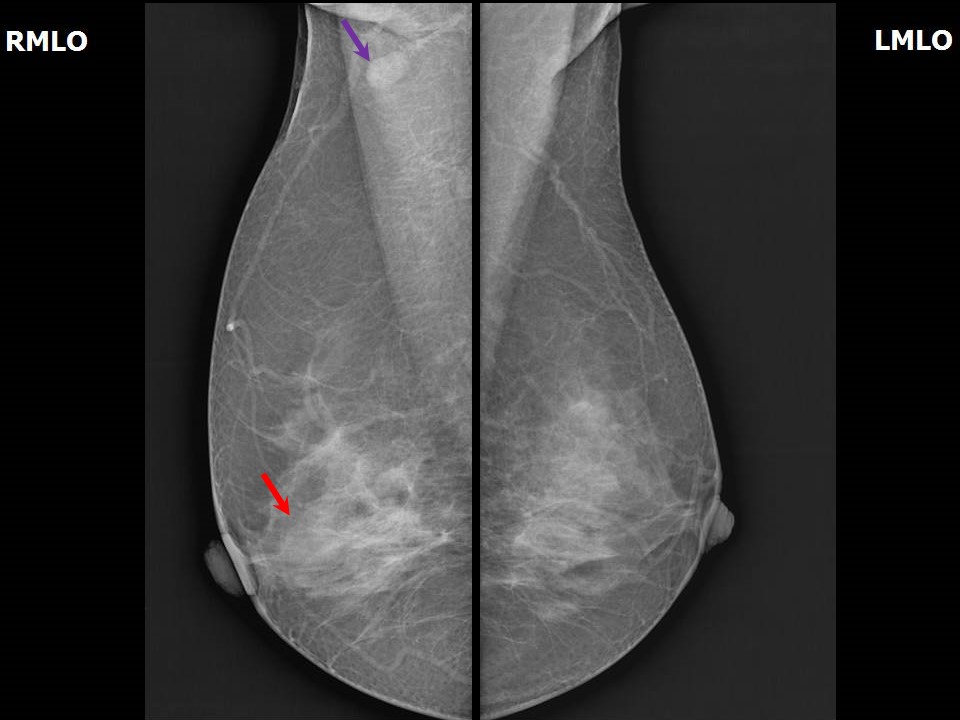 |
| Breast composition: | ACR category c (the breasts are heterogeneously dense, which may obscure small masses) | Mammography features: |
| ‣ Location of the lesion: | Right breast, outer quadrants, central zone, at 9 oclock, middle third |
| ‣ Mass: | |
| • Number: | 1 |
| • Size: | 1.8 × 1.5 cm |
| • Shape: | Oval |
| • Margins: | Obscured |
| • Density: | Equal |
| ‣ Calcifications: | |
| • Typically benign: | None |
| • Suspicious: | None |
| • Distribution: | None |
| ‣ Architectural distortion: | None |
| ‣ Asymmetry: | None |
| ‣ Intramammary node: | None |
| ‣ Skin lesion: | None |
| ‣ Solitary dilated duct: | None |
| ‣ Associated features: | None |
Ultrasound:
 | 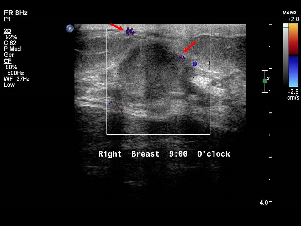 |
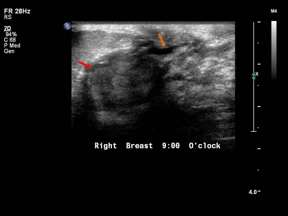 |
| Ultrasound features: Right breast, outer quadrants at 9 oclock, 1.0 cm from nipple and at 0.7 cm skin depth | |
| ‣ Mass | |
| • Location: | Right breast, outer quadrants at 9 oclock, 1.0 cm from nipple and at 0.7 cm skin depth |
| • Number: | 1 |
| • Size: | 1.8 × 1.3 cm |
| • Shape: | Irregular |
| • Orientation: | Parallel |
| • Margins: | Indistinct |
| • Echo pattern: | Hypoechoic |
| • Posterior features: | No posterior features |
| ‣ Calcifications: | None |
| ‣ Associated features: | Duct dilatation and vascularity (vessels in rim) |
| ‣ Special cases: | None |
BI-RADS:
BI-RADS Category: 4B (moderate suspicion of malignancy)Further assessment:
Further assessment advised: Referral for cytologyCytology:
| Cytology features: | |
| ‣ Type of sample: | Nipple discharge |
| ‣ Site of biopsy: | |
| • Laterality: | Right |
| • Quadrant: | |
| • Localization technique: | |
| • Nature of aspirate: | Greenish brown discharge |
| ‣ Cytological description: | Smears reveal cyst macrophages on a proteinaceous background |
| ‣ Reporting category: | Benign |
| ‣ Diagnosis: | Negative for malignant cells |
| ‣ Comments: | None |
| Cytology features: | |
| ‣ Type of sample: | FNAC |
| ‣ Site of biopsy: | |
| • Laterality: | Right |
| • Quadrant: | |
| • Localization technique: | Palpation |
| • Nature of aspirate: | Whitish fluid |
| ‣ Cytological description: | Highly cellular smears reveal numerous highly cohesive sheets of benign ductal epithelial cells with myoepithelial cells. Background shows bare nuclei, foamy macrophages, and blood. A few stromal fragments are also present |
| ‣ Reporting category: | Benign |
| ‣ Diagnosis: | Benign proliferative breast disease |
| ‣ Comments: | None |
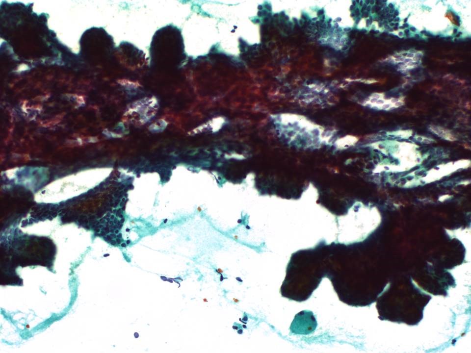 |  |
| Cytology features: | |
| ‣ Type of sample: | FNAC |
| ‣ Site of biopsy: | |
| • Laterality: | Right |
| • Quadrant: | |
| • Localization technique: | Ultrasound-guided FNAC |
| • Nature of aspirate: | Whitish |
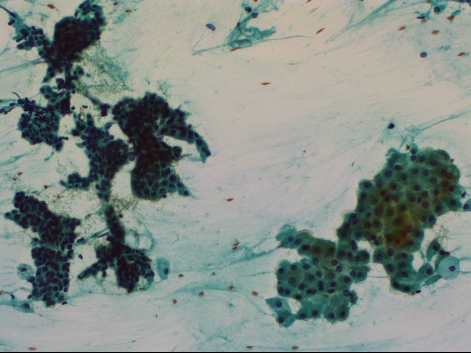
| ‣ Cytological description: | Smears are very cellular. Large, tightly cohesive sheets and clusters of ductal epithelial cells are seen with a few myoepithelial cells. A few papillary clusters and smaller clusters of ductal cells with mild atypia is noted. Many apocrine metaplastic clusters are seen. Background shows many foamy cells and a few bare nuclei |
| ‣ Reporting category: | Atypical, probably benign |
| ‣ Diagnosis: | Proliferative breast lesion with mild atypia, suggestive of papillary neoplasm |
| ‣ Comments: | None |
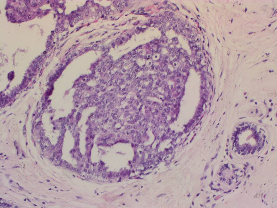
Histopathology:
Lumpectomy
 |
| Histopathology features: | |
| ‣ Specimen type: | Lumpectomy |
| ‣ Laterality: | Right |
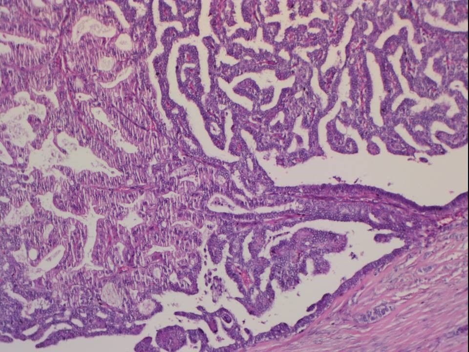
| ‣ Macroscopy: | Lumpectomy specimen (6.5 × 6.0 × 2.2 cm) oriented with long suture laterally and short suture superiorly. Skin flap (3.8 × 1.7 cm). On serial sectioning, a brownish tan, well-circumscribed tumour (1.8 × 1.6 × 0.7 cm) is identified. It is located 1.4 cm from the skin (anterior margin), 3.6 cm from the posterior margin, 1.0 cm from the superior margin, 0.3 cm from the inferior margin, abutting the medial margin, and 4.0 cm from the lateral margin. Adjacent to the tumour a few tiny (< 0.2 cm) firm whitish areas are seen extending from the lateral margin up to the anterolateral margin and abutting the tumour. The remaining breast parenchyma (4.3 × 3.0 × 2.2 cm) is unremarkable. |
| ‣ Histological type: | Complex intraductal papilloma with ADH. On IHC, the luminal cells are ER positive and PR positive in 6080% of cells. Plenty of myoepithelial cells are seen on p63 and smooth muscle myosin heavy chain (SMMHC) |
| ‣ Histological grade: | |
| ‣ Mitosis: | |
| ‣ Maximum invasive tumour size: | |
| ‣ Lymph node status: | |
| ‣ Peritumoural lymphovascular invasion: | |
| ‣ DCIS/EIC: | |
| ‣ Margins: | |
| ‣ Pathological stage: | |
| ‣ Biomarkers: | |
| ‣ Comments: |
Breast revised medial and lateral margin
 |  |
 |  |
| Histopathology features: | |
| ‣ Specimen type: | Breast revised medial and lateral margin |
| ‣ Laterality: | Right |
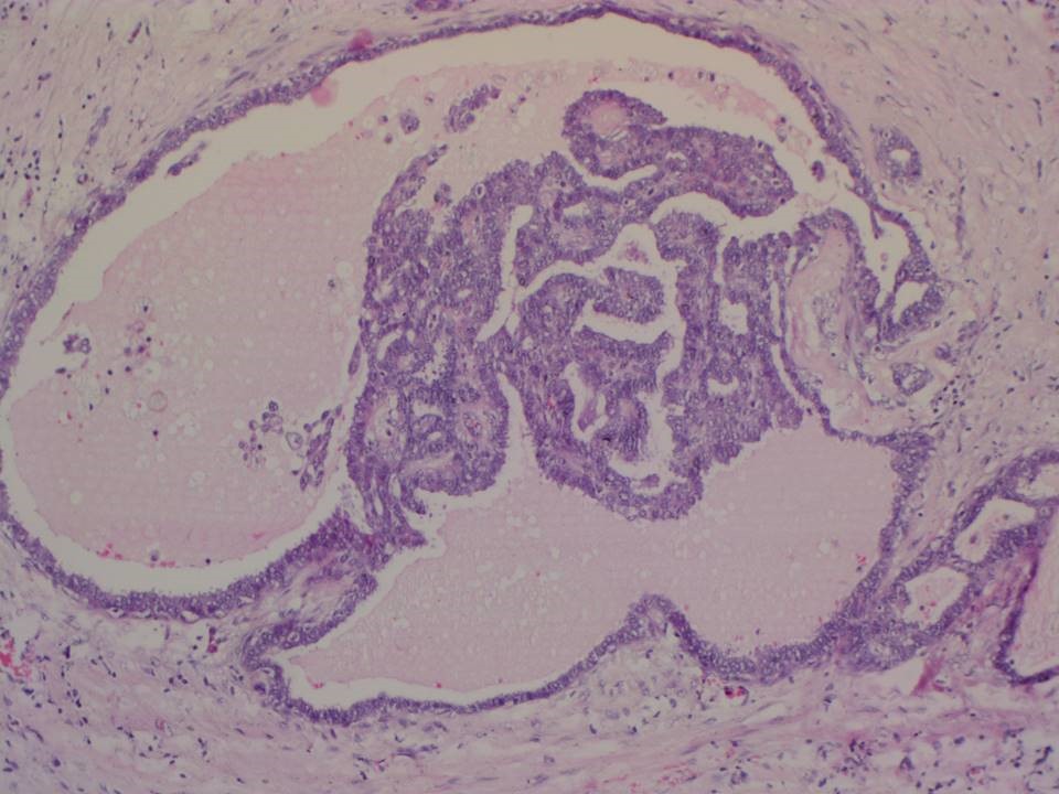
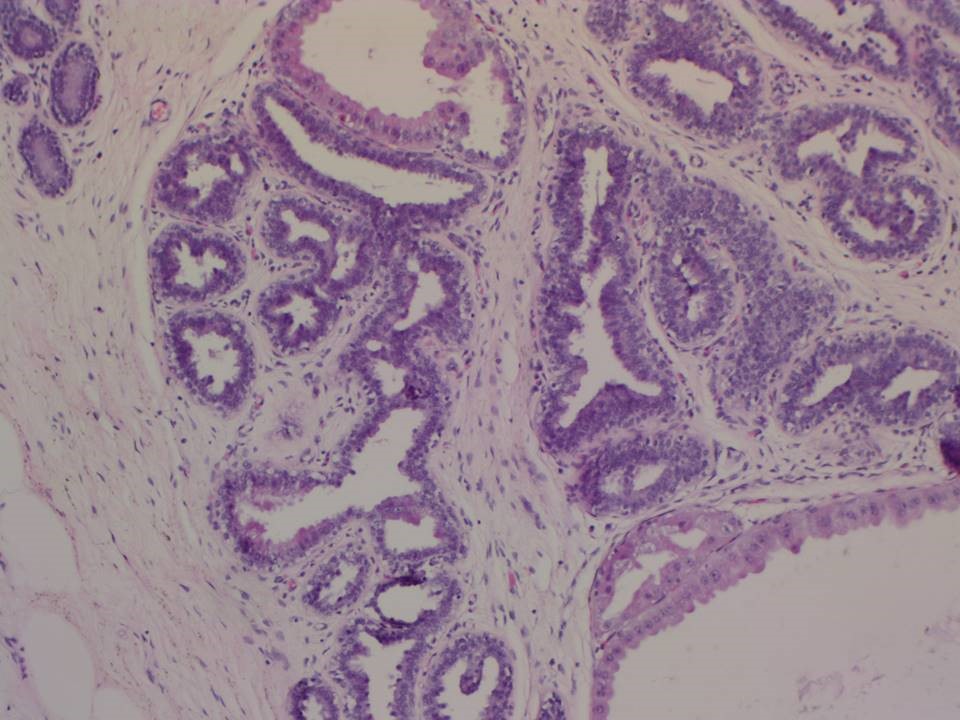
| ‣ Macroscopy: | Revised medial and inferior margin. Single tissue bit (6.0 × 3.5 × 2.0 cm). Cut section shows firm white and fibrosed areas with multiple small whitish nodules (0.20.5 cm) |
| ‣ Histological type: | Shows intraductal papillomas, ADH, fibrocystic changes, columnar cell alteration, and apocrine metaplasia |
| ‣ Histological grade: | |
| ‣ Mitosis: | |
| ‣ Maximum invasive tumour size: | |
| ‣ Lymph node status: | |
| ‣ Peritumoural lymphovascular invasion: | |
| ‣ DCIS/EIC: | |
| ‣ Margins: | |
| ‣ Pathological stage: | |
| ‣ Biomarkers: | |
| ‣ Comments: |
Case summary:
| Postmenopausal woman presented with right breast lump and nipple discharge. Diagnosed as right breast subareolar mass of suspicious morphology with dilated duct, BI-RADS 4B on imaging, as papillary neoplasm on cytology, and as complex intraductal papilloma with ADH on histopathology. |
Learning points:
|