


Home / Training / Manuals / Atlas of breast cancer early detection / Cases
Atlas of breast cancer early detection
Filter by language: English / Русский
Go back to the list of case studies
.png) Click on the pictures to magnify and display the legends
Click on the pictures to magnify and display the legends
| Case number: | 048 |
| Age: | 57 |
| Clinical presentation: | Postmenopausal woman with average risk of developing breast cancer presented with a right breast lump. Examination revealed a 1.7 cm lump in the outer quadrant of the right breast. |
Mammography:
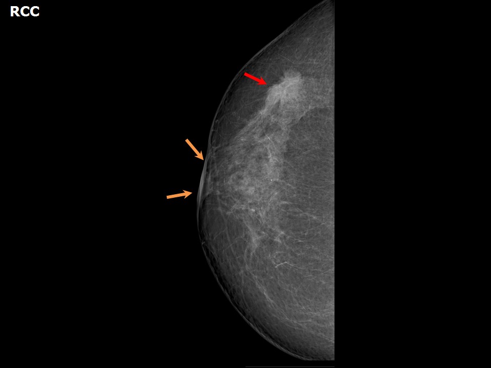 | 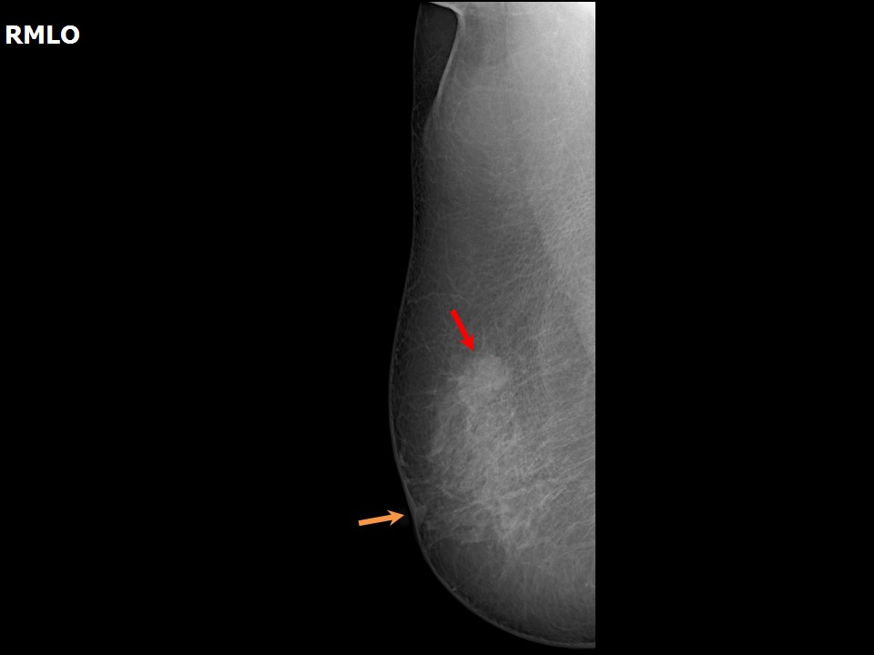 |
| Breast composition: | ACR category b (there are scattered areas of fibroglandular density) | Mammography features: |
| ‣ Location of the lesion: | Right breast, upper outer quadrant at 910 oclock, middle third |
| ‣ Mass: | |
| • Number: | 1 |
| • Size: | 2.9 × 1.5 cm |
| • Shape: | Irregular |
| • Margins: | spiculated |
| • Density: | High |
| ‣ Calcifications: | |
| • Typically benign: | Vascular calcification |
| • Suspicious: | None |
| • Distribution: | None |
| ‣ Architectural distortion: | Present |
| ‣ Asymmetry: | None |
| ‣ Intramammary node: | None |
| ‣ Skin lesion: | None |
| ‣ Solitary dilated duct: | None |
| ‣ Associated features: | Skin thickening, skin retraction, and nipple retraction |
Ultrasound:
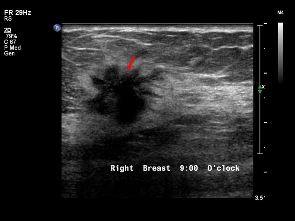 | 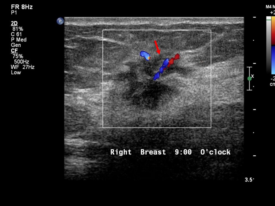 |
| Ultrasound features: Right breast, upper outer quadrant at 9 oclock | |
| ‣ Mass | |
| • Location: | Right breast, upper outer quadrant at 9 oclock |
| • Number: | 1 |
| • Size: | 3.0 × 1.8 × 1.3 cm |
| • Shape: | Irregular |
| • Orientation: | Not parallel |
| • Margins: | Spiculated |
| • Echo pattern: | Hypoechoic |
| • Posterior features: | No posterior features |
| ‣ Calcifications: | None |
| ‣ Associated features: | Rim and internal vascularity in mass |
| ‣ Special cases: | None |
BI-RADS:
BI-RADS Category: 5 (highly suggestive of malignancy)Further assessment:
Further assessment advised: Referral for cytologyCytology:
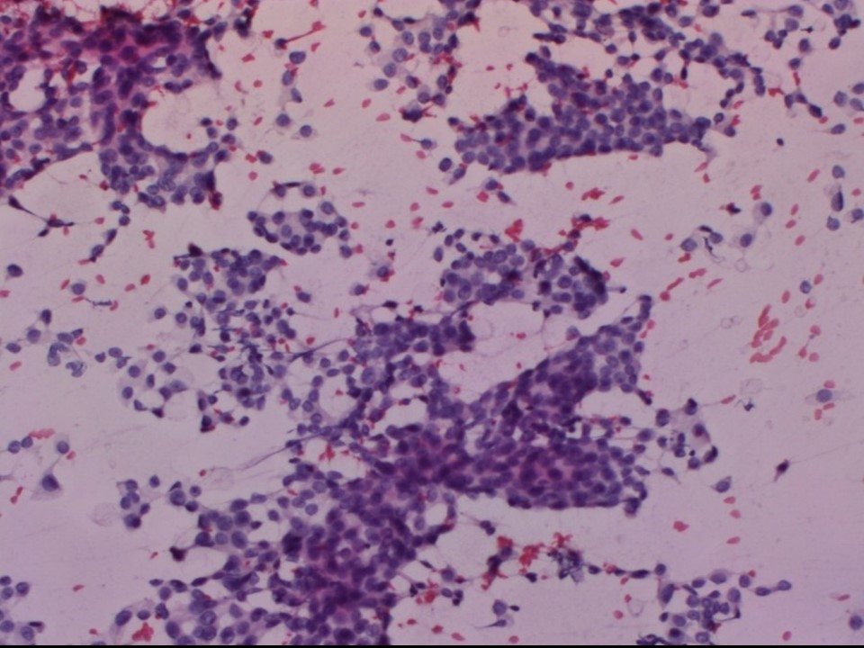 |
| Cytology features: | |
| ‣ Type of sample: | FNAC |
| ‣ Site of biopsy: | |
| • Laterality: | Right |
| • Quadrant: | Upper outer |
| • Localization technique: | Palpation |
| • Nature of aspirate: | Whitish |
| ‣ Cytological description: | Smears are cellular and show many loosely cohesive malignant cell clusters and many isolated single malignant cells. These cells have moderate nuclear pleomorphism with hyperchromatic nuclei and moderate amounts of cytoplasm. Nuclei have inconspicuous nucleoli |
| ‣ Reporting category: | Malignant |
| ‣ Diagnosis: | Carcinoma |
| ‣ Comments: | None |
Histopathology:
Lumpectomy
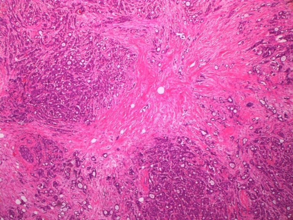 |
| Histopathology features: | |
| ‣ Specimen type: | Lumpectomy |
| ‣ Laterality: | |
| ‣ Macroscopy: | Specimen (6.0 × 4.0 × 4.5 cm) oriented with long suture laterally and short suture superiorly. Skin flap (5.0 × 1.5 cm). On serial sectioning a greyish white tumour (2.0 × 1.5 × 3.0 cm) is identified. Tumour is fused with the base. It is located 2.0 cm from the skin (anterior margin), 0.1 cm from the superior margin, 0.4 cm, from the inferior margin, 2.5 cm from the medial margin, and 1.1 cm from the lateral margin. The posterior margin and superior margin were revised during the same surgery and sent as two additional specimens. |
| ‣ Histological type: | Invasive breast carcinoma |
| ‣ Histological grade: | Grade 2 (3 + 2 + 1 = 6) |
| ‣ Mitosis: | 7 |
| ‣ Maximum invasive tumour size: | 3.0 cm in greatest dimension |
| ‣ Lymph node status: | 1/24 |
| ‣ Peritumoural lymphovascular invasion: | Present |
| ‣ DCIS/EIC: | Absent |
| ‣ Margins: | Free of tumour |
| ‣ Pathological stage: | pT2N1 |
| ‣ Biomarkers: | |
| ‣ Comments: | Section from the medial margin of the lumpectomy specimen shows a focus of atypical epithelial hyperplasia. Sections from specimen labelled hard area superolateral show a sclerosed fibrous area, surrounded by areas of epithelial and myoepithelial hyperplasia |
Case summary:
| Postmenopausal woman presented with right breast lump. Diagnosed as right breast carcinoma, BI-RADS 5 on imaging, as right breast carcinoma on cytology, and as invasive breast carcinoma of no special type, pT2N1 on histopathology. |
Learning points:
|