


Home / Training / Manuals / Atlas of breast cancer early detection / Cases
Atlas of breast cancer early detection
Filter by language: English / Русский
Go back to the list of case studies
.png) Click on the pictures to magnify and display the legends
Click on the pictures to magnify and display the legends
| Case number: | 110 |
| Age: | 60 |
| Clinical presentation: | Postmenopausal woman with average risk of developing breast cancer presented with a lump in the right axilla. Examination revealed normal breasts with axillary lymphadenopathy. |
Mammography:
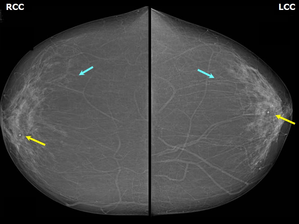 | 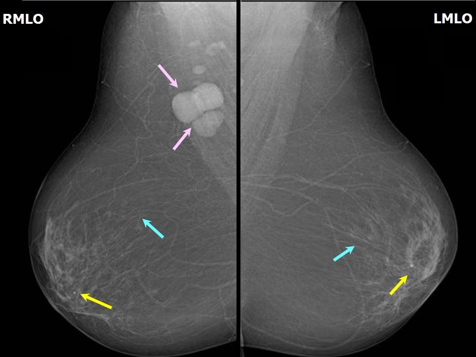 |
| Breast composition: | ACR category a (the breasts are almost entirely fatty) | Mammography features: |
| ‣ Location of the lesion: | Right breast, axillary lymphadenopathy |
| ‣ Mass: | |
| • Number: | 3 |
| • Size: | Largest 2.0 × 1.6 cm |
| • Shape: | None |
| • Margins: | None |
| • Density: | None |
| ‣ Calcifications: | |
| • Typically benign: | Typically benign, round |
| • Suspicious: | None |
| • Distribution: | None |
| ‣ Architectural distortion: | None |
| ‣ Asymmetry: | None |
| ‣ Intramammary node: | None |
| ‣ Skin lesion: | None |
| ‣ Solitary dilated duct: | None |
| ‣ Associated features: | None |
Ultrasound:
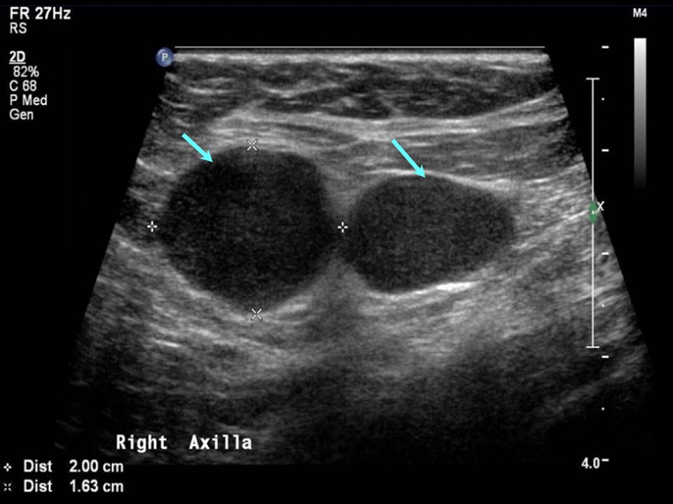 | 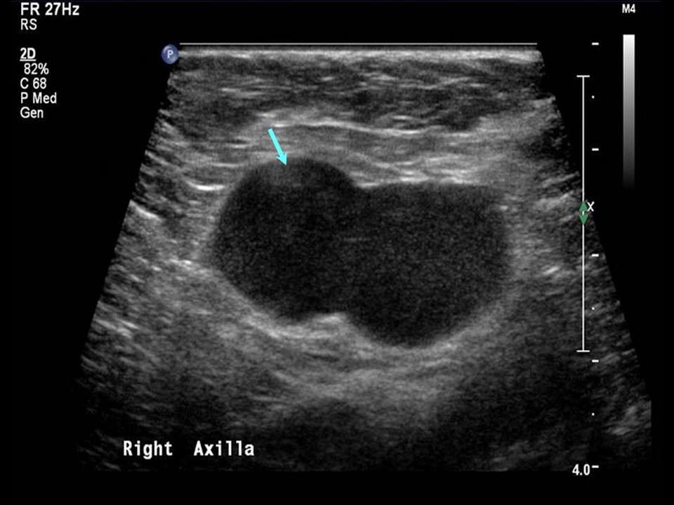 |
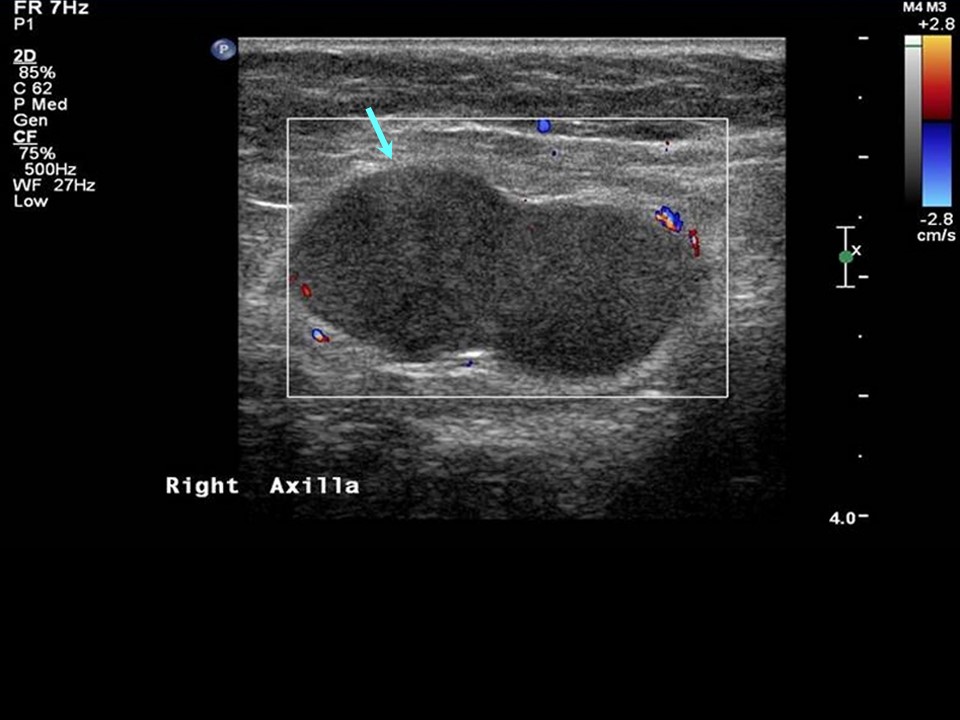 |
| Ultrasound features: Right axillary | |
| ‣ Mass | |
| • Location: | Right axillary |
| • Number: | Multiple |
| • Size: | Largest 2.0 × 1.6 cm |
| • Shape: | None |
| • Orientation: | None |
| • Margins: | None |
| • Echo pattern: | None |
| • Posterior features: | No posterior features |
| ‣ Calcifications: | None |
| ‣ Associated features: | None |
| ‣ Special cases: | None |
BI-RADS:
BI-RADS Category: 4A (low level of suspicion for malignancy)Further assessment:
Further assessment advised: Referral for cytologyCytology:
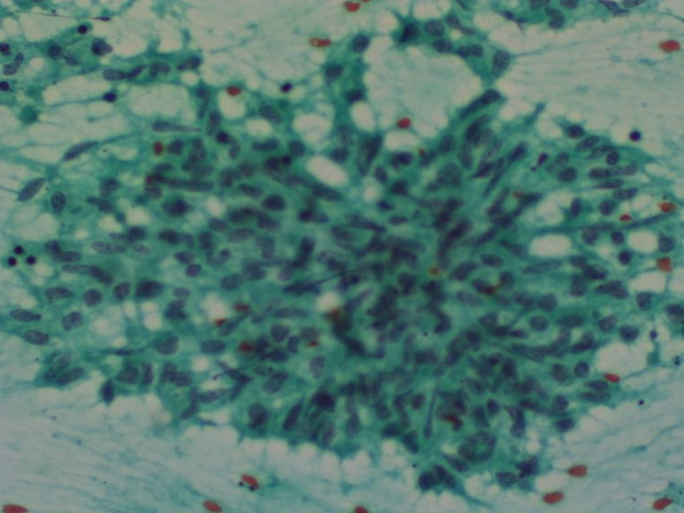 |
| Cytology features: | |
| ‣ Type of sample: | FNAC |
| ‣ Site of biopsy: | |
| • Laterality: | Right |
| • Quadrant: | Axillary nodule |
| • Localization technique: | Ultrasound-guided FNAC |
| • Nature of aspirate: | Whitish |
| ‣ Cytological description: | Epithelioid cell conglomerates with many lymphocytes |
| ‣ Reporting category: | Benign |
| ‣ Diagnosis: | Granulomatous lymphadenitis |
| ‣ Comments: | None |
Histopathology:
Axillary lymph node
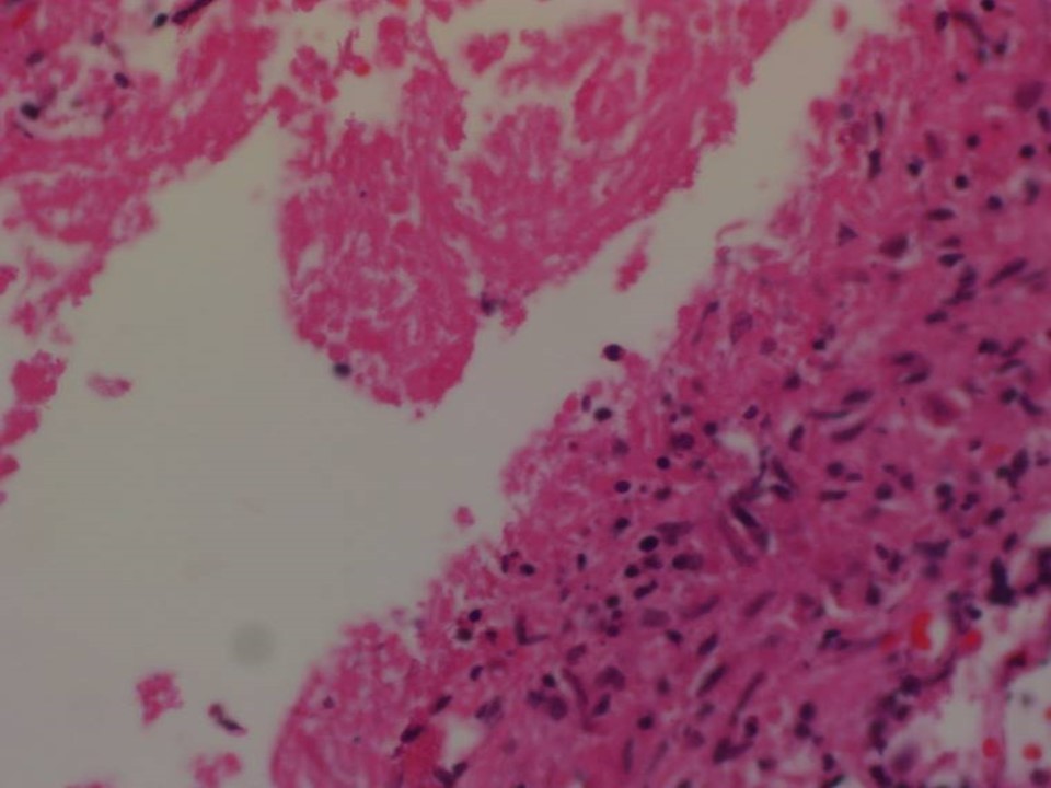 |
| Histopathology features: | |
| ‣ Specimen type: | Axillary lymph node |
| ‣ Laterality: | Right |
| ‣ Macroscopy: | Lymph node (3.5 × 3.5 × 2.0 cm) |
| ‣ Histological type: | Section shows granulomatous inflammation with caseous necrosis, suggestive of tuberculous lymphadenitis |
| ‣ Histological grade: | |
| ‣ Mitosis: | |
| ‣ Maximum invasive tumour size: | |
| ‣ Lymph node status: | |
| ‣ Peritumoural lymphovascular invasion: | |
| ‣ DCIS/EIC: | |
| ‣ Margins: | |
| ‣ Pathological stage: | |
| ‣ Biomarkers: | |
| ‣ Comments: |
Case summary:
| Postmenopausal woman presented with painful lump in right axilla diagnosed as right axillary reactive lymphadenopathy, BI-RADS 4A on imaging, as granulomatous lymphadenitis on cytology, and as tuberculous lymphadenitis on histopathology. |
Learning points:
|