


Home / Training / Manuals / Atlas of breast cancer early detection / Cases
Atlas of breast cancer early detection
Filter by language: English / Русский
Go back to the list of case studies
.png) Click on the pictures to magnify and display the legends
Click on the pictures to magnify and display the legends
| Case number: | 085 |
| Age: | 43 |
| Clinical presentation: | Premenopausal woman presented with a small lump in her left breast. She had no significant family history or risk factors for breast cancer. On examination, a hard 2 cm lump was found in the upper inner quadrant of the left breast. |
Mammography:
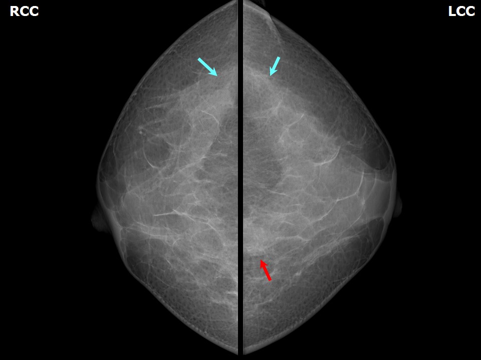 | 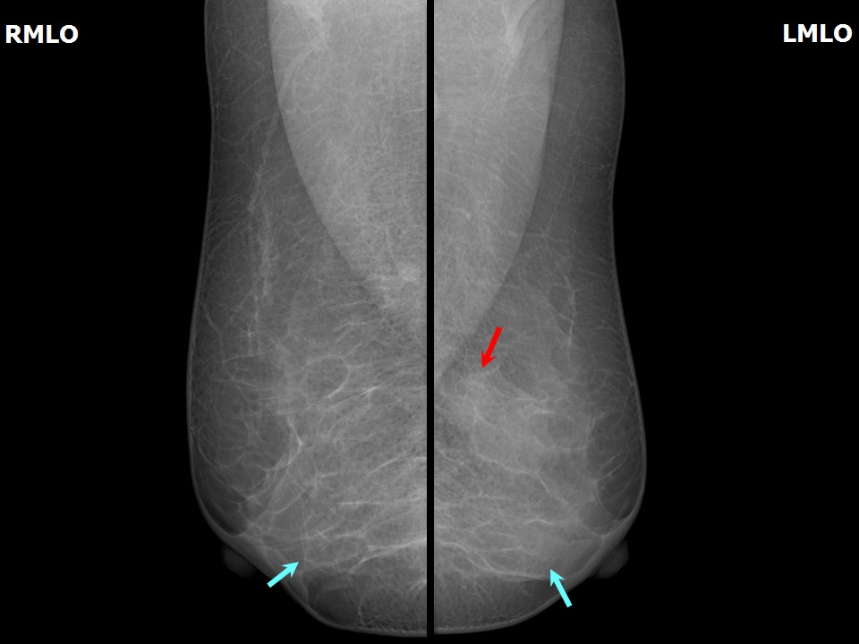 |
| Breast composition: | ACR category c (the breasts are heterogeneously dense, which may obscure small masses) | Mammography features: |
| ‣ Location of the lesion: | Left breast, upper inner quadrant at 11 oclock, posterior third |
| ‣ Mass: | |
| • Number: | 1 |
| • Size: | 2.0 × 1.0 cm |
| • Shape: | Oval |
| • Margins: | Indistinct |
| • Density: | Equal |
| ‣ Calcifications: | |
| • Typically benign: | None |
| • Suspicious: | None |
| • Distribution: | None |
| ‣ Architectural distortion: | None |
| ‣ Asymmetry: | None |
| ‣ Intramammary node: | None |
| ‣ Skin lesion: | None |
| ‣ Solitary dilated duct: | None |
| ‣ Associated features: | Enlarged axillary node with thickened cortex and loss of fatty hilum |
Ultrasound:
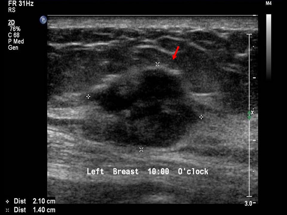 | 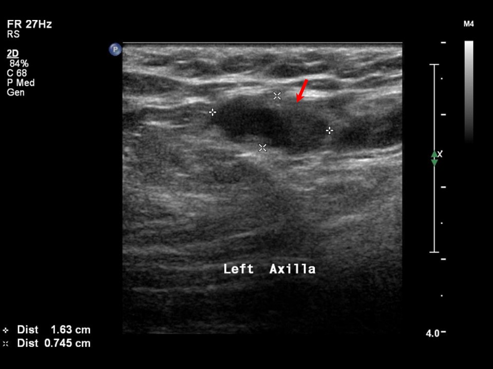 |
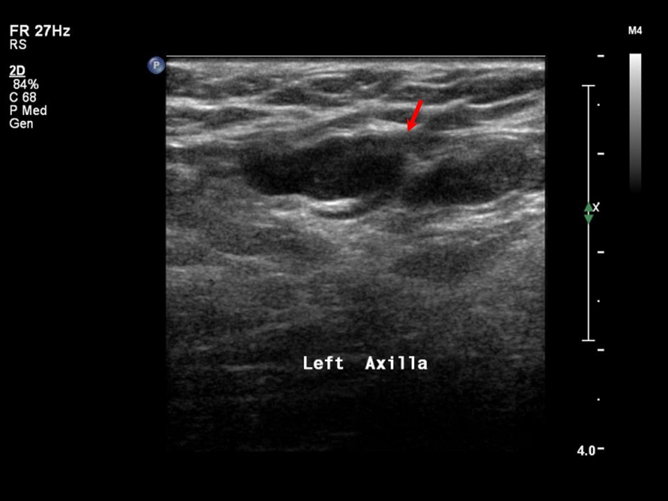 |
| Ultrasound features: Left breast, upper inner quadrant at 1011 oclock | |
| ‣ Mass | |
| • Location: | Left breast, upper inner quadrant at 1011 oclock |
| • Number: | 1 |
| • Size: | 2.1 × 1.4 cm |
| • Shape: | Irregular |
| • Orientation: | Not parallel |
| • Margins: | Angular |
| • Echo pattern: | Hypoechoic |
| • Posterior features: | No posterior features |
| ‣ Calcifications: | None |
| ‣ Associated features: | Enlarged axillary node with thickened cortex and loss of fatty hilum |
| ‣ Special cases: | None |
BI-RADS:
BI-RADS Category: 4C (high suspicion for malignancy)Further assessment:
Further assessment advised: Referral for cytologyCytology:
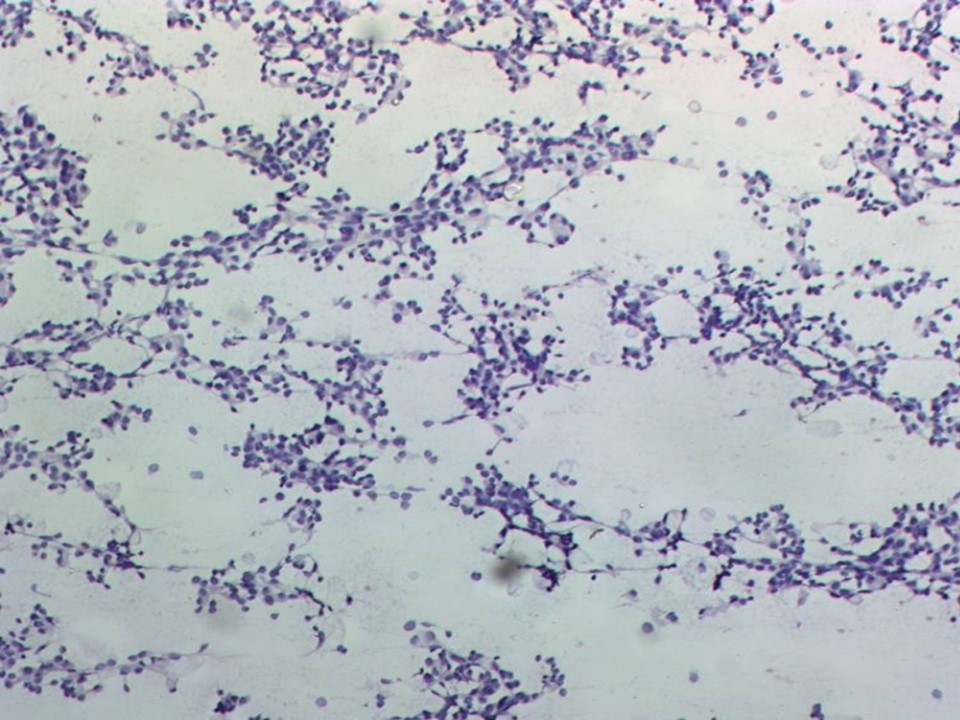 |
| Cytology features: | |
| ‣ Type of sample: | FNAC (solid lesion) |
| ‣ Site of biopsy: | |
| • Laterality: | Left |
| • Quadrant: | Upper inner |
| • Localization technique: | Palpation |
| • Nature of aspirate: | Whitish |
| ‣ Cytological description: | Smear shows a few small clusters and many dissociated malignant cells with moderate amphophilic cytoplasm and large, hyperchromatic and pleomorphic nuclei |
| ‣ Reporting category: | Malignant |
| ‣ Diagnosis: | Carcinoma |
| ‣ Comments: | None |
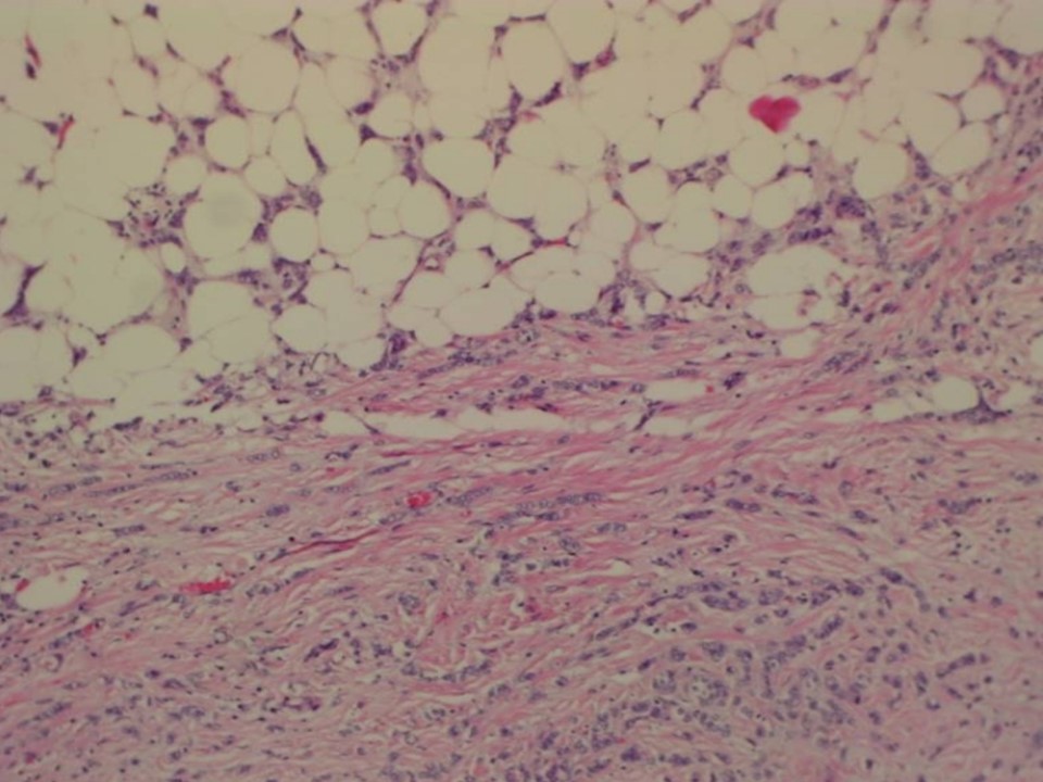
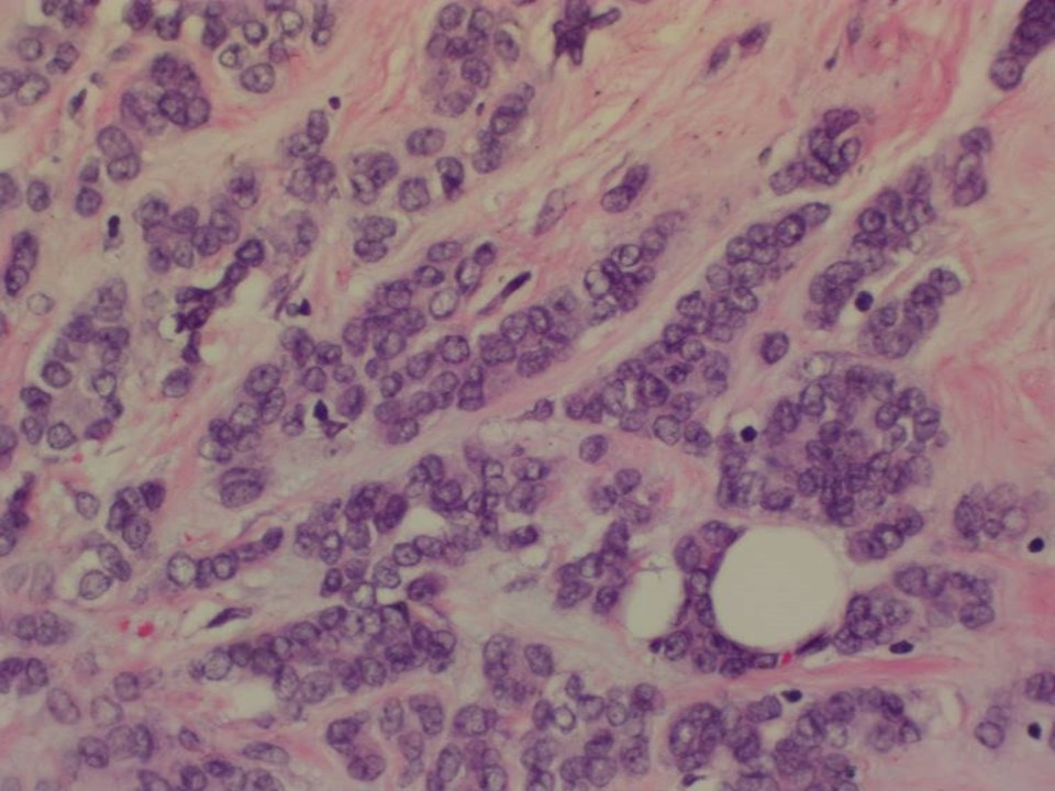
Histopathology:
Breast-conserving surgery
 |  |
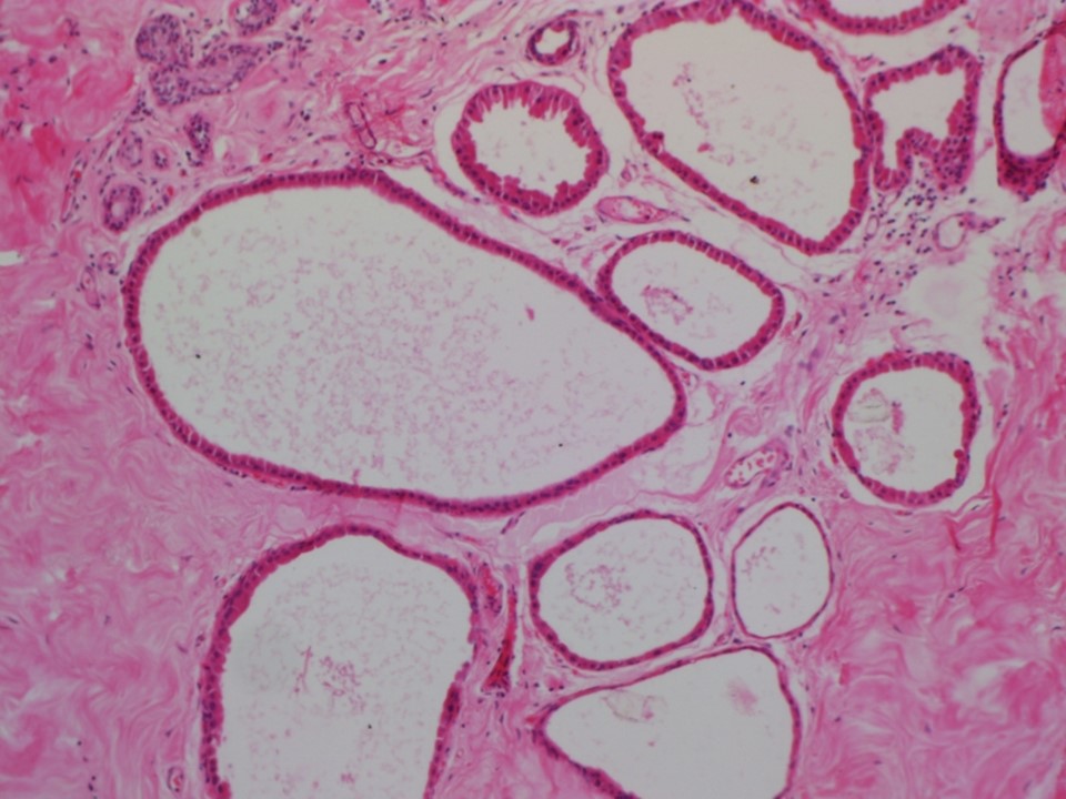 |
| Histopathology features: | |
| ‣ Specimen type: | Breast-conserving surgery |
| ‣ Laterality: | Left |
| ‣ Macroscopy: | The main specimen on serial sectioning showed a greyish white tumour (3.5 × 2.3 × 2.0 cm). Base is 1.5 cm from the tumour. Tumour is located 2.0 cm from the skin, 1.5 cm from the posterior margin, 1.0 cm from the superior margin, 1.0 cm from the inferior margin, 2.0 cm from the medial margin, and 1.0 cm from the lateral margin. A white firm fibrous area seen in the inferolateral margin extends for 1.0 cm from the tumour to the lateral margin. Another tissue labelled Tissue inferolateral to main specimen was a specimen 3.0 × 2.5 × 2.0 cm. Cut surface was firm with a few small cystic areas < 0.5 mm in diameter |
| ‣ Histological type: | Invasive carcinoma of no special type |
| ‣ Histological grade: | Grade 3 (3 + 3 + 2 = 8) |
| ‣ Mitosis: | 14 |
| ‣ Maximum invasive tumour size: | 3.5 cm in greatest dimension |
| ‣ Lymph node status: | 2/14 |
| ‣ Peritumoural lymphovascular invasion: | Present |
| ‣ DCIS/EIC: | Cribriform and solid intermediate grade |
| ‣ Margins: | Free of tumour |
| ‣ Pathological stage: | pT2N1 |
| ‣ Biomarkers: | |
| ‣ Comments: | Sections from the second breast tissue specimen reveal cyst formation with apocrine change of the lining epithelium |
Case summary:
| Premenopausal woman presented with left breast lump. Diagnosed as mass of equal density with indistinct margins and left axillary node of suspicious morphology, BI-RADS 4C on imaging, as left breast carcinoma on cytology, and as invasive carcinoma of no special type on histopathology. |
Learning points:
|