


Home / Training / Manuals / Atlas of breast cancer early detection / Cases
Atlas of breast cancer early detection
Filter by language: English / Русский
Go back to the list of case studies
.png) Click on the pictures to magnify and display the legends
Click on the pictures to magnify and display the legends
BI-RADS Category (September 2014): 6 (known biopsy-proven malignancy)
| Case number: | 081 |
| Age: | 60 |
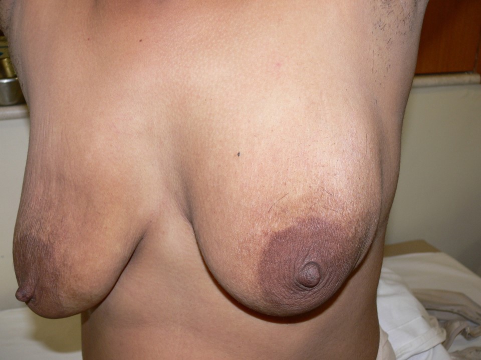
| Clinical presentation: | Postmenopausal woman with average risk of developing breast cancer presented with a lump in the left breast. On clinical examination, she had a hard lump palpable in the upper quadrant of the left breast and another large lump in the left axilla suggestive of palpable axillary nodes. |
 |
Mammography:
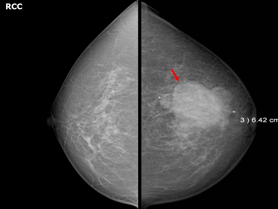 | 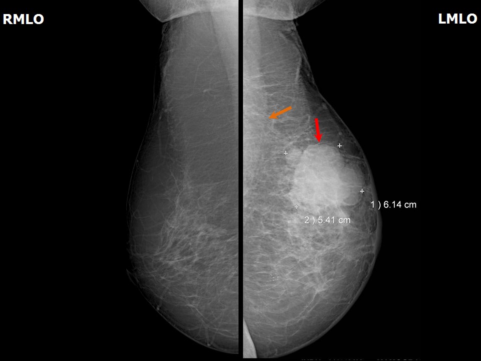 |
| Breast composition: | ACR category b (there are scattered areas of fibroglandular density) | Mammography features: |
| ‣ Location of the lesion: | May 2014: Left breast, upper outer quadrant at 121 oclock, middle third |
| ‣ Mass: | |
| • Number: | 1 |
| • Size: | 6.4 × 6.1 × 5.4 cm |
| • Shape: | Irregular |
| • Margins: | Indistinct |
| • Density: | High |
| ‣ Calcifications: | |
| • Typically benign: | None |
| • Suspicious: | None |
| • Distribution: | None |
| ‣ Architectural distortion: | Present |
| ‣ Asymmetry: | Focal |
| ‣ Intramammary node: | None |
| ‣ Skin lesion: | None |
| ‣ Solitary dilated duct: | None |
| ‣ Associated features: | Trabecular thickening, skin thickening, and enlarged nodes (largest 6 × 2.6 cm) with loss of central fatty hilum |
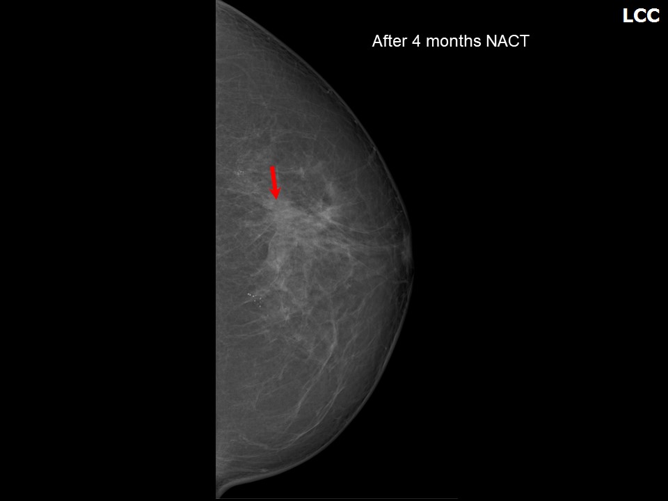 | 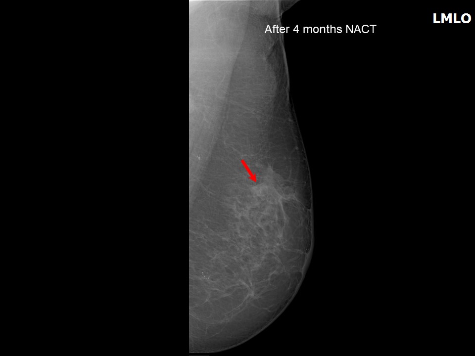 |
| Breast composition: | ACR category b (there are scattered areas of fibroglandular density) | Mammography features: |
| ‣ Location of the lesion: | September 2014: Left breast, upper outer quadrant at 121 oclock, middle third |
| ‣ Mass: | |
| • Number: | 1 |
| • Size: | 2.2 × 1.2 cm |
| • Shape: | Irregular |
| • Margins: | Indistinct |
| • Density: | High |
| ‣ Calcifications: | |
| • Typically benign: | None |
| • Suspicious: | None |
| • Distribution: | None |
| ‣ Architectural distortion: | Present |
| ‣ Asymmetry: | Focal |
| ‣ Intramammary node: | None |
| ‣ Skin lesion: | None |
| ‣ Solitary dilated duct: | None |
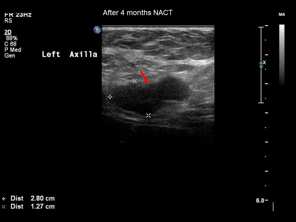
| ‣ Associated features: | Trabecular thickening, skin thickening, and enlarged axillary nodes (largest 2.8 × 1.3 cm) with loss of central fatty hilum |
Ultrasound:
 |  |
 |
| Ultrasound features: May 2014: Left breast, upper outer quadrant at 12 oclock | |
| ‣ Mass | |
| • Location: | May 2014: Left breast, upper outer quadrant at 12 oclock |
| • Number: | 1 |
| • Size: | 5.0 × 3.6 cm |
| • Shape: | Irregular |
| • Orientation: | Not parallel |
| • Margins: | Angular |
| • Echo pattern: | Heterogeneous |
| • Posterior features: | Posterior shadowing |
| ‣ Calcifications: | None |
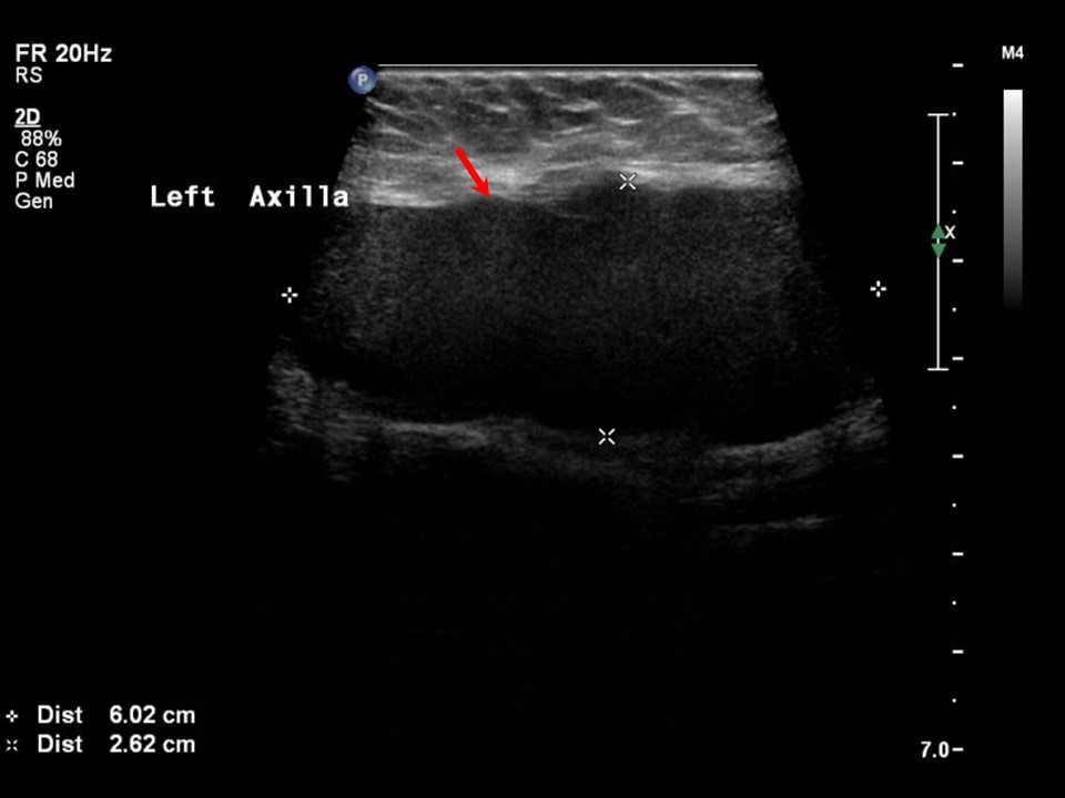
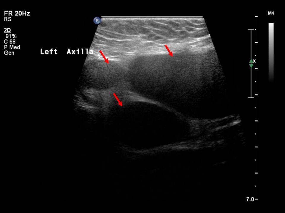
| ‣ Associated features: | Architectural distortion, skin thickening, internal vascularity, and multiple enlarged axillary nodes (largest 6.0 × 2.6 cm) |
| ‣ Special cases: | None |
 |  |
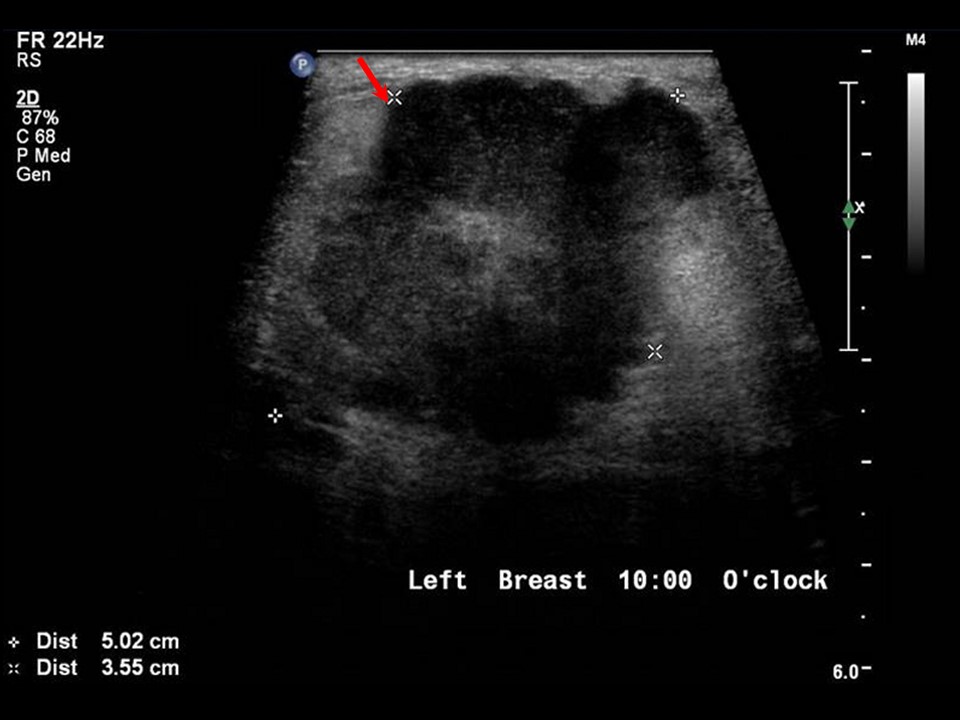
 |
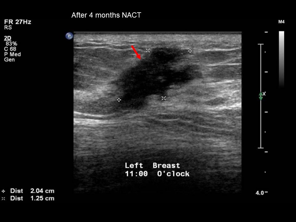
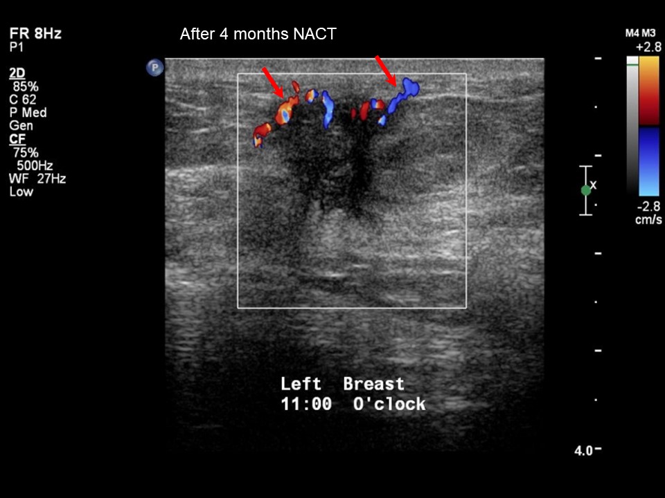
| Ultrasound features: September 2014, neo-adjuvant chemotherapy follow-up: Left breast, upper outer quadrant at 12 oclock | |
| ‣ Mass | |
| • Location: | September 2014, neo-adjuvant chemotherapy follow-up: Left breast, upper outer quadrant at 12 oclock |
| • Number: | 1 |
| • Size: | 2.0 × 1.3 cm |
| • Shape: | Irregular |
| • Orientation: | Not parallel |
| • Margins: | Spiculated |
| • Echo pattern: | Hypoechoic |
| • Posterior features: | No posterior features |
| ‣ Calcifications: | None |
| ‣ Associated features: | Architectural distortion, skin thickening, internal vascularity, and enlarged axillary nodes (largest 2.8 × 1.3 cm) |
| ‣ Special cases: | None |
BI-RADS:
BI-RADS Category (May 2014): 5 (highly suggestive of malignancy)BI-RADS Category (September 2014): 6 (known biopsy-proven malignancy)
Further assessment:
Further assessment advised: Referral for core biopsyCytology:
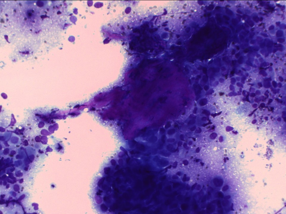 |
| Cytology features: | |
| ‣ Type of sample: | FNAC (solid lesion) |
| ‣ Site of biopsy: | |
| • Laterality: | Left |
| • Quadrant: | Central |
| • Localization technique: | Palpation |
| • Nature of aspirate: | Whitish |
| ‣ Cytological description: | Cellular smears with highly pleomorphic malignant cells seen as single cells and loose clusters |
| ‣ Reporting category: | Malignant |
| ‣ Diagnosis: | Carcinoma high grade |
| ‣ Comments: | None |
Histopathology:
Core needle biopsy
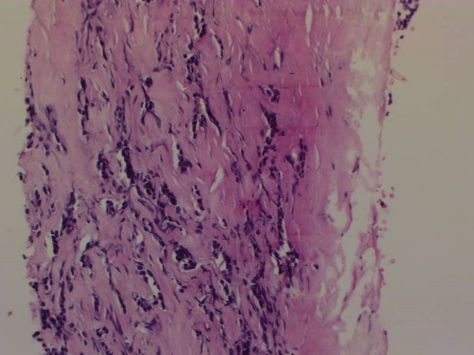 |
| Histopathology features: | |
| ‣ Specimen type: | Core needle biopsy |
| ‣ Laterality: | Left |
| ‣ Macroscopy: | Six cores |
| ‣ Histological type: | Invasive carcinoma of no special type |
| ‣ Histological grade: | Grade 3 (3 + 3 + 2 = 8) |
| ‣ Mitosis: | 12 |
| ‣ Maximum invasive tumour size: | |
| ‣ Lymph node status: | |
| ‣ Peritumoural lymphovascular invasion: | |
| ‣ DCIS/EIC: | |
| ‣ Margins: | |
| ‣ Pathological stage: | |
| ‣ Biomarkers: | |
| ‣ Comments: |
MRM
 |
| Histopathology features: | |
| ‣ Specimen type: | MRM |
| ‣ Laterality: | Left |
| ‣ Macroscopy: | On serial sectioning, residual tumour (3.7 × 2.8 × 3.7 cm) is identified in the upper outer quadrant. It is located 0.6 cm from the skin and 0.7 cm from the base |
| ‣ Histological type: | Invasive carcinoma of no special type |
| ‣ Histological grade: | Grade 3 (3 + 3 + 2 = 8) |
| ‣ Mitosis: | 10 |
| ‣ Maximum invasive tumour size: | 3.7 cm in greatest dimension |
| ‣ Lymph node status: | 9/20 |
| ‣ Peritumoural lymphovascular invasion: | Not identified |
| ‣ DCIS/EIC: | Not identified |
| ‣ Margins: | Free of tumour |
| ‣ Pathological stage: | yT2yN2 |
| ‣ Biomarkers: | |
| ‣ Comments: | After neo-adjuvant chemotherapy, cellular changes were seen. Many tumour cells show degenerative change with smudged appearance. Cytoplasm is abundant, eosinophilic, and granular, with vacuoles in places. There are eosinophilic secretions and mucinous material at many foci. Focal areas show aggregates of lymphocytes. Necrosis is seen in many dilated ducts |
Case summary:
| Postmenopausal woman presented with left breast lump. Diagnosed as left breast carcinoma with enlarged left axillary node, probably metastatic, BI-RADS 5 on imaging, as carcinoma on cytology, and as invasive breast carcinoma of no special type on needle core biopsy. After neo-adjuvant chemotherapy, follow-up shows significant tumour regression, BI-RADS 6 on imaging and an operative specimen revealed residual invasive breast carcinoma of no special type, yT2yN2 on histopathology. |
Learning points:
|