


Home / Training / Manuals / Atlas of breast cancer early detection / Cases
Atlas of breast cancer early detection
Filter by language: English / Русский
Go back to the list of case studies
.png) Click on the pictures to magnify and display the legends
Click on the pictures to magnify and display the legends
| Case number: | 001 |
| Age: | 50 |
| Clinical presentation: | Postmenopausal woman with average risk of developing breast cancer presented with a painless lump of duration 34 days in the upper outer quadrant of the left breast. On examination, she had a firm 3 cm lump in the upper outer quadrant of the left breast with minimal tenderness. |
Mammography:
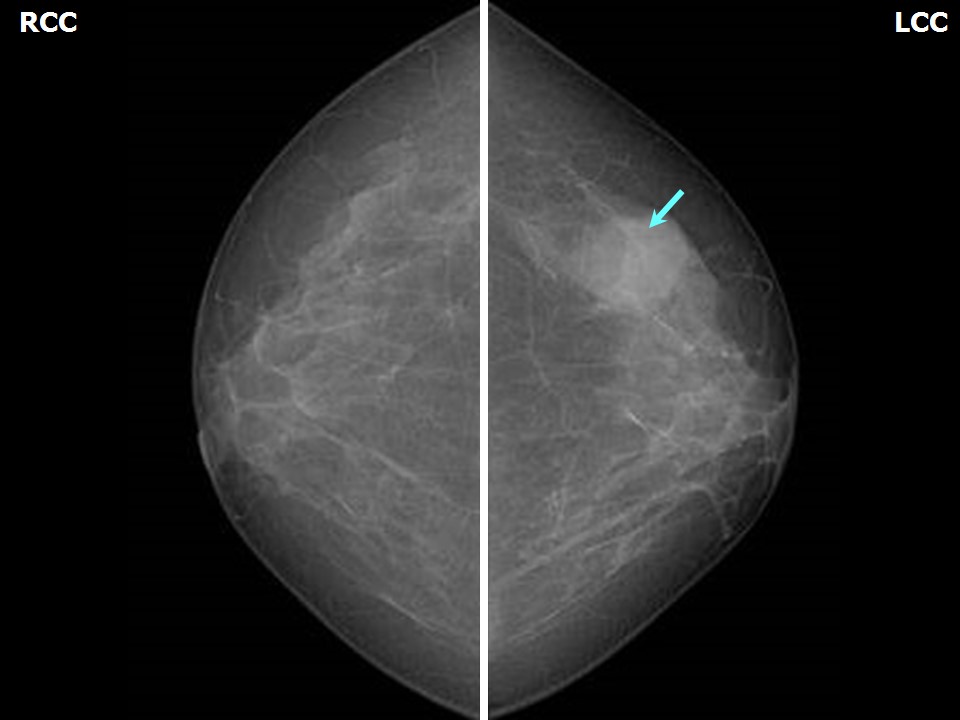 | 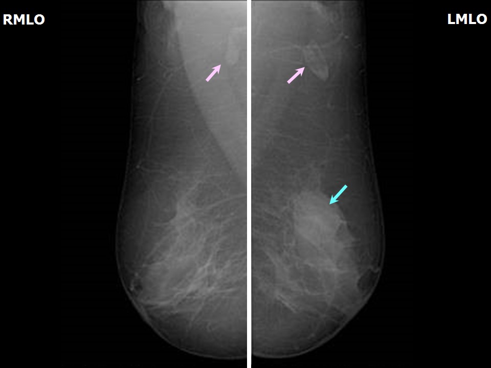 |
| Breast composition: | ACR category b (there are scattered areas of fibroglandular density) | Mammography features: |
| ‣ Location of the lesion: | Left breast, upper outer quadrant at 2 oclock, middle third |
| ‣ Mass: | |
| • Number: | 1 |
| • Size: | 2.8 x 2.2 cm |
| • Shape: | Oval |
| • Margins: | Circumscribed |
| • Density: | Equal |
| ‣ Calcifications: | |
| • Typically benign: | None |
| • Suspicious: | None |
| • Distribution: | None |
| ‣ Architectural distortion: | None |
| ‣ Asymmetry: | None |
| ‣ Intramammary node: | None |
| ‣ Skin lesion: | None |
| ‣ Solitary dilated duct: | None |
| ‣ Associated features: | None |
Ultrasound:
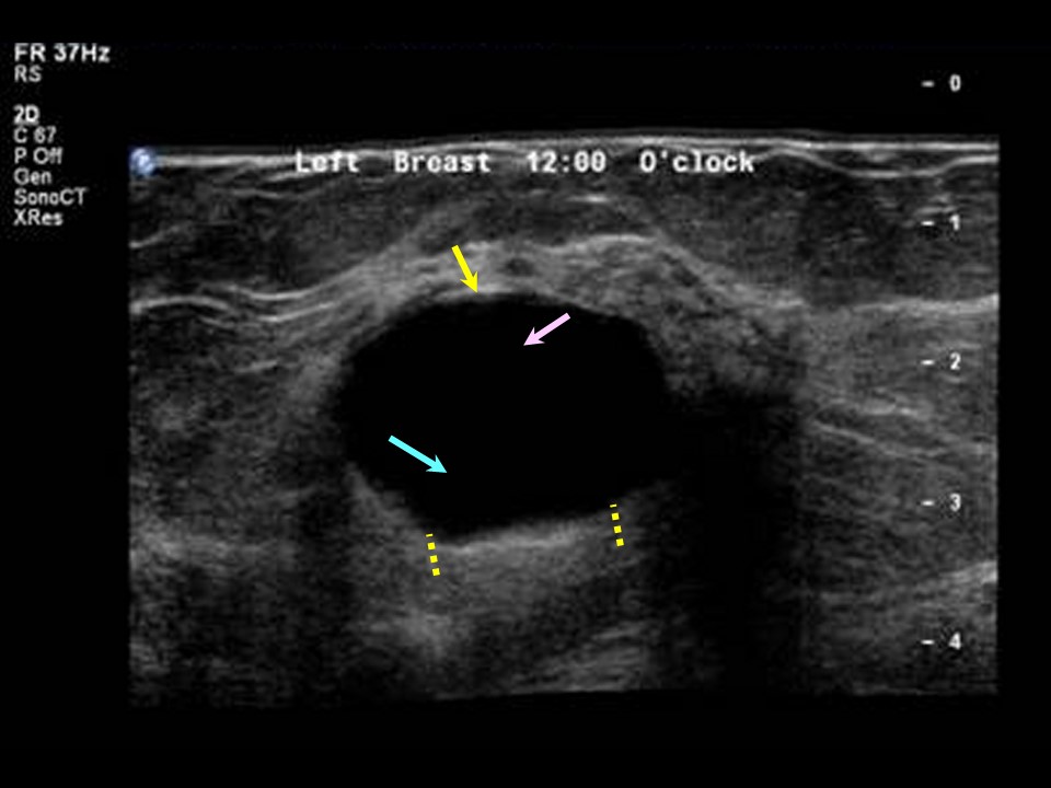 |
| Ultrasound features: Left breast, central portion of the breast | |
| ‣ Mass | |
| • Location: | Left breast, central portion of the breast |
| • Number: | Multiple |
| • Size: | Largest 3.0 × 2.0 cm |
| • Shape: | Oval |
| • Orientation: | Parallel |
| • Margins: | Circumscribed |
| • Echo pattern: | Anechoic |
| • Posterior features: | Posterior shadowing |
| ‣ Calcifications: | None |
| ‣ Associated features: | None |
| ‣ Special cases: | Simple cyst |
| Ultrasound features: Right breast, central portion of the breast at 12 oclock | |
| ‣ Mass | |
| • Location: | Right breast, central portion of the breast at 12 oclock |
| • Number: | Multiple |
| • Size: | Largest 0.7 cm in greatest dimension |
| • Shape: | Oval |
| • Orientation: | Parallel |
| • Margins: | Circumscribed |
| • Echo pattern: | Anechoic |
| • Posterior features: | Posterior shadowing |
| ‣ Calcifications: | None |
| ‣ Associated features: | None |
| ‣ Special cases: | Simple cyst |
BI-RADS:
BI-RADS Category: 2 (benign)Further assessment:
Further assessment advised: Referral for cytologyCytology:
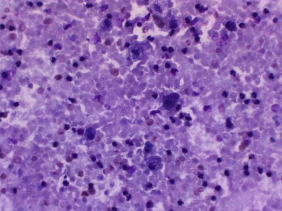 |
| Cytology features: | |
| ‣ Type of sample: | FNAC |
| ‣ Site of biopsy: | |
| • Laterality: | Left |
| • Quadrant: | Upper outer |
| • Localization technique: | Palpation |
| • Nature of aspirate: | 5 mL of yellow turbid fluid |
| ‣ Cytological description: | Smears show large histiocytes (foam cells) with abundant vacuolated cytoplasm and an oval nucleus along with neutrophils on a proteinaceous background |
| ‣ Reporting category: | Benign |
| ‣ Diagnosis: | Non-proliferative fibrocystic change |
| ‣ Comments: | None |
Case summary:
| Postmenopausal woman presented with left breast lump. Diagnosed as simple cyst, BI-RADS 2 on imaging and as non-proliferative fibrocystic change on cytology. |
Learning points:
|