


Home / Training / Manuals / Atlas of breast cancer early detection / Cases
Atlas of breast cancer early detection
Filter by language: English / Русский
Go back to the list of case studies
.png) Click on the pictures to magnify and display the legends
Click on the pictures to magnify and display the legends
| Case number: | 182 |
| Age: | 85 |
| Clinical presentation: | Postmenopausal woman with average risk of developing breast cancer presented with a right breast lump noticed 12 years ago. It has progressively increased in size. On examination, a hard non-tender lump was palpable in the right breast, fixed to the chest wall and skin and fixed to the breast parenchyma. The right nipple was retracted. The left breast was unremarkable. |
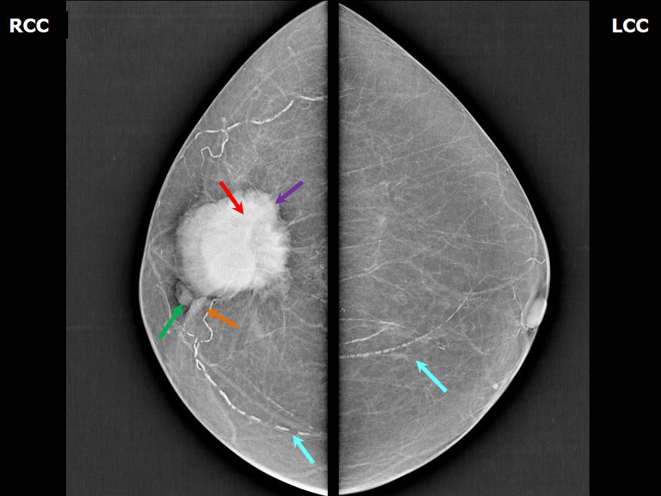
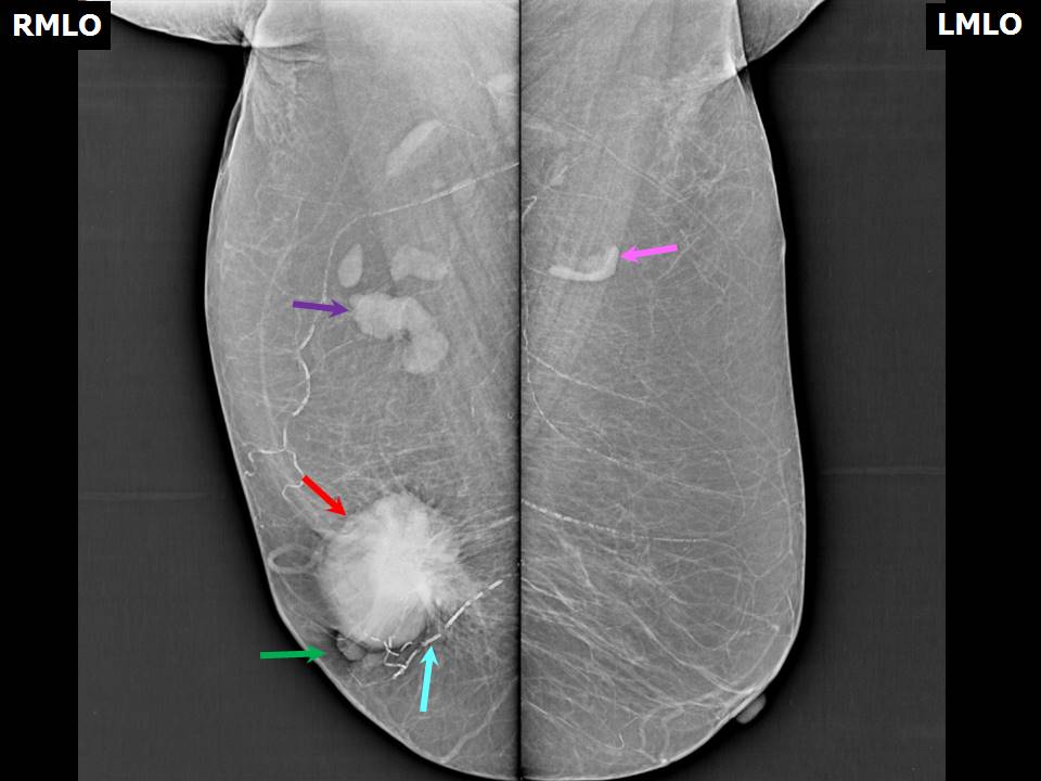
Mammography:
 |  |
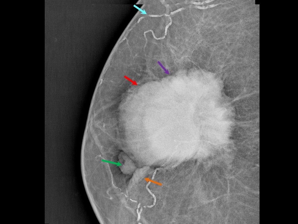 |
| Breast composition: | ACR category a (the breasts are almost entirely fatty) | Mammography features: |
| ‣ Location of the lesion: | Right breast, lower outer quadrant at 79 oclock, anterior and middle thirds |
| ‣ Mass: | |
| • Number: | 1 |
| • Size: | 4.8 × 4.6 cm |
| • Shape: | Irregular |
| • Margins: | Spiculated |
| • Density: | High |
| ‣ Calcifications: | |
| • Typically benign: | Vascular calcification in both breasts |
| • Suspicious: | None |
| • Distribution: | None |
| ‣ Architectural distortion: | None |
| ‣ Asymmetry: | None |
| ‣ Intramammary node: | None |
| ‣ Skin lesion: | None |
| ‣ Solitary dilated duct: | None |
| ‣ Associated features: | Nipple retraction and enlarged right axillary nodes |
Ultrasound:
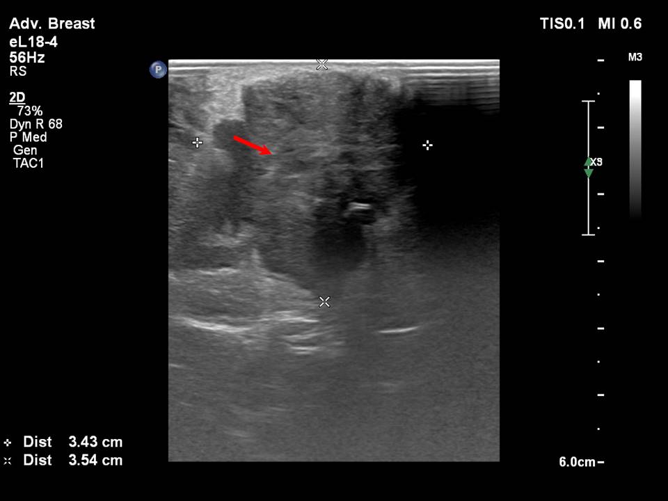 | 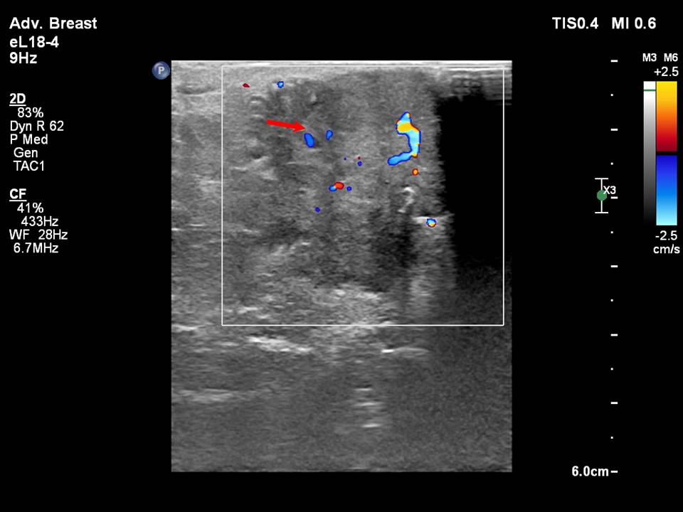 |
| Ultrasound features: Right breast, outer quadrants at 2-9 oclock | |
| ‣ Mass | |
| • Location: | Right breast, outer quadrants at 2-9 oclock |
| • Number: | 1 |
| • Size: | 3.5 × 3.4 cm |
| • Shape: | Irregular |
| • Orientation: | Not parallel |
| • Margins: | Microlobulated |
| • Echo pattern: | Hypoechoic |
| • Posterior features: | No posterior features |
| ‣ Calcifications: | None |
| ‣ Associated features: | Peripheral and central vascularity in mass and right axillary lymphadenopathy |
| ‣ Special cases: | None |
BI-RADS:
BI-RADS Category: 5 (highly suggestive of malignancy)Further assessment:
Further assessment advised: Referral for core biopsyCytology:
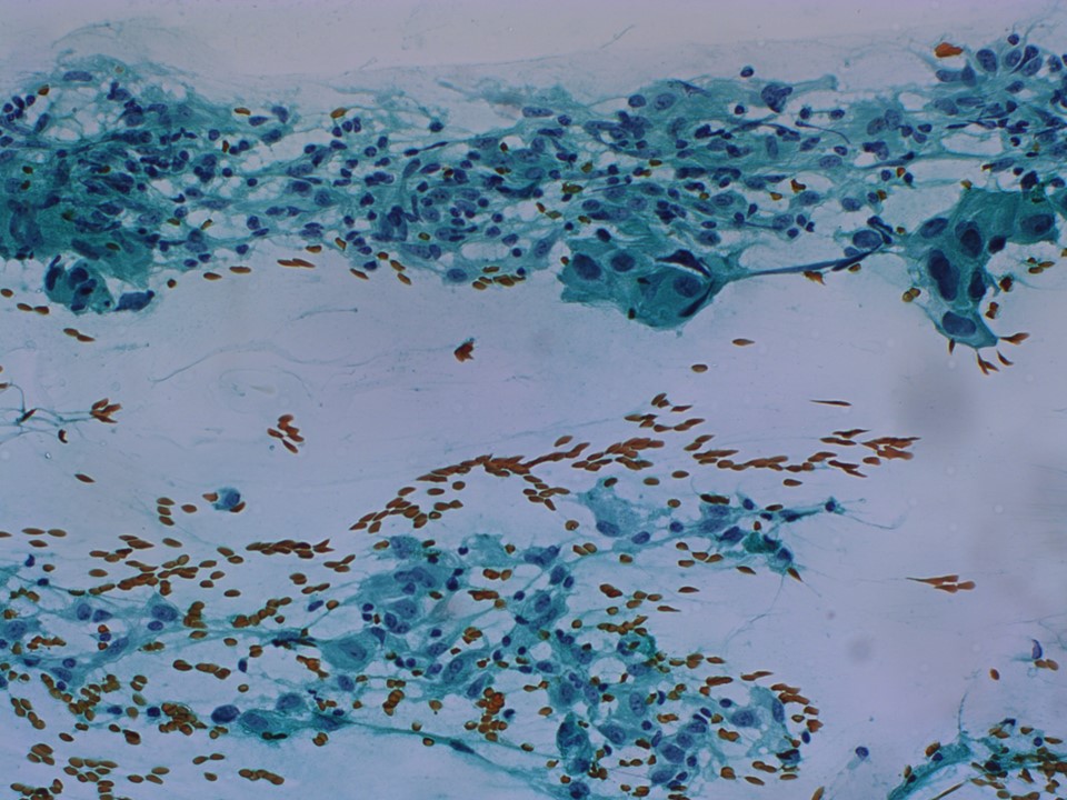 |
| Cytology features: | |
| ‣ Type of sample: | FNAC |
| ‣ Site of biopsy: | |
| • Laterality: | Right |
| • Quadrant: | Lower outer near areolar area |
| • Localization technique: | Palpation |
| • Nature of aspirate: | Whitish |
| ‣ Cytological description: | Smear reveals single isolated and dyscohesive clusters of ductal epithelial cells showing large, variably hyperchromatic, pleomorphic nuclei with prominent nucleoli. Cytoplasm is moderate to abundant |
| ‣ Reporting category: | Malignant |
| ‣ Diagnosis: | Carcinoma breast |
| ‣ Comments: | None |
Histopathology:
MRM
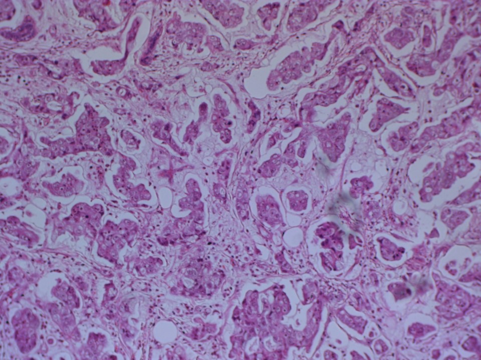 |
| Histopathology features: | |
| ‣ Specimen type: | MRM |
| ‣ Laterality: | Right |
| ‣ Macroscopy: | Right MRM specimen (30 × 18 × 4 cm) with overlying skin flap (13.5 × 7.5 cm). Nippleareolar area is unremarkable. On serial sectioning, a well-circumscribed greyish white tumour (3.5 × 3.0 × 3.5 cm) is identified in the lower outer quadrant. It is located 0.1 cm from the skin and 1.5 cm from the base. The attached axillary tail is 5 cm long. Eighteen lymph nodes were identified, the largest of which is 1.0 × 1.0 cm; the others range from 0.9 to 0.3 cm. Four apical nodes and one interpectoral lymph node were received separately |
| ‣ Histological type: | Invasive breast carcinoma of no special type |
| ‣ Histological grade: | Grade 3 (3 + 3 + 2 = 8) |
| ‣ Mitosis: | 12 |
| ‣ Maximum invasive tumour size: | 3.5 cm |
| ‣ Lymph node status: | 0/23 |
| ‣ Peritumoural lymphovascular invasion: | Present |
| ‣ DCIS/EIC: | Absent |
| ‣ Margins: | The posterior margin of resection (base) is free of tumour |
| ‣ Pathological stage: | pT2N0 |
| ‣ Biomarkers: | ER: Positive (100% of cells show nuclear staining with moderate intensity. Allred score 7/8). PR: Positive (100% of cells show nuclear staining with moderate intensity. Allred score 7/8). HER2: Negative |
| ‣ Comments: | The invasive carcinoma is seen just below the epidermis in the dermis. |
Case summary:
| Postmenopausal woman presented with right breast lump with retracted nipple. Diagnosed as right breast carcinoma with overlying skin thickening, skin retraction, and right nipple retraction, BI-RADS 5 on imaging, as right breast carcinoma on cytology, and as invasive breast carcinoma of no special type, pT2N0 on histopathology. |
Learning points:
|