


Home / Training / Manuals / Atlas of breast cancer early detection / Cases
Atlas of breast cancer early detection
Filter by language: English / Русский
Go back to the list of case studies
.png) Click on the pictures to magnify and display the legends
Click on the pictures to magnify and display the legends
BI-RADS Category (2016): 4C (high suspicion for malignancy)
| Case number: | 115 |
| Age: | 72 |
| Clinical presentation: | Postmenopausal woman with increased risk of developing breast cancer was on surveillance by mammography. She had two first-degree relatives who had been diagnosed with breast cancer. She had also undergone distal pancreatectomy for a pancreatic neuroendocrine tumour. |
Mammography:
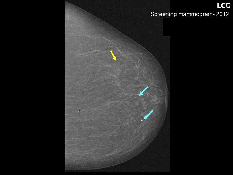 | 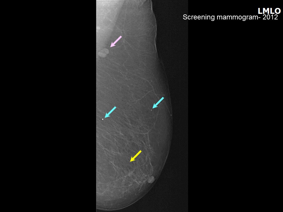 |
| Breast composition: | ACR category a (the breasts are almost entirely fatty) | Mammography features: |
| ‣ Location of the lesion: | 2012: Left breast |
| ‣ Mass: | |
| • Number: | 0 |
| • Size: | None |
| • Shape: | None |
| • Margins: | None |
| • Density: | None |
| ‣ Calcifications: | |
| • Typically benign: | None |
| • Suspicious: | None |
| • Distribution: | None |
| ‣ Architectural distortion: | None |
| ‣ Asymmetry: | None |
| ‣ Intramammary node: | None |
| ‣ Skin lesion: | None |
| ‣ Solitary dilated duct: | None |
| ‣ Associated features: | None |
 |  |
 |
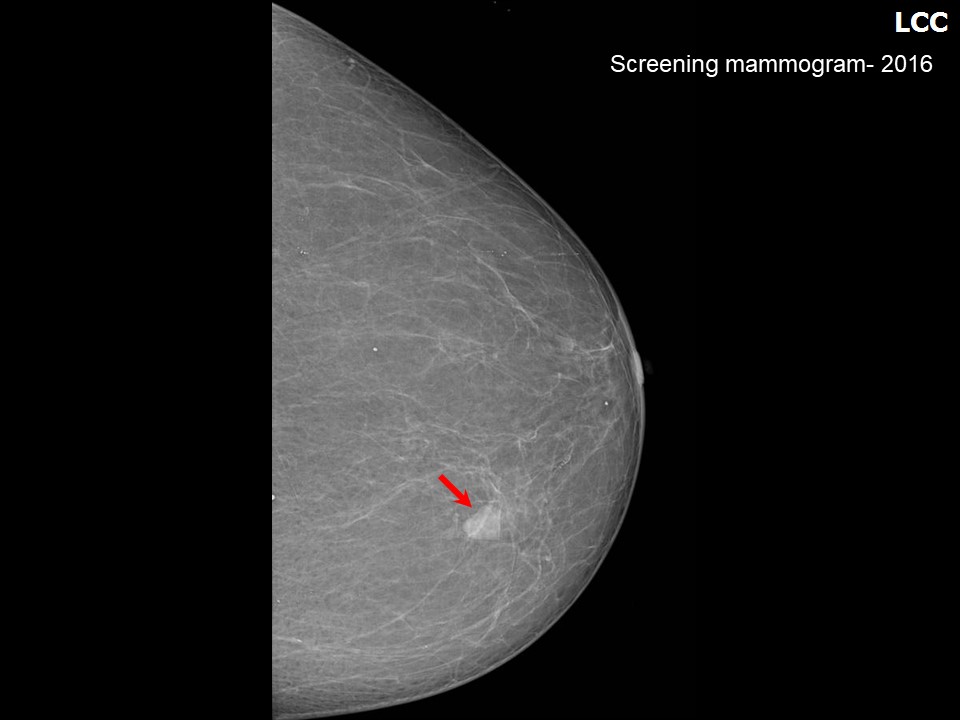
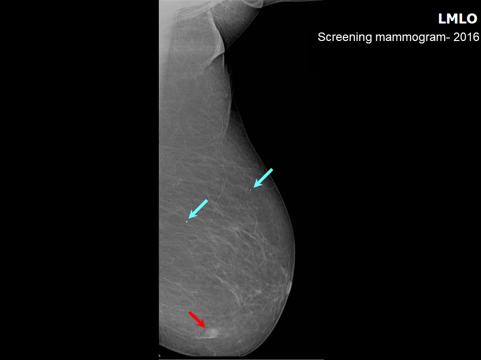
| Breast composition: | ACR category a (the breasts are almost entirely fatty) | Mammography features: |
| ‣ Location of the lesion: | Left breast, lower inner quadrant at 78 oclock, middle third |
| ‣ Mass: | |
| • Number: | 1 (this is the developing asymmetry, interval change seen) |
| • Size: | 1 cm in greatest dimension |
| • Shape: | Irregular |
| • Margins: | Indistinct |
| • Density: | High |
| ‣ Calcifications: | |
| • Typically benign: | Round |
| • Suspicious: | None |
| • Distribution: | Diffuse |
| ‣ Architectural distortion: | None |
| ‣ Asymmetry: | None |
| ‣ Intramammary node: | None |
| ‣ Skin lesion: | None |
| ‣ Solitary dilated duct: | None |
| ‣ Associated features: | None |
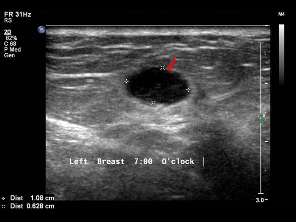
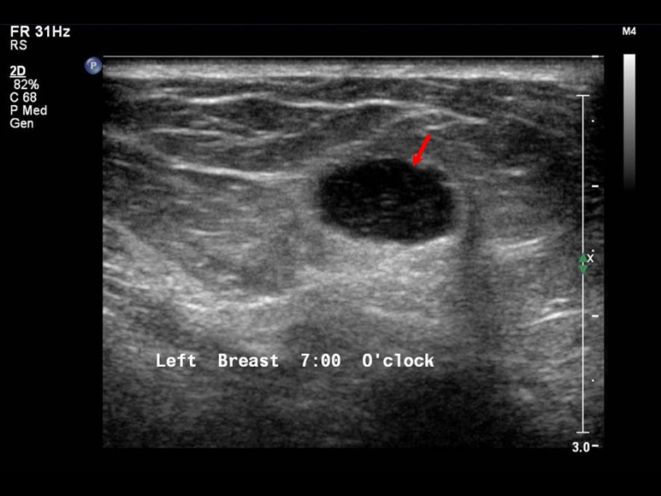
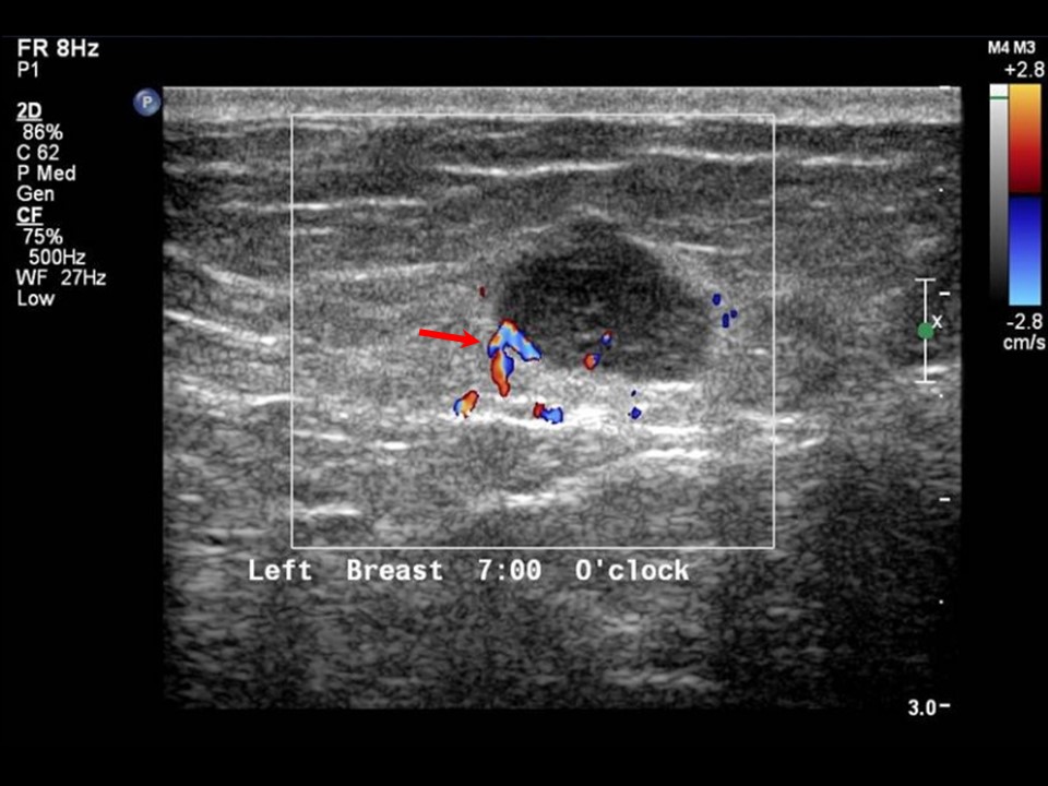
Ultrasound:
 |  |
 |  |
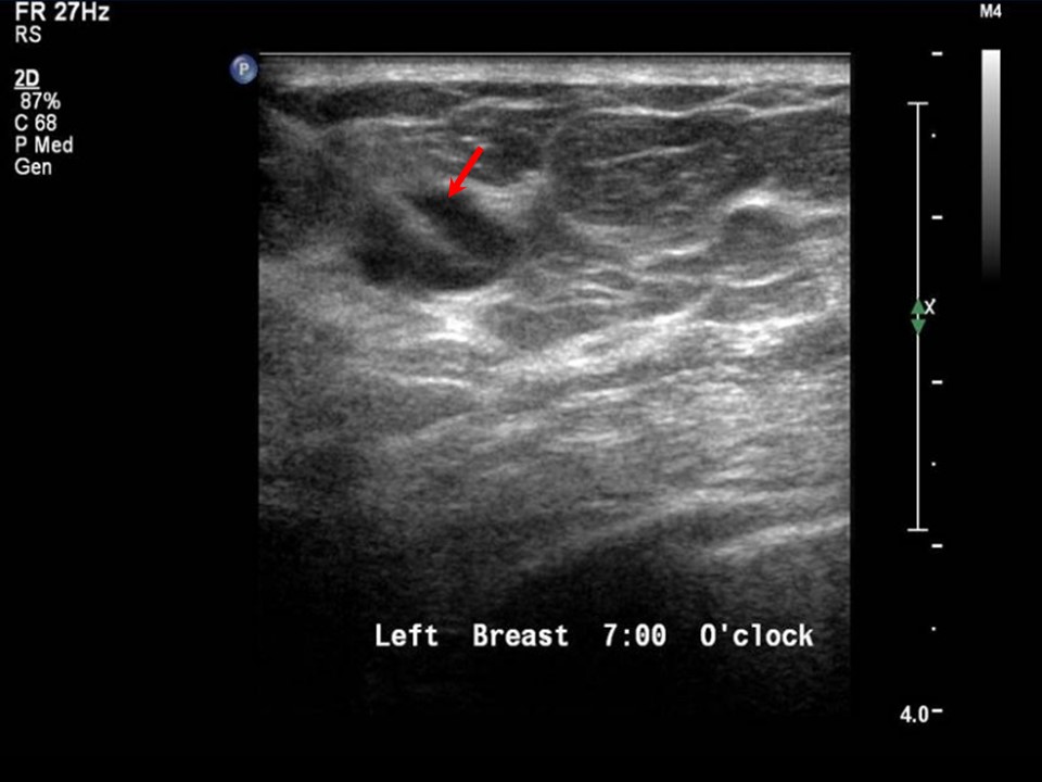 | 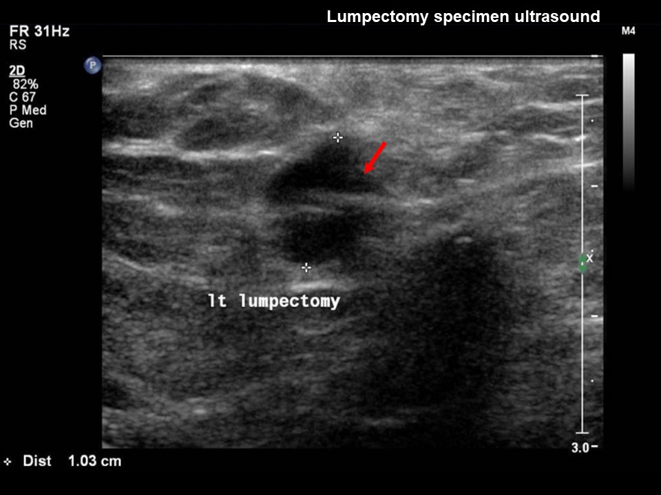 |
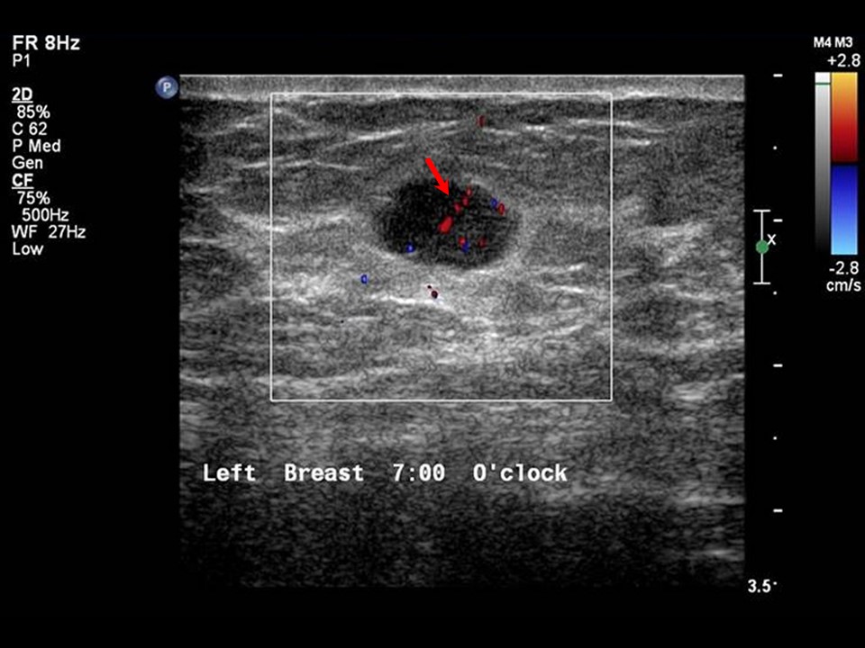
| Ultrasound features: Left breast, lower inner quadrant at 7 oclock | |
| ‣ Mass | |
| • Location: | Left breast, lower inner quadrant at 7 oclock |
| • Number: | 1 |
| • Size: | 1.0 × 0.6 cm |
| • Shape: | Irregular |
| • Orientation: | Parallel |
| • Margins: | Indistinct |
| • Echo pattern: | Hypoechoic |
| • Posterior features: | No posterior features |
| ‣ Calcifications: | None |
| ‣ Associated features: | Internal vascularity |
| ‣ Special cases: | None |
BI-RADS:
BI-RADS Category (2012): 2 (benign)BI-RADS Category (2016): 4C (high suspicion for malignancy)
Further assessment:
Further assessment advised: Referral for cytologyCytology:
 |
| Cytology features: | |
| ‣ Type of sample: | FNAC |
| ‣ Site of biopsy: | |
| • Laterality: | Left |
| • Quadrant: | Lower inner |
| • Localization technique: | Ultrasound-guided FNAC |
| • Nature of aspirate: | Whitish |
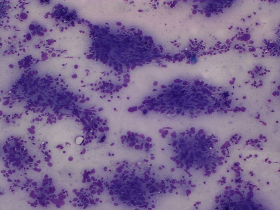
| ‣ Cytological description: | Smears show many clusters of pleomorphic malignant cells. These cells have prominent nucleoli. Many lymphocytes and plasma cells are visible in background |
| ‣ Reporting category: | Malignant |
| ‣ Diagnosis: | Carcinoma |
| ‣ Comments: | None |
Histopathology:
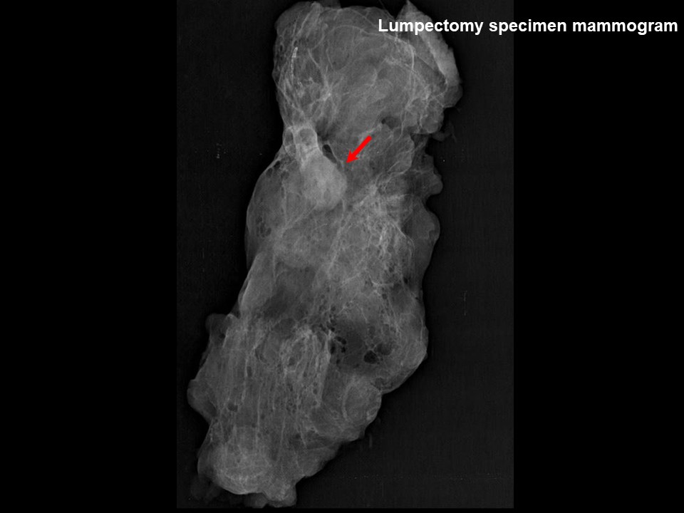
Breast-conserving surgery
 |  |
 |
| Histopathology features: | |
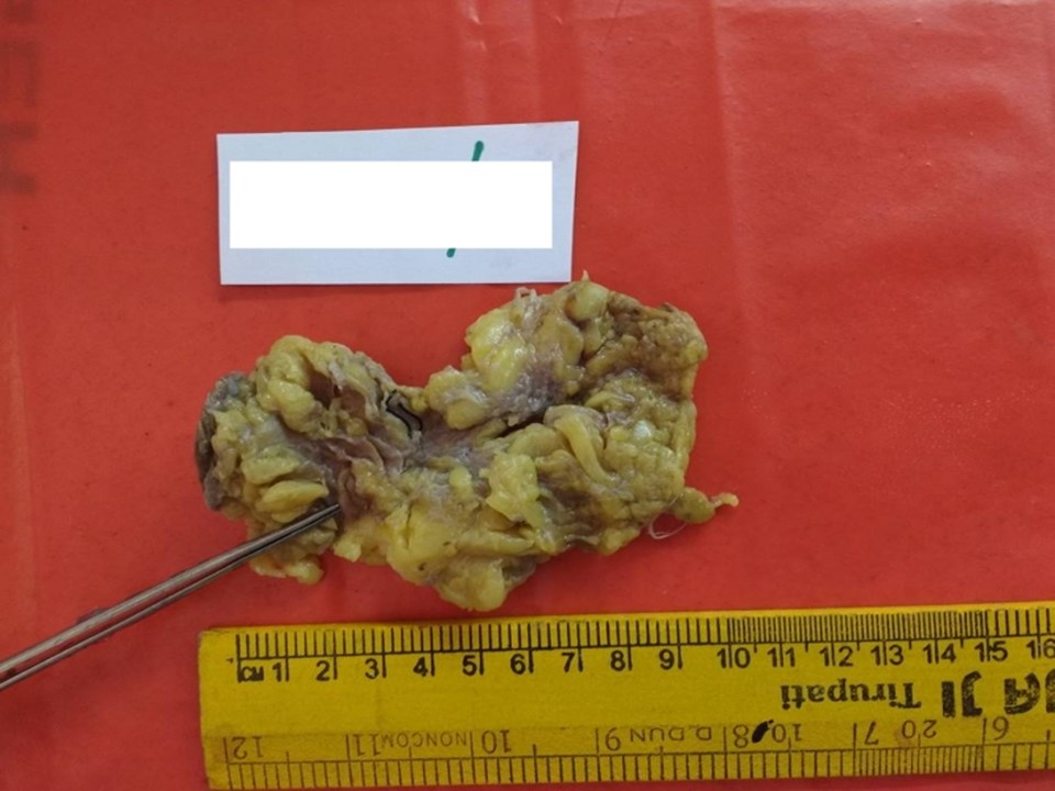
| ‣ Specimen type: | Breast-conserving surgery |
| ‣ Laterality: | Left |
| ‣ Macroscopy: | On serial sectioning, a soft to firm tan-coloured nodule (1.7 × 1.3 × 0.5 cm) is identified very close to the inferior margin. It is located 3.0 cm from the skin (anterior margin), 1.5 cm from the base (posterior margin), 1.5 cm from the superior margin, 1.4 cm from the medial margin, and 3.0 cm from the lateral margin. The remaining breast parenchyma is unremarkable |
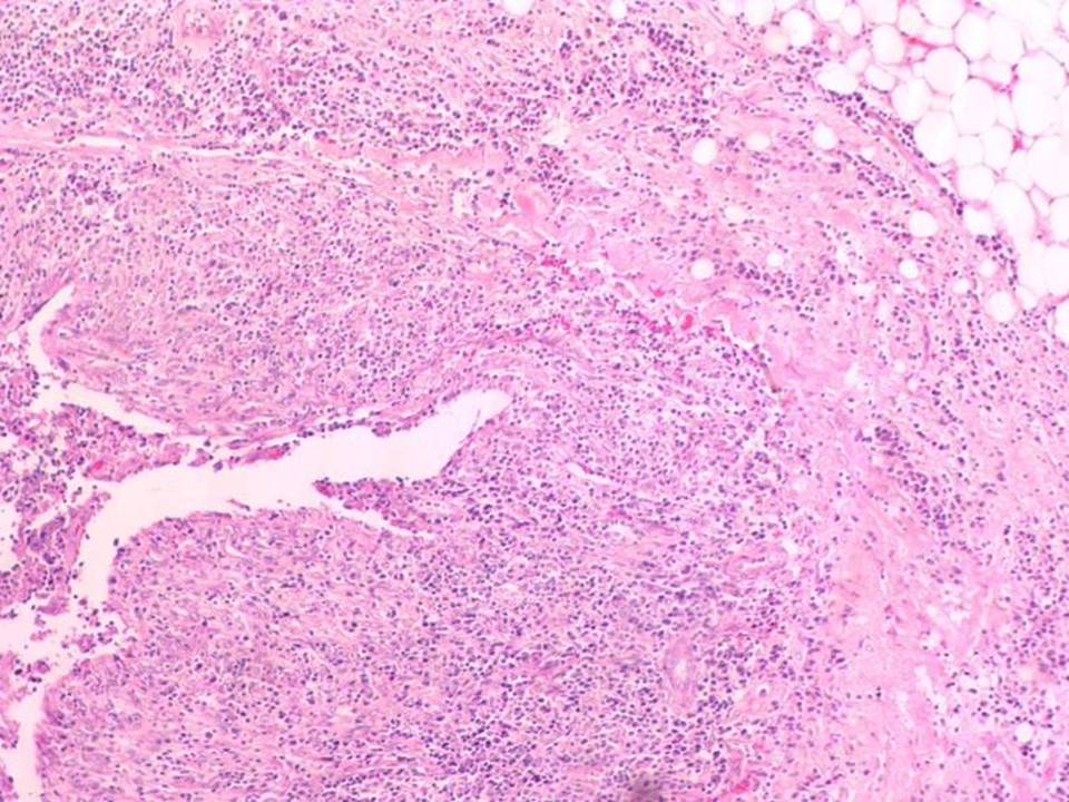
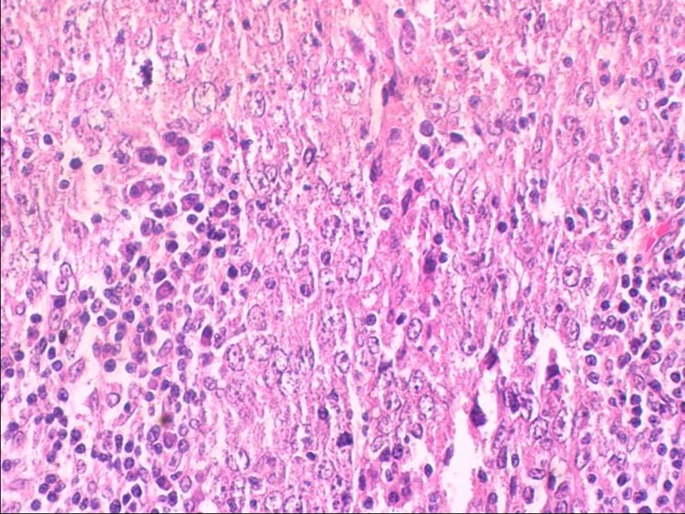
| ‣ Histological type: | Breast carcinoma of no special type with medullary pattern |
| ‣ Histological grade: | Grade 3 (3 + 3 + 3 = 9) |
| ‣ Mitosis: | 22 |
| ‣ Maximum invasive tumour size: | 1.7 cm in greatest dimension |
| ‣ Lymph node status: | 0/17 |
| ‣ Peritumoural lymphovascular invasion: | Not identified |
| ‣ DCIS/EIC: | Not identified |
| ‣ Margins: | Free of tumour |
| ‣ Pathological stage: | pT1cN0 |
| ‣ Biomarkers: | |
| ‣ Comments: |
Case summary:
| Postmenopausal woman with increased risk of developing breast cancer presented for follow-up mammography screening. Examination did not reveal any lump in either of the breasts or axillae. Mammography and breast ultrasound dated 2016 revealed new suspicious irregular lesion with indistinct margins in the left breast. Compared with previous mammograms dated 2012, follow-up screening revealed developing asymmetry in the left breast. Interval change is seen. Diagnosed as highly suspicious for malignancy, BI-RADS 4C on imaging, as carcinoma on cytology, and as invasive breast carcinoma of no special type with medullary pattern, pT1cN0 on histopathology. |
Learning points:
|