


Home / Training / Manuals / Atlas of breast cancer early detection / Cases
Atlas of breast cancer early detection
Filter by language: English / Русский
Go back to the list of case studies
.png) Click on the pictures to magnify and display the legends
Click on the pictures to magnify and display the legends
| Case number: | 079 |
| Age: | 41 |
| Clinical presentation: | Premenopausal woman with increased risk of developing breast cancer presented with history of right breast lump noticed 1 day ago on breast self-examination. She has family history of cervical carcinoma and tongue cancer. |
Mammography:
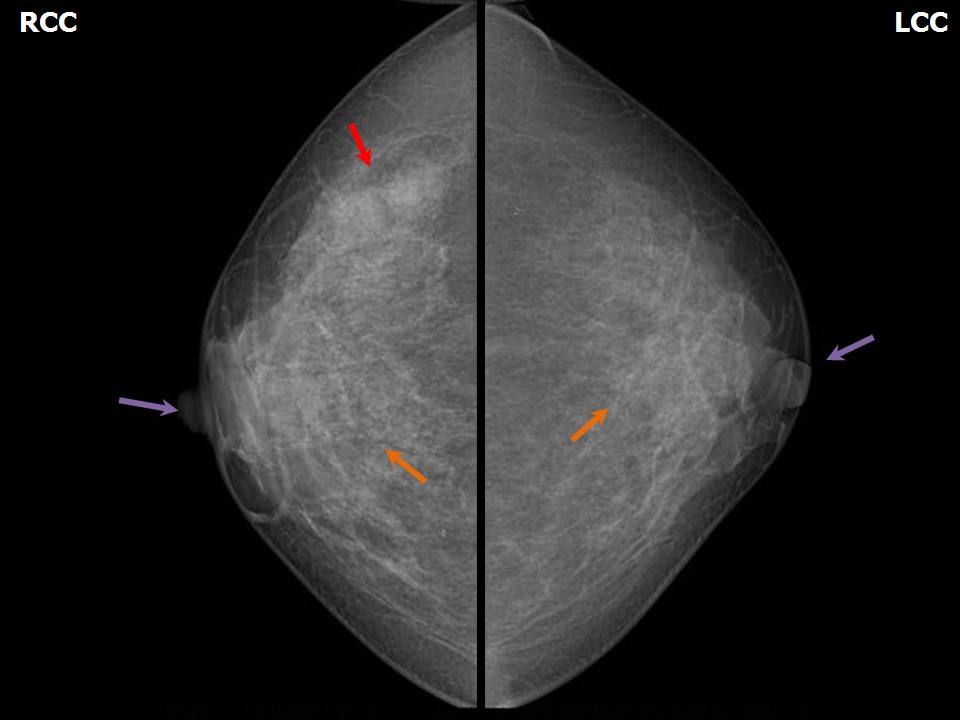 | 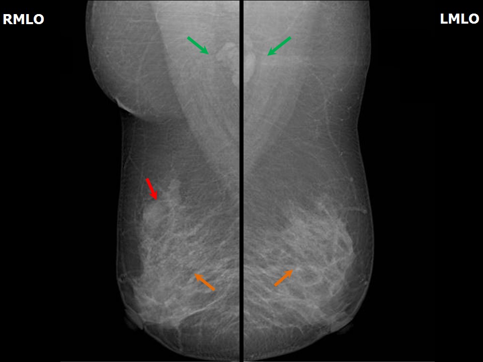 |
| Breast composition: | ACR category b (there are scattered areas of fibroglandular density) | Mammography features: |
| ‣ Location of the lesion: | Right breast, upper outer quadrant at 10 oclock, middle third |
| ‣ Mass: | |
| • Number: | 3 |
| • Size: | 1.5 cm, 1.0 cm, and 0.9 cm in greatest dimension |
| • Shape: | Irregular |
| • Margins: | Obscured |
| • Density: | Equal |
| ‣ Calcifications: | |
| • Typically benign: | None |
| • Suspicious: | None |
| • Distribution: | None |
| ‣ Architectural distortion: | None |
| ‣ Asymmetry: | None |
| ‣ Intramammary node: | None |
| ‣ Skin lesion: | None |
| ‣ Solitary dilated duct: | None |
| ‣ Associated features: | None |
Ultrasound:
 | 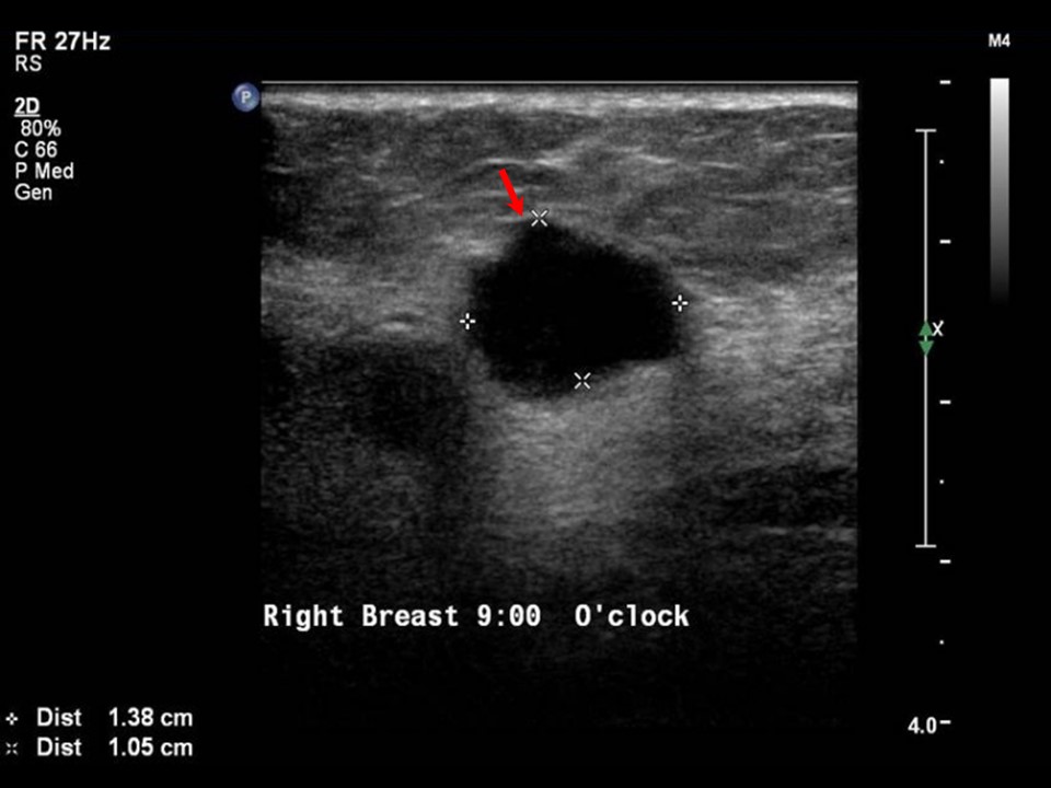 |
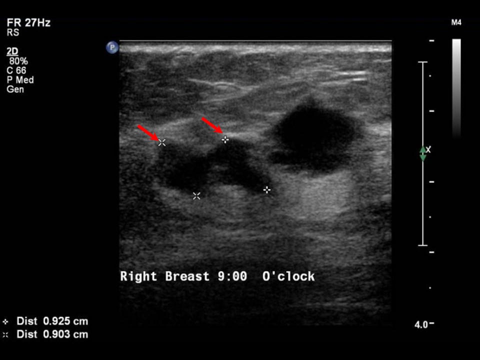 | 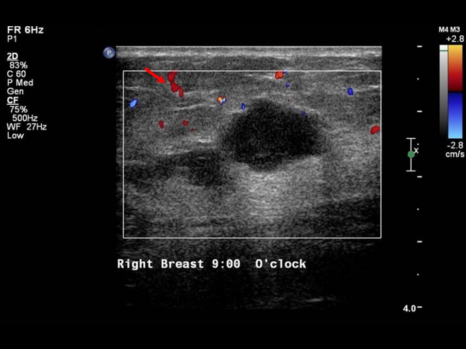 |
| Ultrasound features: Right breast, upper outer quadrant at 10 oclock | |
| ‣ Mass | |
| • Location: | Right breast, upper outer quadrant at 10 oclock |
| • Number: | 3 |
| • Size: | 1.4 × 1.0 cm, 1.0 cm, and 0.9 cm |
| • Shape: | Irregular |
| • Orientation: | Not parallel |
| • Margins: | Angular |
| • Echo pattern: | Hypoechoic |
| • Posterior features: | No posterior features |
| ‣ Calcifications: | None |
| ‣ Associated features: | Vessels in rim |
| ‣ Special cases: | None |
BI-RADS:
BI-RADS Category: 5 (highly suggestive of malignancy)Further assessment:
Further assessment advised: Referral for cytologyCytology:
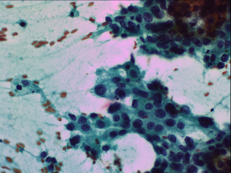 |
| Cytology features: | |
| ‣ Type of sample: | FNAC (solid lesion) |
| ‣ Site of biopsy: | |
| • Laterality: | Right |
| • Quadrant: | Upper outer |
| • Localization technique: | palpation |
| • Nature of aspirate: | whitish |
| ‣ Cytological description: | Very cellular smears with highly pleomorphic malignant cells in loose clusters. |
| ‣ Reporting category: | Malignant |
| ‣ Diagnosis: | Carcinoma high grade |
| ‣ Comments: | None |
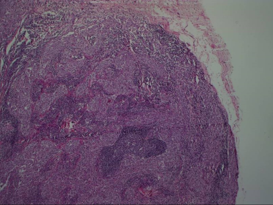
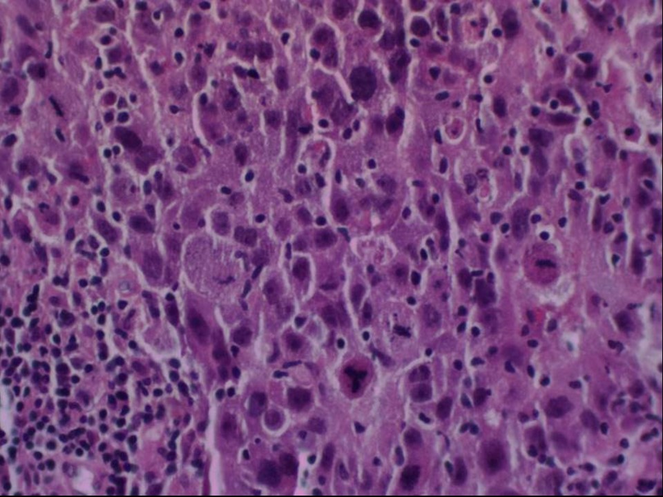
Histopathology:
Breast-conserving surgery
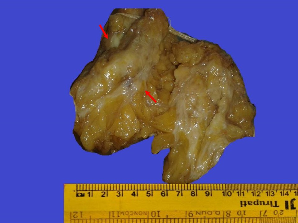 |  |
 |
| Histopathology features: | |
| ‣ Specimen type: | Breast-conserving surgery |
| ‣ Laterality: | Right |
| ‣ Macroscopy: | On serial sectioning, the following four firm areas are identified:
|
| ‣ Histological type: | Invasive breast cancer of no special type with medullary pattern |
| ‣ Histological grade: | Grade 3 (3 + 3 + 3 = 9) |
| ‣ Mitosis: | 25 |
| ‣ Maximum invasive tumour size: | Largest 1.6 cm in greatest dimension, two other nodules 0.5 cm and 0.4 |
| ‣ Lymph node status: | 1/32 |
| ‣ Peritumoural lymphovascular invasion: | Not identified |
| ‣ DCIS/EIC: | Not identified |
| ‣ Margins: | Free |
| ‣ Pathological stage: | pT1(3)N1 |
| ‣ Biomarkers: | |
| ‣ Comments: | Sections from the first firm area (1), fourth firm area (4), and focal parts of second firm area (2) show features of medullary carcinoma with dense lymphocytic infiltrate and tumour circumscription with rounded tumour margins. The largest tumour (1) is 1.6 cm in greatest dimension, microscopically. Multiple sections from the third firm area (3) show features of benign proliferative breast disease and sclerosis |
Case summary:
| Premenopausal woman presented with a lump in the right breast. Diagnosed as right breast carcinoma (multifocal), BI-RADS 5 on imaging, as high-grade breast carcinoma on cytology, and as medullary carcinoma, pT1(3)N1 on histopathology. |
Learning points:
|