


Home / Training / Manuals / Atlas of breast cancer early detection / Cases
Atlas of breast cancer early detection
Filter by language: English / Русский
Go back to the list of case studies
.png) Click on the pictures to magnify and display the legends
Click on the pictures to magnify and display the legends
| Case number: | 033 |
| Age: | 42 |
| Clinical presentation: | Premenopausal woman with average risk of developing breast cancer presented with a right breast lump noticed 3 days ago. |
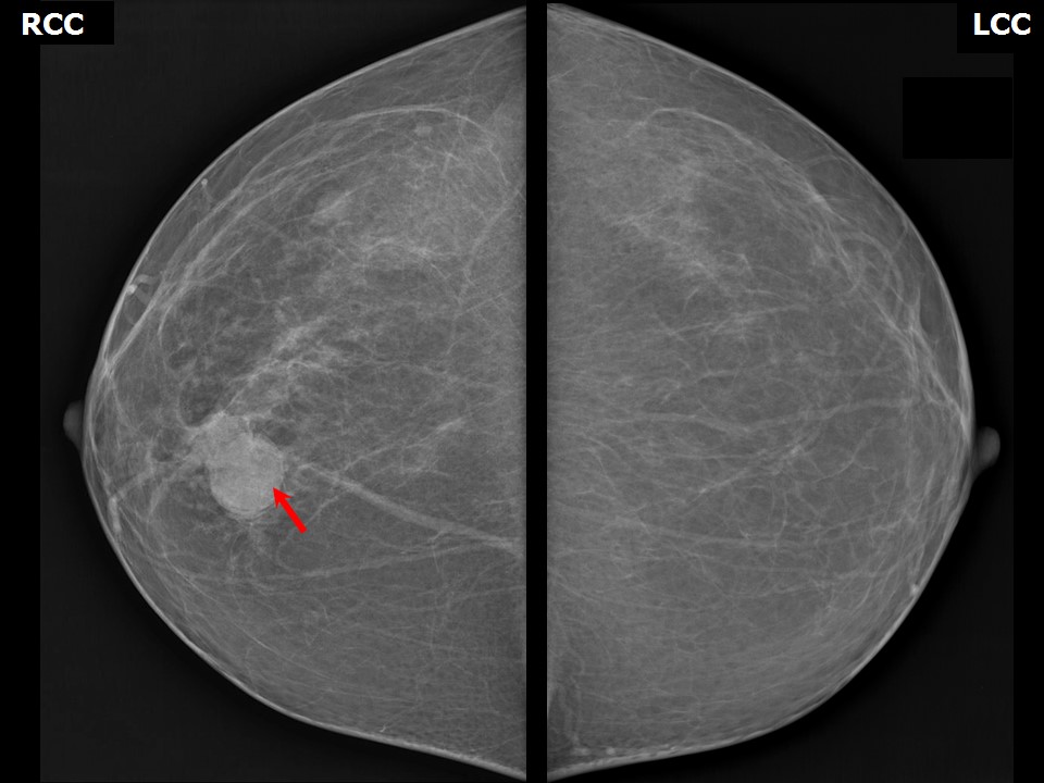
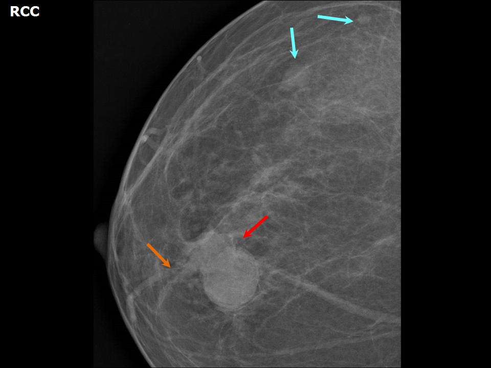
Mammography:
 |  |
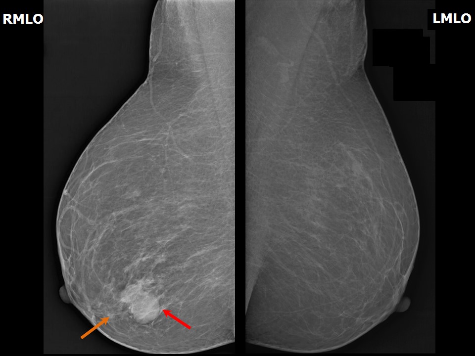 |
| Breast composition: | ACR category a (the breasts are almost entirely fatty) | Mammography features: |
| ‣ Location of the lesion: | Right breast, lower inner quadrant at 5 oclock, anterior and middle thirds, 3.5 cm from nipple and 1.5 cm from skin |
| ‣ Mass: | |
| • Number: | 2 |
| • Size: | 2.5 × 2.2 cm and 1.9 × 1.5 cm |
| • Shape: | Round |
| • Margins: | Indistinct |
| • Density: | Equal |
| ‣ Calcifications: | |
| • Typically benign: | None |
| • Suspicious: | None |
| • Distribution: | None |
| ‣ Architectural distortion: | Focal |
| ‣ Asymmetry: | None |
| ‣ Intramammary node: | None |
| ‣ Skin lesion: | None |
| ‣ Solitary dilated duct: | Present |
| ‣ Associated features: | Dilated duct |
| Breast composition: | ACR category a (the breasts are almost entirely fatty) | Mammography features: |
| ‣ Location of the lesion: | Right breast, upper outer quadrant at 1011 oclock, middle and posterior thirds |
| ‣ Mass: | |
| • Number: | 2 |
| • Size: | 1.6 × 1.0 cm and 0.6 cm in greatest dimension |
| • Shape: | Oval |
| • Margins: | Circumscribed |
| • Density: | Equal |
| ‣ Calcifications: | |
| • Typically benign: | None |
| • Suspicious: | None |
| • Distribution: | None |
| ‣ Architectural distortion: | None |
| ‣ Asymmetry: | None |
| ‣ Intramammary node: | None |
| ‣ Skin lesion: | None |
| ‣ Solitary dilated duct: | None |
| ‣ Associated features: | None |
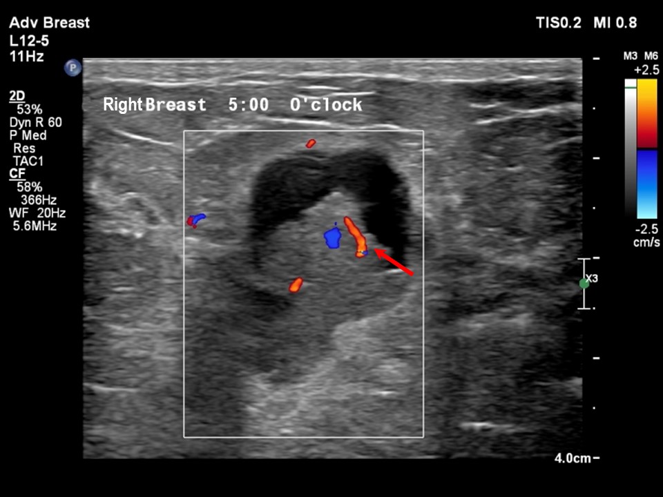
Ultrasound:
 |  |
 |
| Ultrasound features: Right breast, lower inner quadrant at 5 oclock | |
| ‣ Mass | |
| • Location: | Right breast, lower inner quadrant at 5 oclock |
| • Number: | 1 |
| • Size: | 3.0 × 2.6 × 1.6 cm |
| • Shape: | Oval |
| • Orientation: | Not parallel |
| • Margins: | Circumscribed |
| • Echo pattern: | Complex cystic and solid |
| • Posterior features: | No posterior features |
| ‣ Calcifications: | None |
| ‣ Associated features: | Duct changes, internal vascularity |
| ‣ Special cases: | None |
BI-RADS:
BI-RADS Category: 5 (highly suggestive of malignancy)Further assessment:
Further assessment advised: Further imaging with breast MRIMRI:
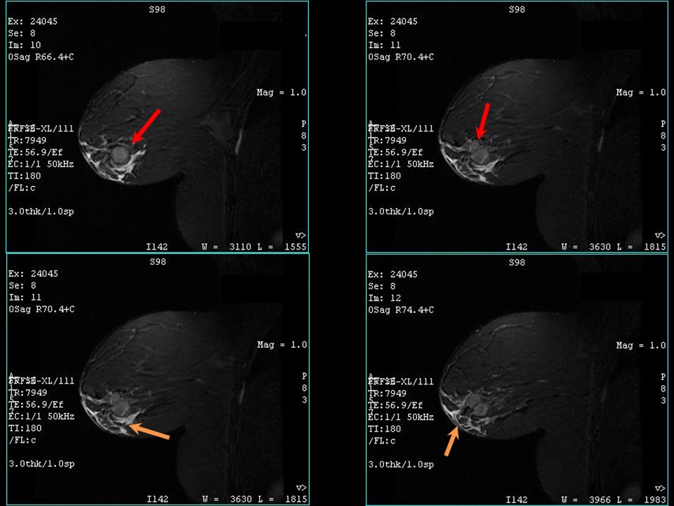 | 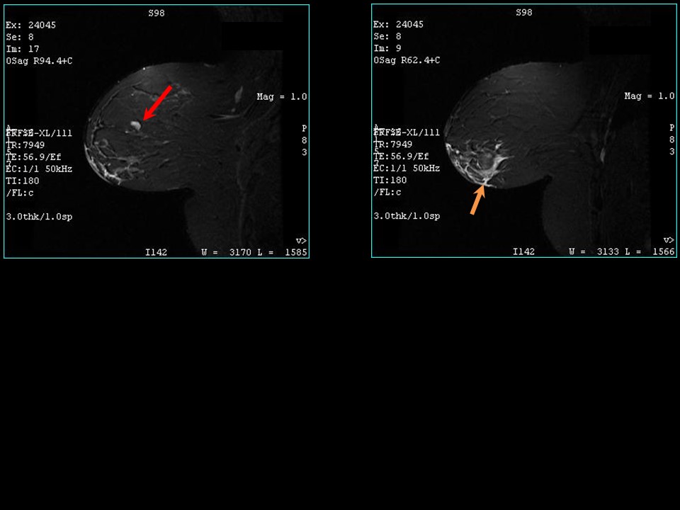 |
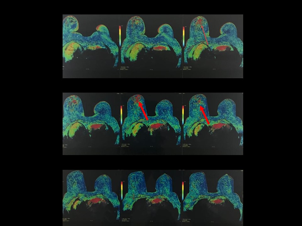 | 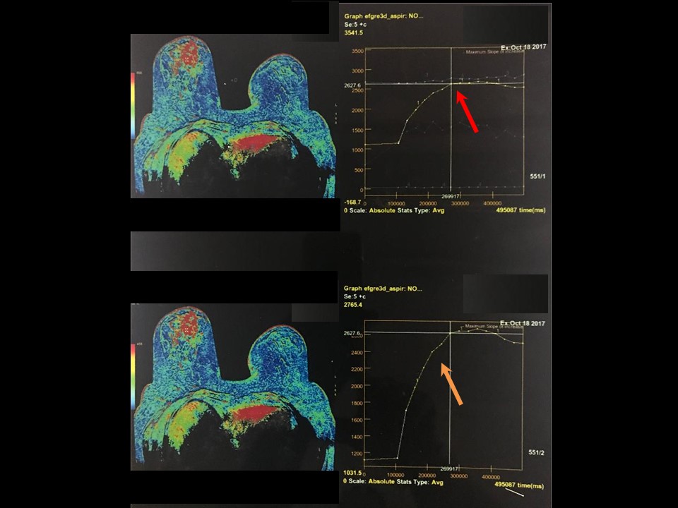 |
| MRI features: | ||
| ‣ MRI features: | MRI features: Amount of fibroglandular tissue (FGT): category a. Almost entirely fatty. Background parenchymal enhancement: Minimal (< 25%), symmetrical. Both Kuhl curves are for lower inner quadrant | |
| ‣ Location: | At 6 oclock | |
| ‣ Focus: | No | |
| ‣ Mass: | ||
| • Shape: | Bilobed | |
| • Margin: | Circumscribed | |
| • Internal enhancement: | Heterogeneous | |
| • Kinetic curve: | Intense fast initial phase with delayed phase plateau | |
| ‣ Non-mass enhancement: | ||
| • Distribution: | Multiple regions: In inferior medial and inferior lateral in lower inner and lower outer quadrants | |
| • Internal enhancement: | Heterogeneous | |
| ‣ Non-enhancing findings: | No | |
| ‣ Associated features: | Skin thickening of nipple and areola | |
| ‣ Axillary nodes: | No | |
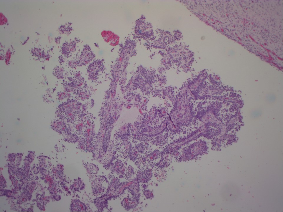
Cytology:
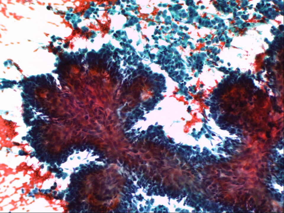 | 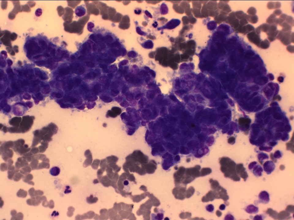 |
| Cytology features: | |
| ‣ Type of sample: | FNAC |
| ‣ Site of biopsy: | |
| • Laterality: | Right |
| • Quadrant: | Lump inferior to areola |
| • Localization technique: | Palpation |
| • Nature of aspirate: | 2 mL of bloody fluid |
| ‣ Cytological description: | Smears are very cellular and reveal many papillary clusters with a fibrovascular core as well-rounded three-dimensional epithelial groups. Many dispersed and loosely cohesive clusters are also seen. Tumour cells show atypical round nucleus with coarse chromatin and moderate cytoplasm. Myoepithelial cells are seen in occasional clusters. A few multinucleate cells are noted. Background shows haemorrhage and macrophages |
| ‣ Reporting category: | Suspicious, probably in situ or invasive carcinoma |
| ‣ Diagnosis: | Papillary neoplasm favours malignancy. Frozen section or biopsy advised |
| ‣ Comments: | None |
Histopathology:
MRM
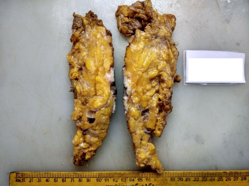 |  |
 | 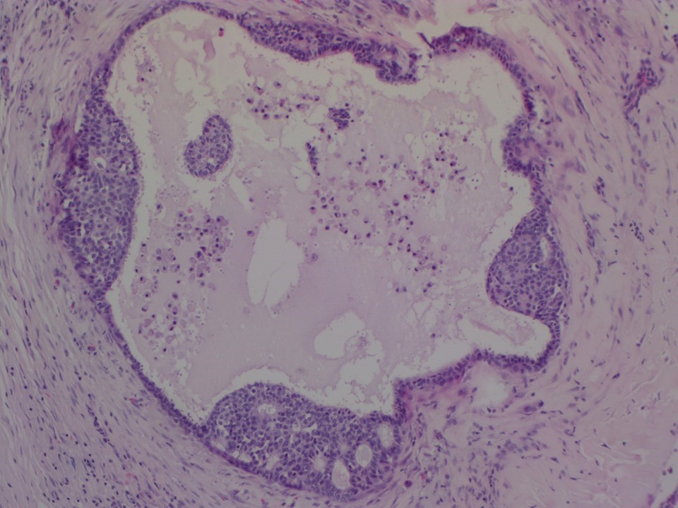 |
| Histopathology features: | |
| ‣ Specimen type: | MRM |
| ‣ Laterality: | Right |
| ‣ Macroscopy: | Right MRM specimen (28 × 25 × 5.5 cm) with overlying skin flap (13.5 × 5.0 cm). The nipple and areola are unremarkable. On serial sectioning a cyst and a surrounding irregular, firm, whitish area was seen. The cyst was well circumscribed with a thick fibrous wall (1.5 cm in greatest dimension) and a brownish, firm, friable mass (0.5 × 0.5 × 0.7 cm) within the cavity. The surrounding firm whitish area (3.6 × 3.7 × 2.5 cm) adjacent to the cyst and extending anteriorly towards the skin was located 0.6 cm from the skin and 2.4 cm from the base. Another cystic area (1.0 cm in diameter) with brownish fluid was noted 4.0 cm from the tumour in the lower outer quadrant |
| ‣ Histological type: | Encapsulated papillary carcinoma |
| ‣ Histological grade: | Encapsulated papillary carcinoma of intermediate nuclear grade |
| ‣ Mitosis: | |
| ‣ Maximum invasive tumour size: | There is no invasive tumour in the sections studied |
| ‣ Lymph node status: | 0/39 |
| ‣ Peritumoural lymphovascular invasion: | Absent |
| ‣ DCIS/EIC: | Extensive DCIS of papillary, comedo, solid, and cribriform pattern with low to intermediate nuclear grade seen in the sections from the firm whitish areas seen grossly |
| ‣ Margins: | Free of tumour |
| ‣ Pathological stage: | Encapsulated papillary carcinoma should be staged and managed as Tis disease (according to the WHO Working Group 2011) |
| ‣ Biomarkers: | ER positive, PR positive, and HER2 negative |
| ‣ Comments: | The adjacent breast shows focus of apocrine metaplasia, fibrocystic changes, and fibrosis |
Case summary:
| Premenopausal woman presented with a right breast lump, diagnosed as complex cystic-solid bilobed mass lesion in the right breast, BI-RADS category 5 on imaging, as papillary neoplasm suspicious for malignancy on cytology, and as encapsulated papillary carcinoma, pTisN0 on histopathology. |
Learning points:
|