


Home / Training / Manuals / Atlas of breast cancer early detection / Cases
Atlas of breast cancer early detection
Filter by language: English / Русский
Go back to the list of case studies
.png) Click on the pictures to magnify and display the legends
Click on the pictures to magnify and display the legends
| Case number: | 083 |
| Age: | 56 |
| Clinical presentation: | Postmenopausal woman, a known operated case of left breast cancer, now presented with a lump in the left breast at the operated site. |
Mammography:
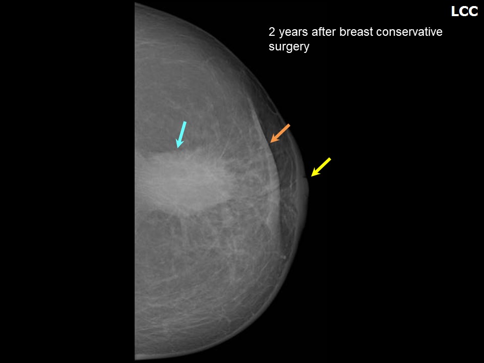 | 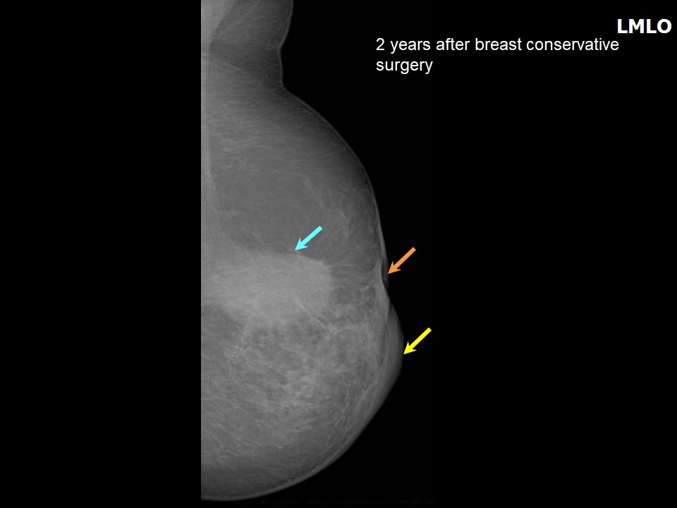 |
| Breast composition: | ACR category a (the breasts are almost entirely fatty) | Mammography features: |
| ‣ Location of the lesion: | Left breast, central portion of the breast, central zone, posterior third, beneath surgical scar |
| ‣ Mass: | |
| • Number: | 1 |
| • Size: | 3.8 × 2.2 cm |
| • Shape: | Oval |
| • Margins: | Indistinct |
| • Density: | Equal |
| ‣ Calcifications: | |
| • Typically benign: | None |
| • Suspicious: | None |
| • Distribution: | None |
| ‣ Architectural distortion: | Present |
| ‣ Asymmetry: | Focal |
| ‣ Intramammary node: | None |
| ‣ Skin lesion: | None |
| ‣ Solitary dilated duct: | None |
| ‣ Associated features: | Skin retraction, nipple retraction, skin thickening, trabecular thickening, and architectural distortion in postoperative breast with seroma |
Ultrasound:
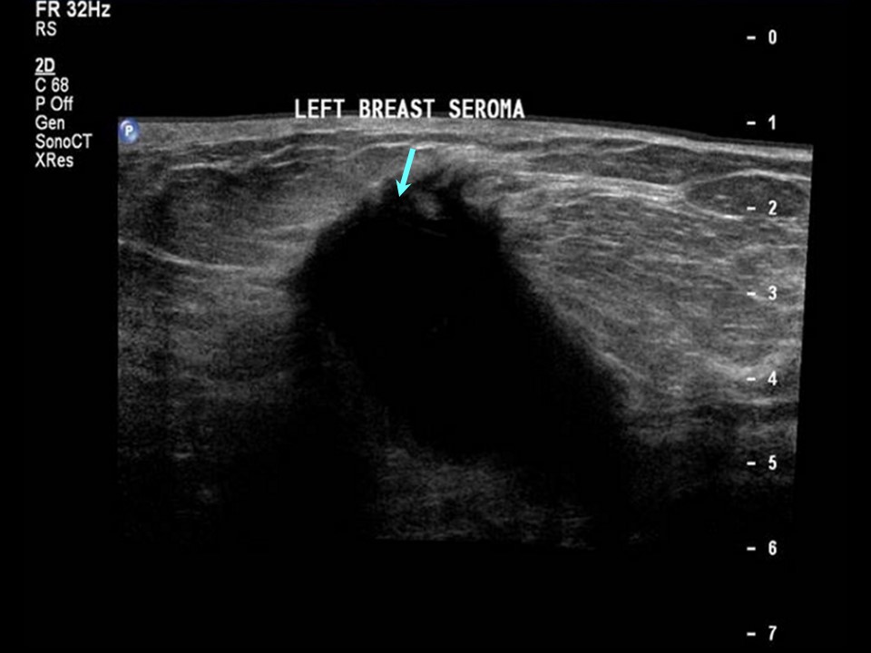 |
| Ultrasound features: Left breast, central portion of the breast along surgical scar | |
| ‣ Mass | |
| • Location: | Left breast, central portion of the breast along surgical scar |
| • Number: | 1 |
| • Size: | 4.1 × 2.8 × 3.7 cm |
| • Shape: | Irregular |
| • Orientation: | Not parallel |
| • Margins: | Indistinct |
| • Echo pattern: | Anechoic |
| • Posterior features: | Posterior shadowing |
| ‣ Calcifications: | None |
| ‣ Associated features: | Postoperative fluid collection, architectural distortion, skin thickening, and absent vascularity |
| ‣ Special cases: | Postoperative fluid collection |
BI-RADS:
BI-RADS Category (Left BCS, post operative breast): 2 (benign)Further assessment:
Further assessment advised: Referral for cytologyCytology:
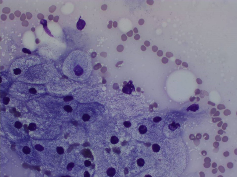 |
| Cytology features: | |
| ‣ Type of sample: | FNAC (cystic lesion) |
| ‣ Site of biopsy: | |
| • Laterality: | left |
| • Quadrant: | Upper outer |
| • Localization technique: | Palpation |
| • Nature of aspirate: | Yellowish fluid |
| ‣ Cytological description: | Smears show only foamy histiocytes on a proteinaceous background. |
| ‣ Reporting category: | Benign |
| ‣ Diagnosis: | Consistent with seroma |
| ‣ Comments: | None |
Case summary:
| Postmenopausal woman on postoperative surveillance after surgery for left breast cancer presented with left breast lump at site of surgery. Diagnosed as organized seroma in left breast with surgical scar, BI-RADS 2 on imaging and as left breast seroma collection on cytology. |
Learning points:
|