


Home / Training / Manuals / Atlas of breast cancer early detection / Cases
Atlas of breast cancer early detection
Filter by language: English / Русский
Go back to the list of case studies
.png) Click on the pictures to magnify and display the legends
Click on the pictures to magnify and display the legends
| Case number: | 009 |
| Age: | 41 |
| Clinical presentation: | Premenopausal woman with average risk of developing breast cancer presented with breast pain. She did not report a lump or nipple discharge. Examination revealed a 5 cm lump in the right breast. |
Mammography:
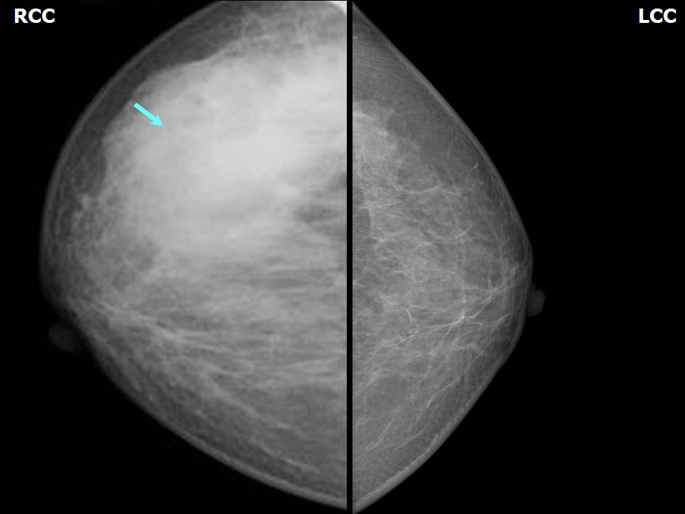 | 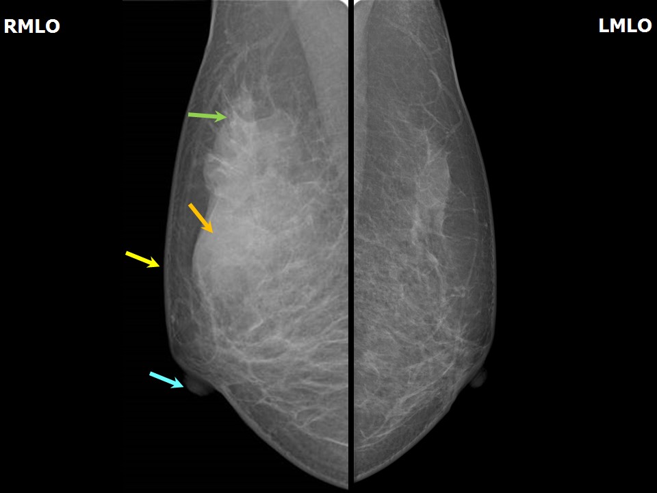 |
| Breast composition: | ACR category c (the breasts are heterogeneously dense, which may obscure small masses) | Mammography features: |
| ‣ Location of the lesion: | Right breast, upper outer quadrant at 1012 oclock, anterior, middle, and posterior thirds |
| ‣ Mass: | |
| • Number: | 1 |
| • Size: | 6.5 × 3.5 × 3.0 cm |
| • Shape: | Irregular |
| • Margins: | Obscured |
| • Density: | High |
| ‣ Calcifications: | |
| • Typically benign: | None |
| • Suspicious: | None |
| • Distribution: | None |
| ‣ Architectural distortion: | None |
| ‣ Asymmetry: | None |
| ‣ Intramammary node: | None |
| ‣ Skin lesion: | None |
| ‣ Solitary dilated duct: | None |
| ‣ Associated features: | Trabecular thickening |
Ultrasound:
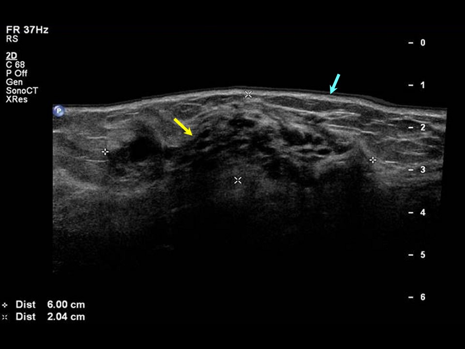 | 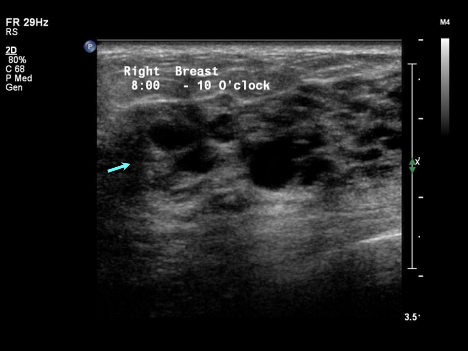 |
| Ultrasound features: Right breast, upper outer quadrant at 810 oclock | |
| ‣ Mass | |
| • Location: | Right breast, upper outer quadrant at 810 oclock |
| • Number: | 1 |
| • Size: | 6.0 × 2.0 cm |
| • Shape: | Irregular |
| • Orientation: | Parallel |
| • Margins: | Indistinct |
| • Echo pattern: | Heterogeneous |
| • Posterior features: | Combined pattern |
| ‣ Calcifications: | None |
| ‣ Associated features: | None |
| ‣ Special cases: | Clustered microcysts |
BI-RADS:
BI-RADS Category: 2 (benign)Further assessment:
Further assessment advised: Referral for cytologyCytology:
 |
| Cytology features: | |
| ‣ Type of sample: | FNAC: 3 mL of greenish clear fluid was aspirated; swelling completely subsided after aspiration |
| ‣ Site of biopsy: | |
| • Laterality: | Right |
| • Quadrant: | Upper outer quadrant |
| • Localization technique: | |
| • Nature of aspirate: | 3 mL of clear greenish fluid; swelling completely subsided after aspiration |
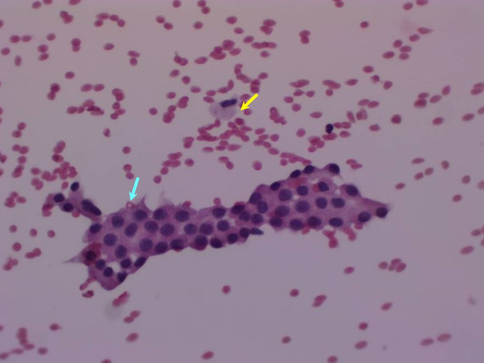
| ‣ Cytological description: | A few ductal epithelial cells, some with apocrine metaplasia seen with foamy histiocytes on a proteinaceous background |
| ‣ Reporting category: | Benign |
| ‣ Diagnosis: | Proliferative fibrocystic change |
| ‣ Comments: | None |
Case summary:
| Premenopausal woman presented with painful right breast lump. Diagnosed as fibrocystic change with clustered microcysts in right breast, BI-RADS 2 on imaging and as proliferative fibrocystic change on cytology. |
Learning points:
|