


Home / Training / Manuals / Atlas of breast cancer early detection / Cases
Atlas of breast cancer early detection
Filter by language: English / Русский
Go back to the list of case studies
.png) Click on the pictures to magnify and display the legends
Click on the pictures to magnify and display the legends
| Case number: | 063 |
| Age: | 39 |
| Clinical presentation: | Premenopausal woman with average risk of developing breast cancer presented with a rapidly growing left breast lump. On examination, she was found to have a large firm to hard lump in her left breast. |
Mammography:
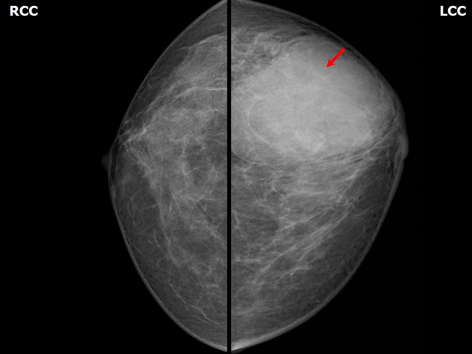 | 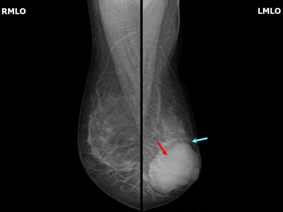 |
| Breast composition: | ACR category b (there are scattered areas of fibroglandular density) | Mammography features: |
| ‣ Location of the lesion: | Left breast, lower outer quadrant at 5 oclock, anterior, middle, and posterior thirds |
| ‣ Mass: | |
| • Number: | 1 |
| • Size: | 5.5 × 4.0 cm |
| • Shape: | Oval |
| • Margins: | Circumscribed |
| • Density: | High |
| ‣ Calcifications: | |
| • Typically benign: | None |
| • Suspicious: | None |
| • Distribution: | None |
| ‣ Architectural distortion: | None |
| ‣ Asymmetry: | None |
| ‣ Intramammary node: | None |
| ‣ Skin lesion: | None |
| ‣ Solitary dilated duct: | None |
| ‣ Associated features: | None |
Ultrasound:
 |  |
 |  |
 |
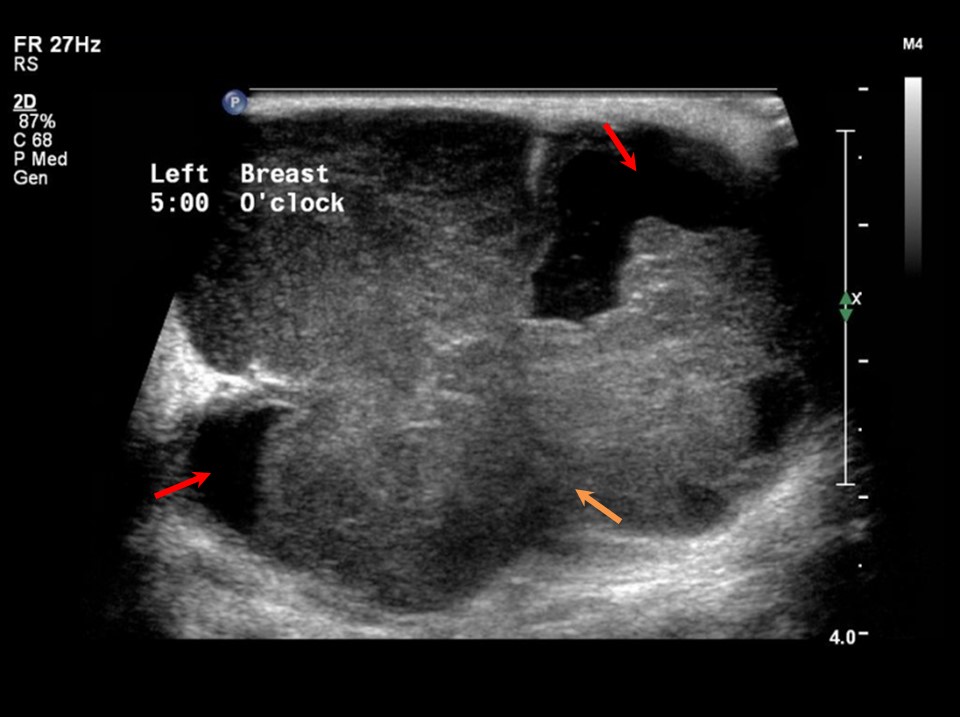
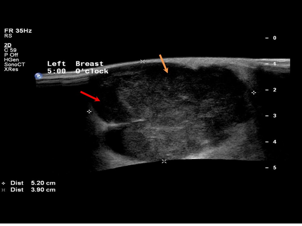
| Ultrasound features: Left breast, lower outer quadrant at 5 oclock | |
| ‣ Mass | |
| • Location: | Left breast, lower outer quadrant at 5 oclock |
| • Number: | 1 |
| • Size: | 5.2 × 4.0 cm |
| • Shape: | Irregular |
| • Orientation: | Parallel |
| • Margins: | Indistinct |
| • Echo pattern: | Heterogeneous with areas of breakdown |
| • Posterior features: | No posterior features |
| ‣ Calcifications: | None |
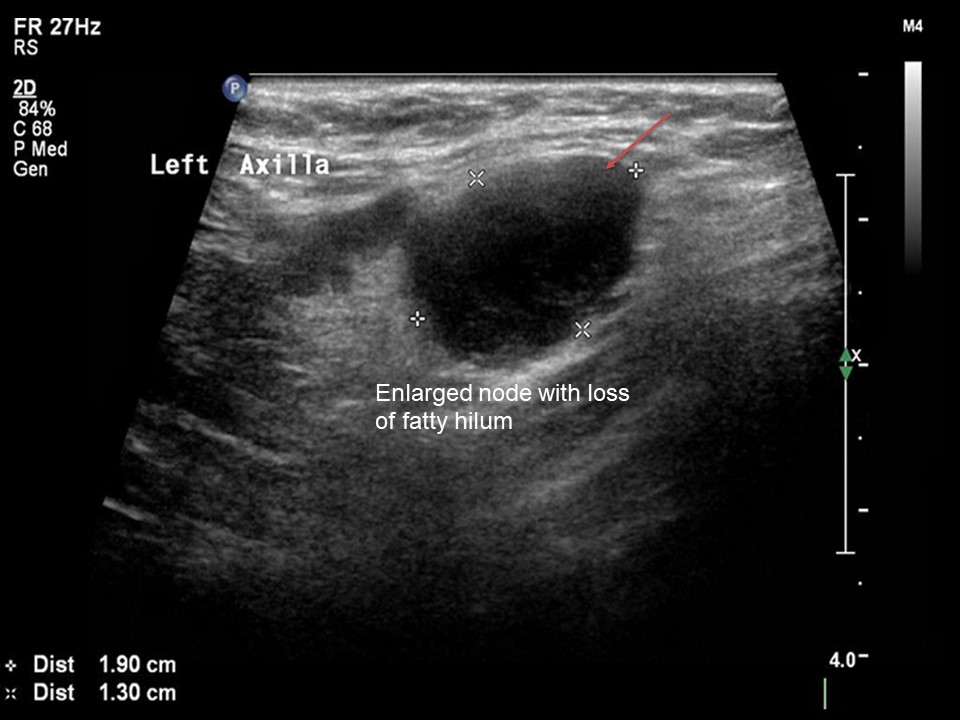
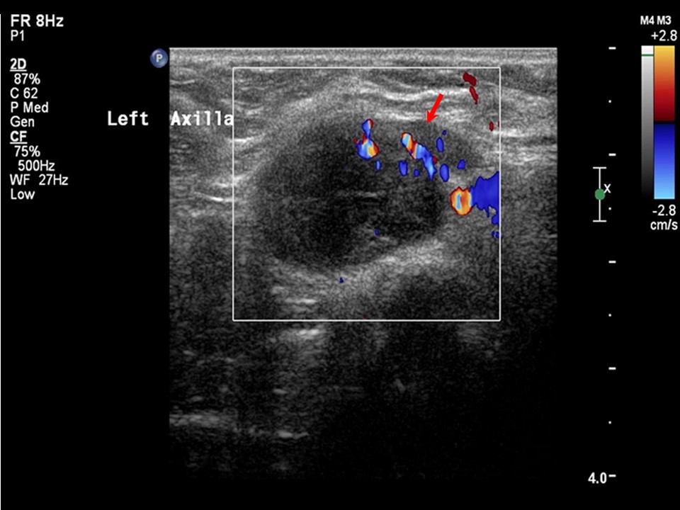
| ‣ Associated features: | Internal vascularity in mass. Enlarged lymph nodes (2.0 × 1.3 cm and 2.0 × 0.7 cm) with loss of central fatty hilum |
| ‣ Special cases: | None |
BI-RADS:
BI-RADS Category: 4C (high suspicion for malignancy)Further assessment:
Further assessment advised: Referral for cytologyCytology:
 |
| Cytology features: | |
| ‣ Type of sample: | FNAC (solid lesion) |
| ‣ Site of biopsy: | |
| • Laterality: | Left |
| • Quadrant: | Lower outer |
| • Localization technique: | Palpation |
| • Nature of aspirate: | Whitish |
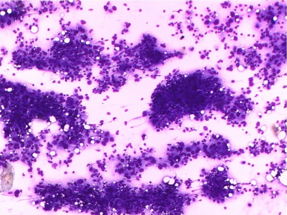
| ‣ Cytological description: | Highly pleomorphic malignant cells seen with lymphocytes in the background |
| ‣ Reporting category: | Malignant |
| ‣ Diagnosis: | Carcinoma high grade |
| ‣ Comments: | None |
Histopathology:
Breast-conserving surgery
 | 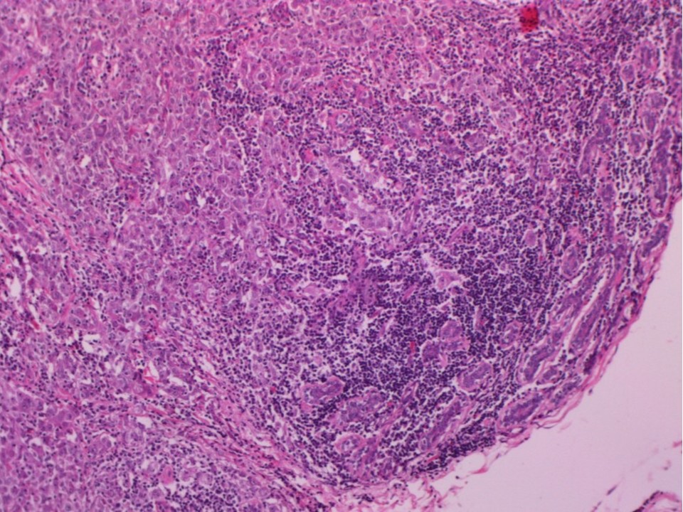 |
| Histopathology features: | |
| ‣ Specimen type: | Breast-conserving surgery |
| ‣ Laterality: | Left |
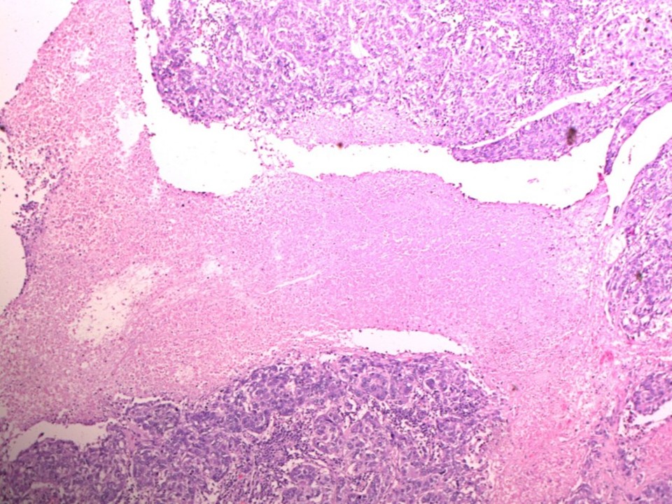
| ‣ Macroscopy: | Cut surface shows a large, greyish white, well-circumscribed tumour (5.5 × 5.0 × 4.0 cm) (T3) with a soft central necrotic area |
| ‣ Histological type: | Invasive breast carcinoma of no special type with medullary pattern |
| ‣ Histological grade: | Grade 3 (3 + 2 + 2 = 7) |
| ‣ Mitosis: | 15 |
| ‣ Maximum invasive tumour size: | 5.5 cm in greatest dimension |
| ‣ Lymph node status: | 1/24 |
| ‣ Peritumoural lymphovascular invasion: | Not identified |
| ‣ DCIS/EIC: | Not identified |
| ‣ Margins: | Free of tumour |
| ‣ Pathological stage: | pT3N1 |
| ‣ Biomarkers: | |
| ‣ Comments: | Extensive areas of tumour necrosis seen |
Case summary:
| Premenopausal woman presented with lump in the left breast. Diagnosed as circumscribed high-density mass with perilesional halo, with left axillary enlarged node of altered morphology, BI-RADS 4C on imaging, as breast carcinoma on cytology, and as invasive breast carcinoma of no specific type with medullary pattern, pT3N1 on histopathology. |
Learning points:
|