


Home / Training / Manuals / Atlas of breast cancer early detection / Cases
Atlas of breast cancer early detection
Filter by language: English / Русский
Go back to the list of case studies
.png) Click on the pictures to magnify and display the legends
Click on the pictures to magnify and display the legends
| Case number: | 124 |
| Age: | 38 |
| Clinical presentation: | Premenopausal woman with average risk of developing breast cancer presented with nipple discharge on the right breast along with left breast lump. Examination revealed a lump (4 × 4 cm) in the upper outer quadrant of the left breast, which was firm in consistency, mildly tender, and fixed to breast tissue. |
Mammography:
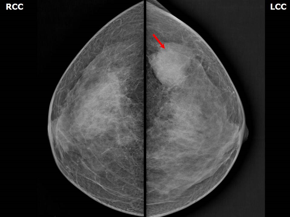 | 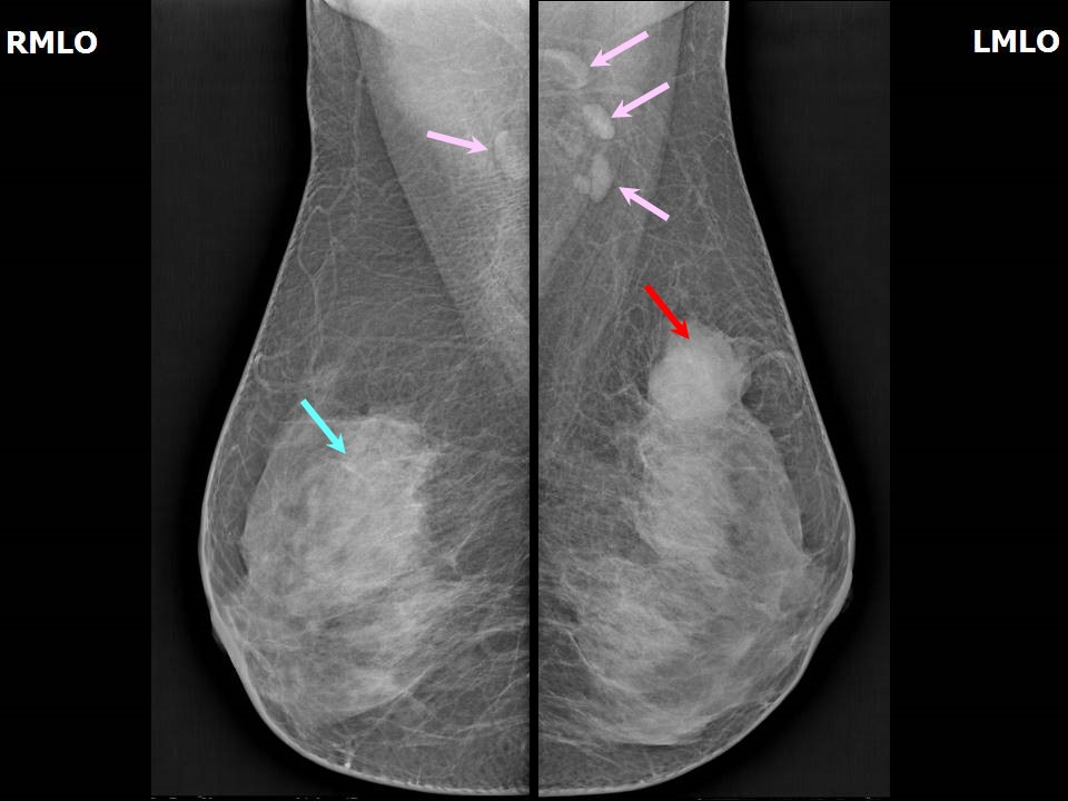 |
| Breast composition: | ACR category c (the breasts are heterogeneously dense, which may obscure small masses) | Mammography features: |
| ‣ Location of the lesion: | Left breast, upper outer quadrant at 2 oclock, middle and posterior thirds |
| ‣ Mass: | |
| • Number: | 1 |
| • Size: | 3.6 × 3.5 × 3.3 cm |
| • Shape: | Irregular |
| • Margins: | Obscured |
| • Density: | High |
| ‣ Calcifications: | |
| • Typically benign: | None |
| • Suspicious: | None |
| • Distribution: | None |
| ‣ Architectural distortion: | None |
| ‣ Asymmetry: | None |
| ‣ Intramammary node: | None |
| ‣ Skin lesion: | None |
| ‣ Solitary dilated duct: | None |
| ‣ Associated features: | None |
Ultrasound:
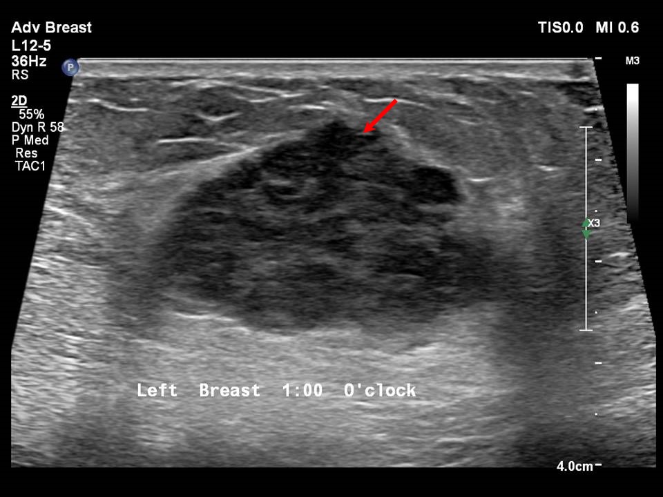 | 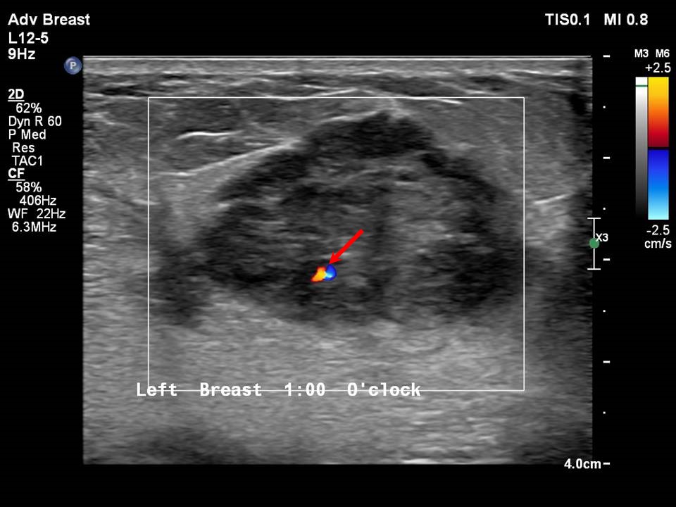 |
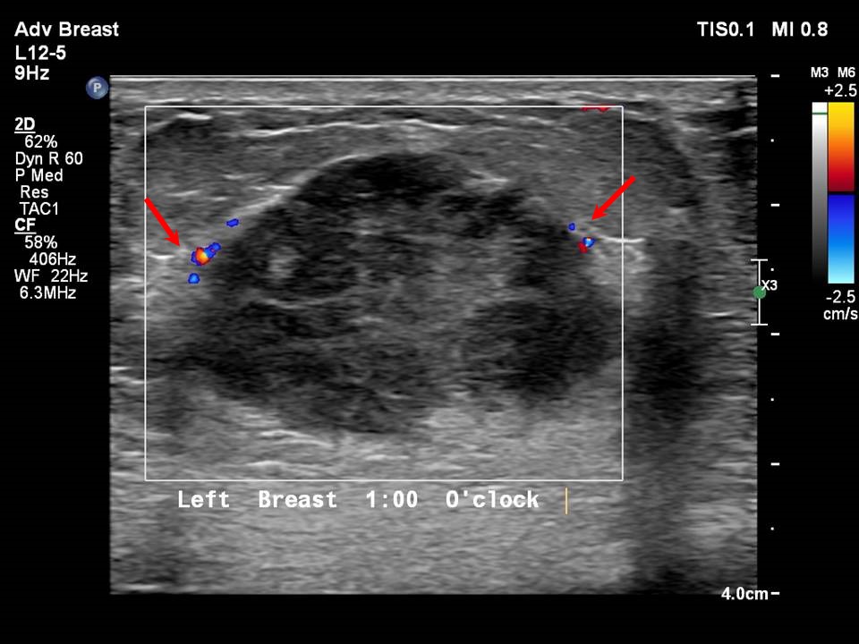 |
| Ultrasound features: Left breast, upper outer quadrant at 12 oclock, 5.0 cm from nipple and 1.7 cm skin depth | |
| ‣ Mass | |
| • Location: | Left breast, upper outer quadrant at 12 oclock, 5.0 cm from nipple and 1.7 cm skin depth |
| • Number: | 1 |
| • Size: | 3.7 × 2.5 cm |
| • Shape: | Irregular |
| • Orientation: | Parallel |
| • Margins: | Indistinct |
| • Echo pattern: | Heteroechoic |
| • Posterior features: | No posterior features |
| ‣ Calcifications: | None |
| ‣ Associated features: | Oedema, internal vascularity, and vessels in rim |
| ‣ Special cases: | None |
BI-RADS:
BI-RADS Category: 5 (highly suggestive of malignancy)Further assessment:
Further assessment advised: Referral for cytologyCytology:
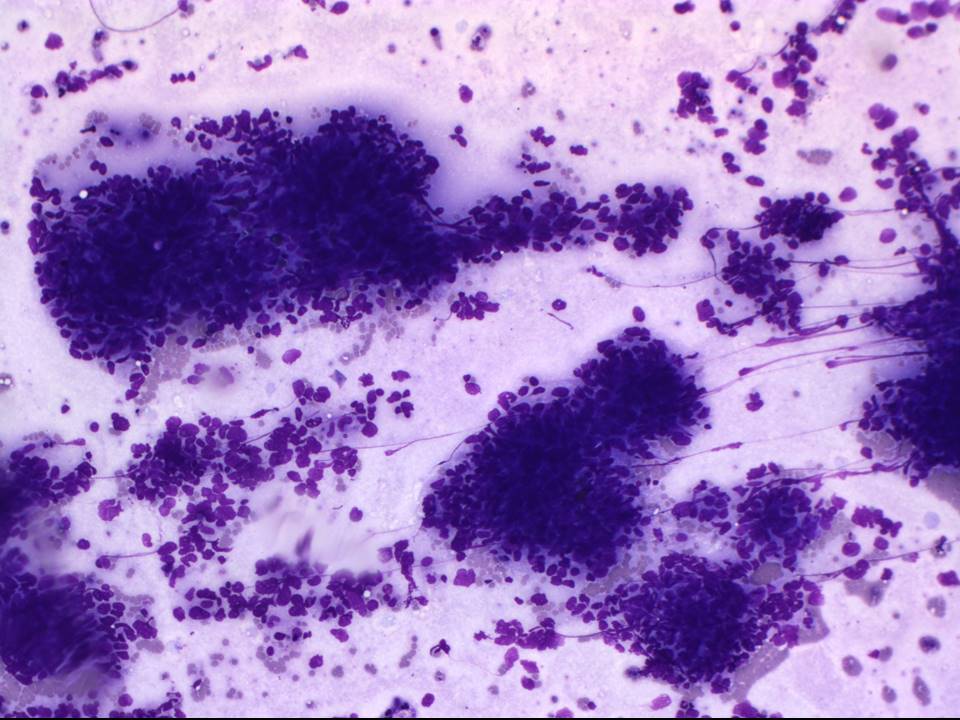 | 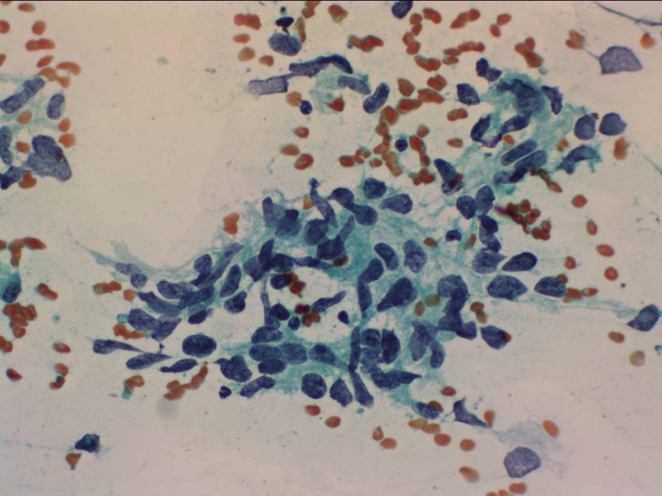 |
| Cytology features: | |
| ‣ Type of sample: | FNAC |
| ‣ Site of biopsy: | |
| • Laterality: | Left |
| • Quadrant: | Upper outer |
| • Localization technique: | Palpation |
| • Nature of aspirate: | Whitish, blood tinged |
| ‣ Cytological description: | Smears are cellular and reveal many crowded clusters, many loosely cohesive clusters, and many scattered large, hyperchromatic, and pleomorphic nuclei with irregular nuclear margins and inconspicuous nucleoli. Cytoplasm is moderate to abundant. Many clusters show plump spindled morphology. Background shows scanty necrosis and fibroadipose stroma |
| ‣ Reporting category: | Malignant |
| ‣ Diagnosis: | Carcinoma |
| ‣ Comments: | None |
Histopathology:
Breast-conserving surgery
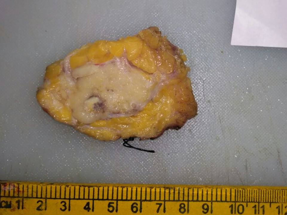 |  |
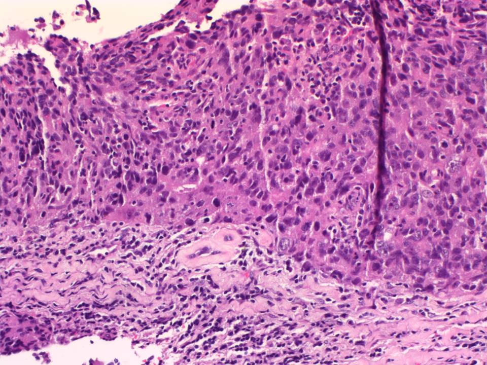 | 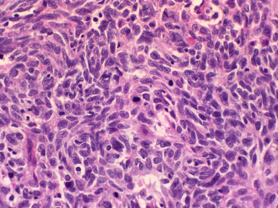 |
| Histopathology features: | |
| ‣ Specimen type: | Breast-conserving surgery |
| ‣ Laterality: | Left |
| ‣ Macroscopy: | Lumpectomy specimen (7.0 × 6.8 × 3.0 cm) oriented with long suture laterally and short suture superiorly. Skin flap is 4.0 × 1.5 cm. On serial sectioning, a firm whitish tumour (3.5 × 2.4 × 2.5 cm) with areas of haemorrhage is identified. It is located 0.8 cm from the skin (anterior margin), 0.6 cm from the posterior margin, 1.7 cm from the superior margin, 1.1 cm from the inferior margin, 2.0 cm from the medial margin, and 1.0 cm from the lateral margin. The remaining breast parenchyma shows areas of fibrosis |
| ‣ Histological type: | Metaplastic carcinoma |
| ‣ Histological grade: | Grade 3 (3 + 3 + 3 = 9) |
| ‣ Mitosis: | 28 |
| ‣ Maximum invasive tumour size: | 3.5 cm in greatest dimension |
| ‣ Lymph node status: | 0/18 |
| ‣ Peritumoural lymphovascular invasion: | Absent |
| ‣ DCIS/EIC: | Absent |
| ‣ Margins: | Free of tumour |
| ‣ Pathological stage: | pT2N0 |
| ‣ Biomarkers: | ER negative, PR negative, and HER2 negative (score 0) |
| ‣ Comments: |
Case summary:
| Premenopausal woman presented with right nipple discharge and left breast lump. Diagnosed as left breast carcinoma, BI-RADS 5 on imaging, as left breast carcinoma on cytology, and as metaplastic breast carcinoma, pT2N0 on histopathology. |
Learning points:
|