


Home / Training / Manuals / Atlas of breast cancer early detection / Cases
Atlas of breast cancer early detection
Filter by language: English / Русский
Go back to the list of case studies
.png) Click on the pictures to magnify and display the legends
Click on the pictures to magnify and display the legends
| Case number: | 129 |
| Age: | 51 |
| Clinical presentation: | Perimenopausal woman with average risk of developing breast cancer presented with a lump in the right breast and mastalgia of duration 15 days. Examination revealed a 4 cm lump in the right breast. |
Mammography:
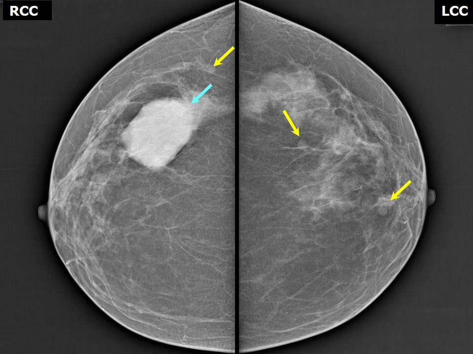 | 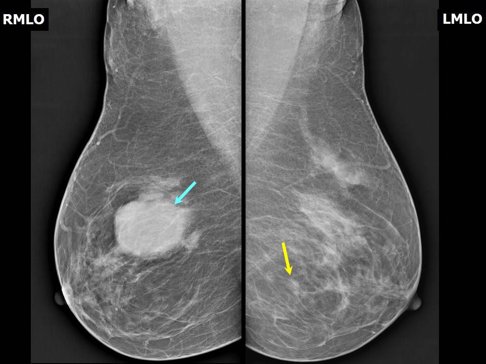 |
| Breast composition: | ACR category b (there are scattered areas of fibroglandular density) | Mammography features: |
| ‣ Location of the lesion: | Right breast, upper outer quadrant at 10 oclock, middle third, 2.0 cm from the nipple and at 1.0 cm skin depth |
| ‣ Mass: | |
| • Number: | Multiple |
| • Size: | Largest 4.5 × 3.5 cm. This is the clinically palpable lump of concern |
| • Shape: | Oval |
| • Margins: | Circumscribed |
| • Density: | Equal |
| ‣ Calcifications: | |
| • Typically benign: | None |
| • Suspicious: | None |
| • Distribution: | None |
| ‣ Architectural distortion: | None |
| ‣ Asymmetry: | None |
| ‣ Intramammary node: | None |
| ‣ Skin lesion: | None |
| ‣ Solitary dilated duct: | None |
| ‣ Associated features: | None |
| Breast composition: | ACR category b (there are scattered areas of fibroglandular density) | Mammography features: |
| ‣ Location of the lesion: | Left breast, lower inner quadrant at 69 oclock, anterior and middle thirds |
| ‣ Mass: | |
| • Number: | Multiple |
| • Size: | Largest 0.5 cm |
| • Shape: | Round |
| • Margins: | Circumscribed |
| • Density: | Equal |
| ‣ Calcifications: | |
| • Typically benign: | None |
| • Suspicious: | None |
| • Distribution: | None |
| ‣ Architectural distortion: | None |
| ‣ Asymmetry: | None |
| ‣ Intramammary node: | None |
| ‣ Skin lesion: | None |
| ‣ Solitary dilated duct: | None |
| ‣ Associated features: | None |
Ultrasound:
 |
| Ultrasound features: Right breast, central portion of the breast, multiple cysts in para-areolar region, the largest in the upper outer quadrant at 10 oclock position, 2.0 cm from the nipple and at 1.0 cm skin depth | |
| ‣ Mass | |
| • Location: | Right breast, central portion of the breast, multiple cysts in para-areolar region, the largest in the upper outer quadrant at 10 oclock position, 2.0 cm from the nipple and at 1.0 cm skin depth |
| • Number: | Multiple |
| • Size: | Largest 4.0 × 2.1 cm |
| • Shape: | Oval |
| • Orientation: | Parallel |
| • Margins: | Circumscribed |
| • Echo pattern: | Anechoic |
| • Posterior features: | Posterior shadowing |
| ‣ Calcifications: | None |
| ‣ Associated features: | None |
| ‣ Special cases: | Simple cyst |
| Ultrasound features: Left breast, upper outer quadrant at 10 o'clock, a few subcentimetre-sized cysts are seen in the para-areolar region | |
| ‣ Mass | |
| • Location: | Left breast, upper outer quadrant at 10 o'clock, a few subcentimetre-sized cysts are seen in the para-areolar region |
| • Number: | 2 to 3 |
| • Size: | Largest 0.6 cm |
| • Shape: | Round |
| • Orientation: | Parallel |
| • Margins: | Circumscribed |
| • Echo pattern: | Anechoic |
| • Posterior features: | No posterior features |
| ‣ Calcifications: | None |
| ‣ Associated features: | None |
| ‣ Special cases: | Simple cyst |
BI-RADS:
BI-RADS Category: 2 (benign)Further assessment:
Further assessment advised: Referral for cytologyCytology:
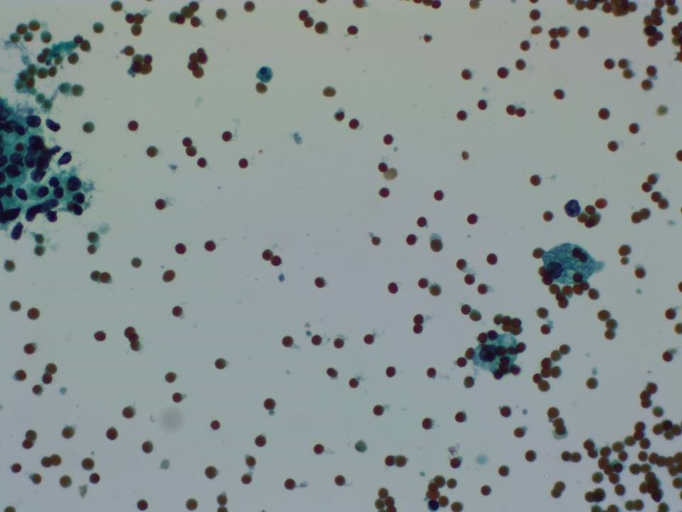 | 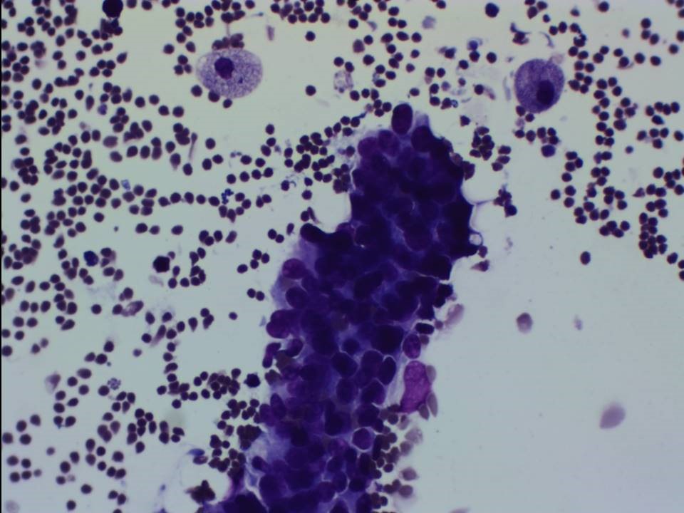 |
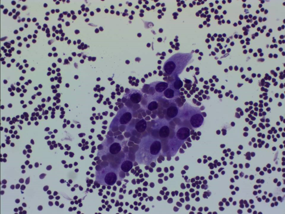 |
| Cytology features: | |
| ‣ Type of sample: | FNAC |
| ‣ Site of biopsy: | |
| • Laterality: | Right |
| • Quadrant: | |
| • Localization technique: | Palpation |
| • Nature of aspirate: | 14 mL of yellowish fluid aspirated |
| ‣ Cytological description: | Smears reveal a few benign ductal cell clusters, foamy macrophages, occasional cluster of apocrine cells, against a background of proteinaceous material. Smears from residual lump reveal a few benign fibroadipose fragments |
| ‣ Reporting category: | Benign |
| ‣ Diagnosis: | Fibrocystic lesion, non-proliferative |
| ‣ Comments: | None |
Case summary:
| Perimenopausal woman presented with a right breast lump and mastalgia. Diagnosed as multiple simple cysts in both breasts, BI-RADS 2 on imaging and as non-proliferative fibrocystic change on cytology. |
Learning points:
|