


Home / Training / Manuals / Atlas of breast cancer early detection / Cases
Atlas of breast cancer early detection
Filter by language: English / Русский
Go back to the list of case studies
.png) Click on the pictures to magnify and display the legends
Click on the pictures to magnify and display the legends
| Case number: | 166 |
| Age: | 41 |
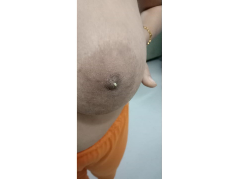
| Clinical presentation: | Premenopausal woman with average risk of breast cancer presented with a left breast lump first noticed 68 months earlier. |
 |
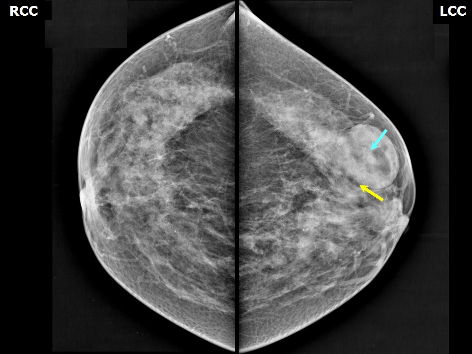
Mammography:
 |  |
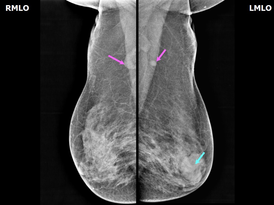 | 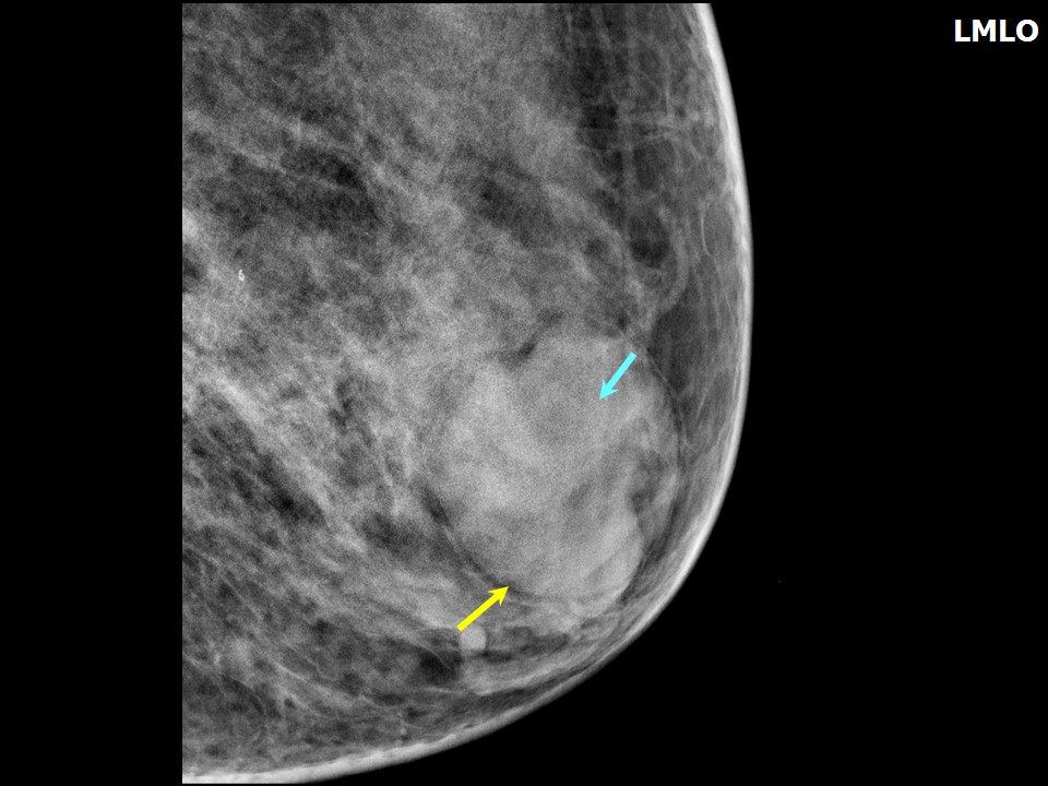 |
| Breast composition: | ACR category c (the breasts are heterogeneously dense, which may obscure small masses) | Mammography features: |
| ‣ Location of the lesion: | Left breast, lower outer quadrant at 5 oclock, anterior third |
| ‣ Mass: | |
| • Number: | 1 |
| • Size: | 4.4 × 3.4 cm |
| • Shape: | Oval |
| • Margins: | Circumscribed with perilesional halo |
| • Density: | Equal with fat-containing areas |
| ‣ Calcifications: | |
| • Typically benign: | None |
| • Suspicious: | None |
| • Distribution: | None |
| ‣ Architectural distortion: | None |
| ‣ Asymmetry: | None |
| ‣ Intramammary node: | None |
| ‣ Skin lesion: | None |
| ‣ Solitary dilated duct: | None |
| ‣ Associated features: | None |
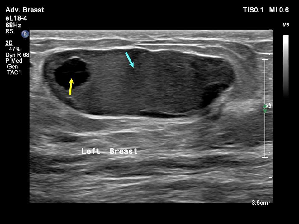
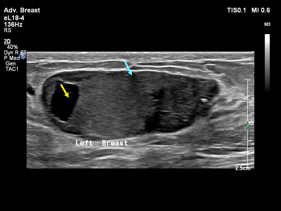
Ultrasound:
 |  |
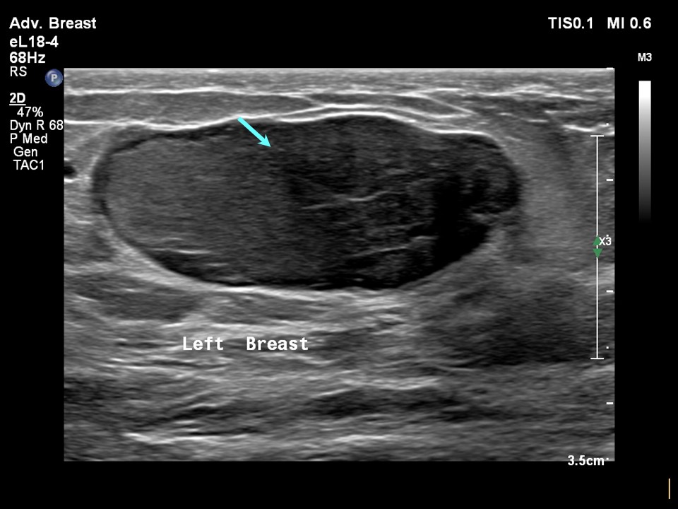 |
| Ultrasound features: Left breast, lower outer quadrant at 5 oclock, 5.0 cm from the nipple and at 0.7 cm skin depth | |
| ‣ Mass | |
| • Location: | Left breast, lower outer quadrant at 5 oclock, 5.0 cm from the nipple and at 0.7 cm skin depth |
| • Number: | 1 |
| • Size: | 4.0 × 1.5 cm |
| • Shape: | Oval |
| • Orientation: | Parallel |
| • Margins: | Circumscribed, 23 lobulations present |
| • Echo pattern: | Hypoechoic |
| • Posterior features: | No posterior features |
| ‣ Calcifications: | None |
| ‣ Associated features: | None |
| ‣ Special cases: | None |
BI-RADS:
BI-RADS Category: 2 (benign)Further assessment:
Further assessment advised: Referral for cytologyCytology:
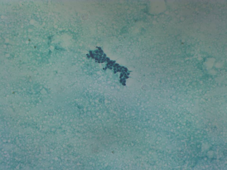 |
| Cytology features: | |
| ‣ Type of sample: | FNAC |
| ‣ Site of biopsy: | |
| • Laterality: | Left |
| • Quadrant: | Lower outer |
| • Localization technique: | Palpation |
| • Nature of aspirate: | 1.5 mL of milky white fluid |
| ‣ Cytological description: | Aspirates show predominantly cystic fluid background with thick degenerated proteinaceous material intermixed with fat droplets, RBCs, occasional macrophages, and inflammatory cells. Occasional clusters of benign epithelial and myoepithelial cells are seen. A few epithelial cells show apocrine change |
| ‣ Reporting category: | Benign |
| ‣ Diagnosis: | Consistent with galactocele advice: correlate with radiological findings and a guided FNAC sampling to be done if any solid areas are seen. In view of the milky aspirate in non-lactating status, causes of hyperprolactinaemia may also be investigated |
| ‣ Comments: | None |
Case summary:
| Premenopausal woman presented with a left breast lump diagnosed as galactocele, BI-RADS 2 on imaging and as benign, galactocele on cytology. |
Learning points:
|