Home / Training / Manuals / Atlas of breast cancer early detection / Cases
Atlas of breast cancer early detection
Filter by language: English / Русский
Go back to the list of case studies
.png) Click on the pictures to magnify and display the legends
Click on the pictures to magnify and display the legends
| Case number: | 127 |
| Age: | 50 |
| Clinical presentation: | Premenopausal woman with average risk of developing breast cancer presented with left breast lump with mastalgia. |
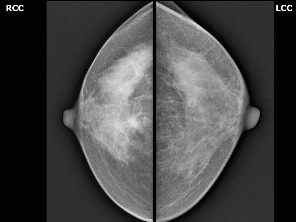
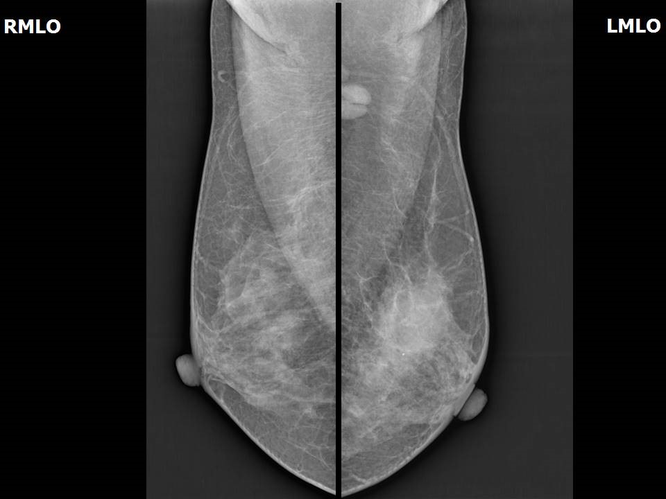
Mammography:
| Breast composition: | ACR category c (the breasts are heterogeneously dense, which may obscure small masses) | Mammography features: |
| ‣ Location of the lesion: | Left breast, upper outer quadrant at 2 o’clock, middle third, 3.0 cm from nipple and at 1.5 cm skin depth |
| ‣ Mass: | |
| • Number: | 1 |
| • Size: | 3.0 cm |
| • Shape: | Round |
| • Margins: | Partly circumscribed and partly obscured |
| • Density: | Equal |
| ‣ Calcifications: | |
| • Typically benign: | None |
| • Suspicious: | None |
| • Distribution: | None |
| ‣ Architectural distortion: | None |
| ‣ Asymmetry: | None |
| ‣ Intramammary node: | None |
| ‣ Skin lesion: | None |
| ‣ Solitary dilated duct: | None |
| ‣ Associated features: | None |
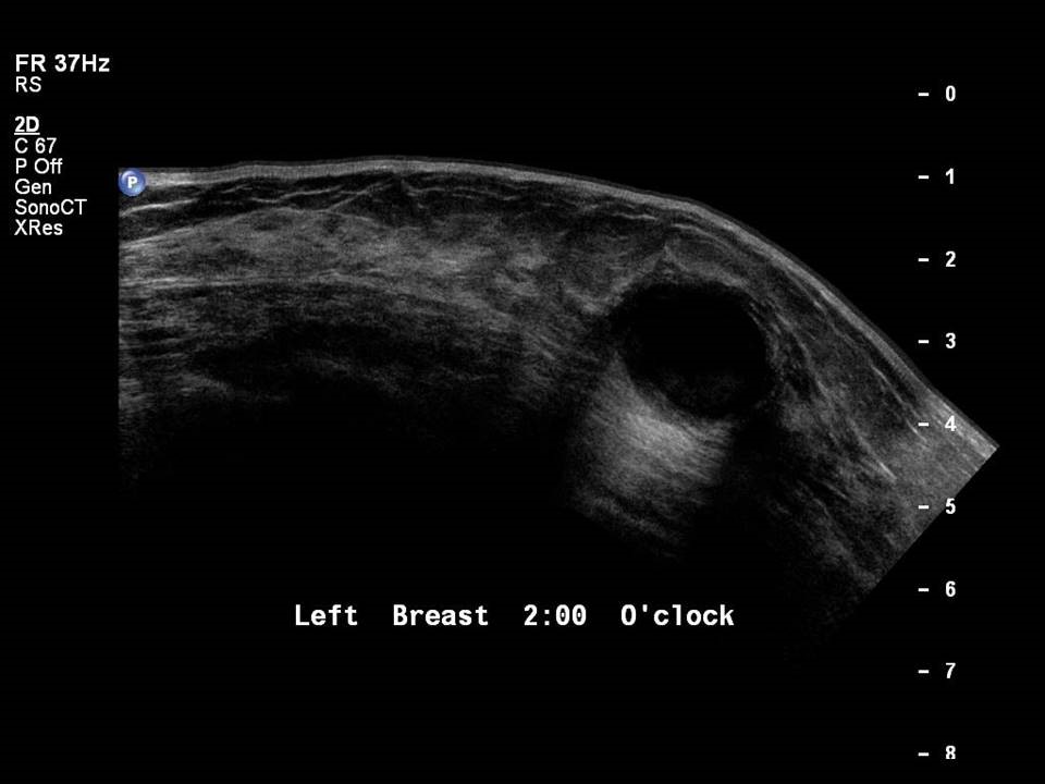
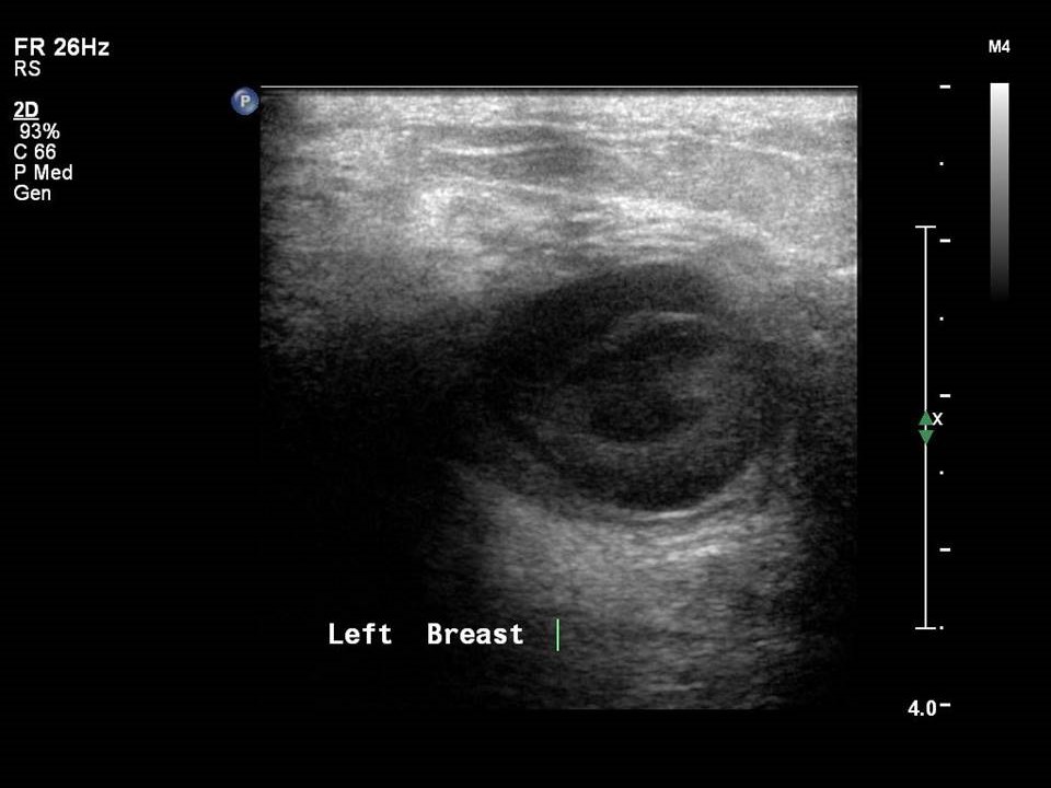
Ultrasound:
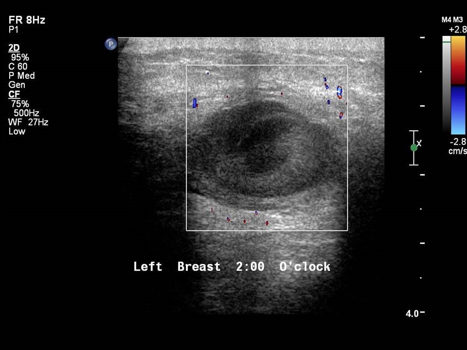
| Ultrasound features: Left breast, upper outer quadrant at 2 o’clock | |
| ‣ Mass | |
| • Location: | Left breast, upper outer quadrant at 2 o’clock |
| • Number: | 1 |
| • Size: | 3.0 × 2.0 cm |
| • Shape: | Oval |
| • Orientation: | Parallel |
| • Margins: | Indistinct |
| • Echo pattern: | Heteroechoic |
| • Posterior features: | No posterior features |
| ‣ Calcifications: | None |
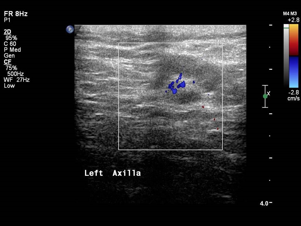
| ‣ Associated features: | Minimal peripheral vascularity and axillary lymphadenopathy |
| ‣ Special cases: | Complicated cyst |
BI-RADS:
BI-RADS Category: 2 (benign)Further assessment:
Further assessment advised: Referral for cytologyCytology:
| Cytology features: | |
| ‣ Type of sample: | FNAC |
| ‣ Site of biopsy: | |
| • Laterality: | Left |
| • Quadrant: | |
| • Localization technique: | Palpation |
| • Nature of aspirate: | 0.2 mL of yellowish pus-like material |
| ‣ Cytological description: | Smears reveal neutrophilic exudate entrapped in fibrin along with sheets of benign ductal epithelial cells, stromal fragments, and foamy macrophages. Background shows blood |
| ‣ Reporting category: | Benign |
| ‣ Diagnosis: | Acute mastitis |
| ‣ Comments: | None |
Histopathology:
Lumpectomy
| Histopathology features: | |
| ‣ Specimen type: | Lumpectomy |
| ‣ Laterality: | |
| ‣ Macroscopy: | Lumpectomy specimen (4.0 × 3.5 × 2.8 cm). Cut surface shows a central cavity (1.8 cm in diameter) with shaggy walls and scant purulent material in the lumen |
| ‣ Histological type: | Sections reveal benign proliferative and fibrocystic lesions composed of adenosis and fibrosis epitheliosis focally. There is chronic inflammatory infiltrate composed of lymphocytes and histiocytes involving the ducts and in the stroma. Many cystically dilated glands and glands lined by metaplastic apocrine cells are seen. Granulation tissue and sheets of histiocytic cells are seen focally. There are no granulomas. Impression: Benign fibrocystic and proliferative breast disease. There is no evidence of malignancy in the sections studied |
| ‣ Histological grade: | |
| ‣ Mitosis: | |
| ‣ Maximum invasive tumour size: | |
| ‣ Lymph node status: | |
| ‣ Peritumoural lymphovascular invasion: | |
| ‣ DCIS/EIC: | |
| ‣ Margins: | |
| ‣ Pathological stage: | |
| ‣ Biomarkers: | |
| ‣ Comments: |
Case summary:
| Premenopausal woman presented with left breast lump and mastalgia. Diagnosed as complicated cyst, BI-RADS 2 on imaging, as acute mastitis on cytology, and as benign proliferative breast lesion on histopathology. |
Learning points:
|


