


Home / Training / Manuals / Atlas of breast cancer early detection / Cases
Atlas of breast cancer early detection
Filter by language: English / Русский
Go back to the list of case studies
.png) Click on the pictures to magnify and display the legends
Click on the pictures to magnify and display the legends
| Case number: | 015 |
| Age: | 42 |
| Clinical presentation: | Premenopausal woman with average risk of breast cancer presented with a right breast lump. Examination revealed a 5 cm lump in the right breast. |
Mammography:
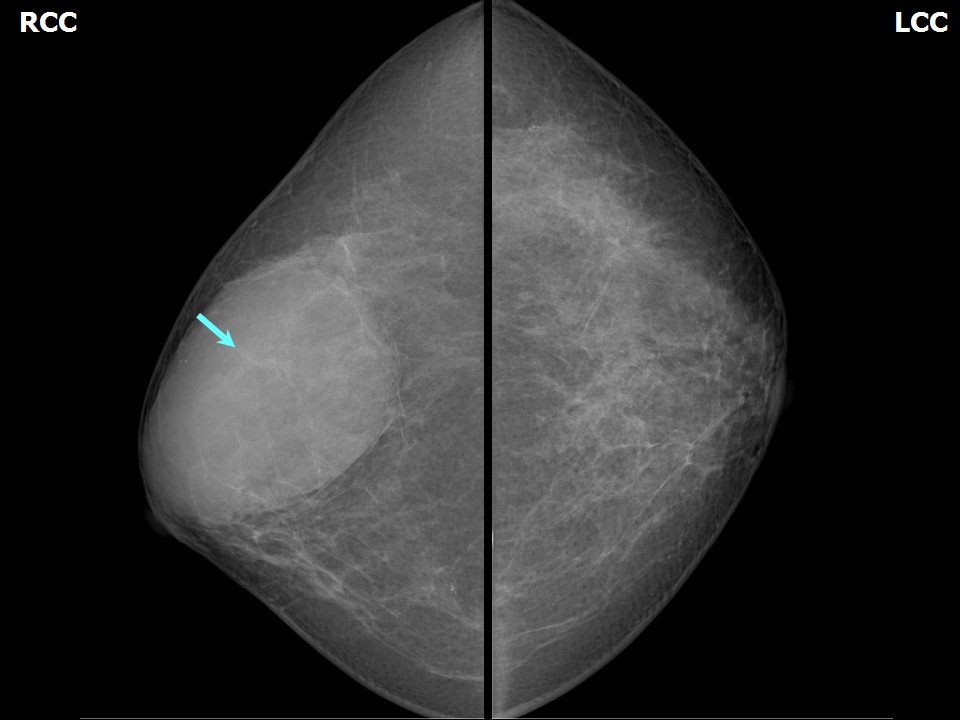 |  |
| Breast composition: | ACR category b (there are scattered areas of fibroglandular density) | Mammography features: |
| ‣ Location of the lesion: | Right breast, upper outer quadrant at 10 oclock, anterior and middle thirds |
| ‣ Mass: | |
| • Number: | 1 |
| • Size: | 9.0 × 8.5 × 5.5 cm |
| • Shape: | Oval |
| • Margins: | Circumscribed |
| • Density: | Equal |
| ‣ Calcifications: | |
| • Typically benign: | None |
| • Suspicious: | None |
| • Distribution: | None |
| ‣ Architectural distortion: | None |
| ‣ Asymmetry: | None |
| ‣ Intramammary node: | None |
| ‣ Skin lesion: | None |
| ‣ Solitary dilated duct: | None |
| ‣ Associated features: | None |
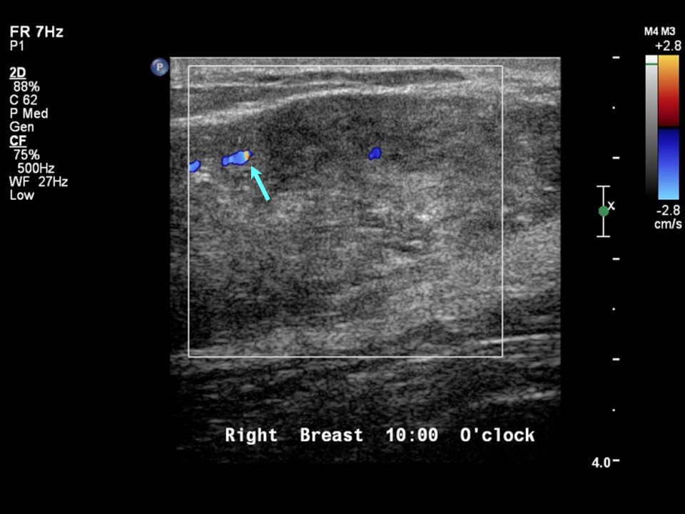
Ultrasound:
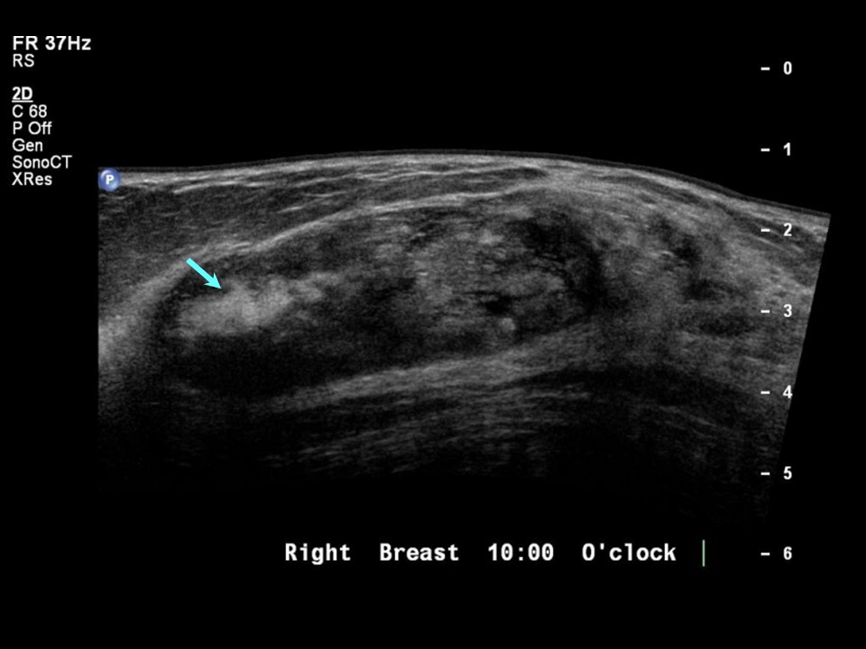 |  |
| Ultrasound features: Right breast upper outer quadrant at 10 oclock position | |
| ‣ Mass | |
| • Location: | Right breast upper outer quadrant at 10 oclock position |
| • Number: | 1 |
| • Size: | 8.0 × 2.5 cm |
| • Shape: | Oval |
| • Orientation: | Parallel |
| • Margins: | Circumscribed |
| • Echo pattern: | Heteroechoic |
| • Posterior features: | No posterior features |
| ‣ Calcifications: | None |
| ‣ Associated features: | Minimum internal vascularity |
| ‣ Special cases: | None |
BI-RADS:
BI-RADS Category: 2 (benign)Further assessment:
Further assessment advised: Referral for cytologyCytology:
 |
| Cytology features: | |
| ‣ Type of sample: | FNAC |
| ‣ Site of biopsy: | |
| • Laterality: | Right |
| • Quadrant: | Upper outer |
| • Localization technique: | Palpation, rubbery consistency felt through the needle |
| • Nature of aspirate: | Whitish material |
| ‣ Cytological description: | Cell-rich smear of elongated, branching fragments of ductal epithelium and numerous single bipolar nuclei in the background. Fragments of fibromyxoid stroma were seen in the background |
| ‣ Reporting category: | Benign |
| ‣ Diagnosis: | Fibroadenoma |
| ‣ Comments: | None |
Histopathology:
Lumpectomy
 |
| Histopathology features: | |
| ‣ Specimen type: | Lumpectomy |
| ‣ Laterality: | Right |
| ‣ Macroscopy: | Single whitish tissue piece (7.5 × 5.5 × 3.5 cm). On cut section shows firm whitish areas and mucoid areas |
| ‣ Histological type: | Histological features of fibroadenoma with extensive sclerosis of stromal tissue. Some of the ducts are cystically dilated |
| ‣ Histological grade: | |
| ‣ Mitosis: | |
| ‣ Maximum invasive tumour size: | |
| ‣ Lymph node status: | |
| ‣ Peritumoural lymphovascular invasion: | |
| ‣ DCIS/EIC: | |
| ‣ Margins: | |
| ‣ Pathological stage: | |
| ‣ Biomarkers: | |
| ‣ Comments: |
Case summary:
| Premenopausal woman presented with right breast lump. Diagnosed as fibroadenoma, BI-RADS 2 on imaging and as fibroadenoma on cytology and histopathology. |
Learning points:
|