


Home / Training / Manuals / Atlas of breast cancer early detection / Cases
Atlas of breast cancer early detection
Filter by language: English / Русский
Go back to the list of case studies
.png) Click on the pictures to magnify and display the legends
Click on the pictures to magnify and display the legends
| Case number: | 167 |
| Age: | 63 |
| Clinical presentation: | Postmenopausal woman who had undergone breast-conserving surgery in 2003 for right breast cancer presented for regular surveillance check-up. On clinical examination, a firm nodularity was noted at the scar site in the right breast. |
 |
Mammography:
 |
| Breast composition: | ACR category a (the breasts are almost entirely fatty) | Mammography features: |
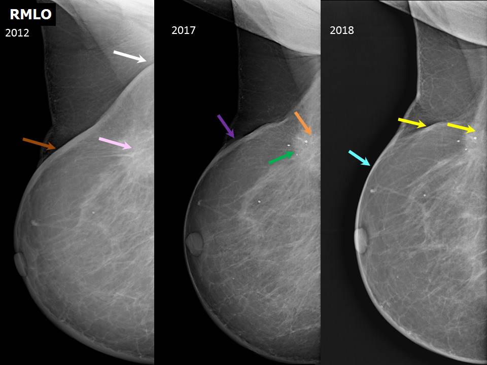
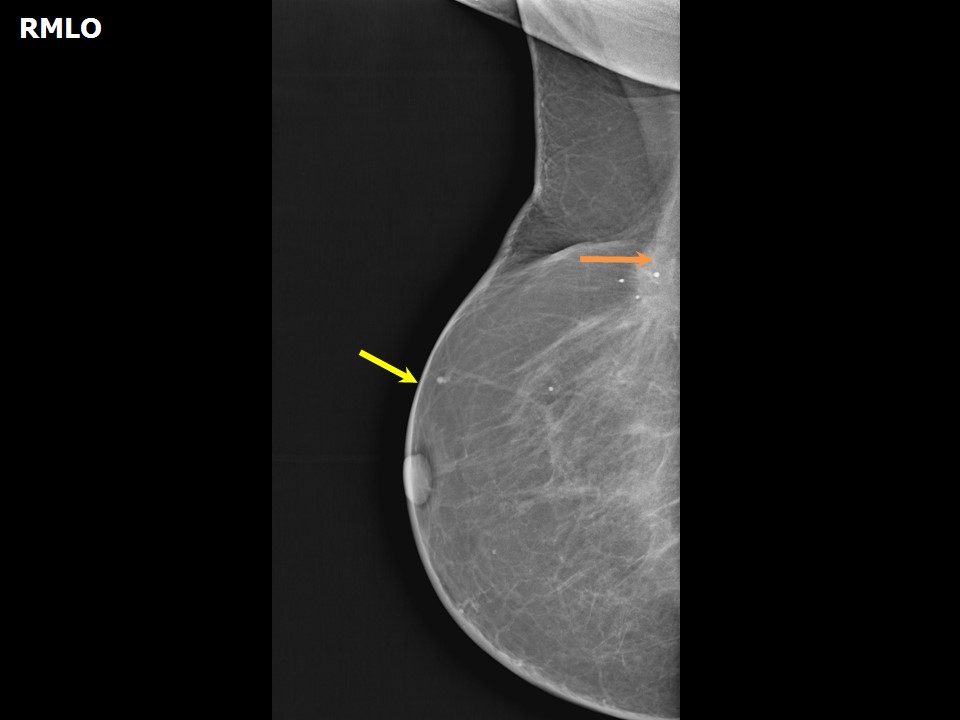
| ‣ Location of the lesion: | 2012: Right breast, upper outer quadrant at 12 oclock, posterior third, beneath surgical scar |
| ‣ Mass: | |
| • Number: | 1 |
| • Size: | |
| • Shape: | Irregular |
| • Margins: | Spiculated |
| • Density: | Equal |
| ‣ Calcifications: | |
| • Typically benign: | None |
| • Suspicious: | None |
| • Distribution: | None |
| ‣ Architectural distortion: | Present |
| ‣ Asymmetry: | None |
| ‣ Intramammary node: | None |
| ‣ Skin lesion: | None |
| ‣ Solitary dilated duct: | None |
| ‣ Associated features: | Scar tissue with skin thickening |
| Mammography features: | |
| ‣ Location of the lesion: | 2017: Right breast, upper outer quadrant at 12 oclock, posterior third, beneath surgical scar |
| ‣ Mass: | |
| • Number: | 1 |
| • Size: | |
| • Shape: | Irregular |
| • Margins: | Spiculated |
| • Density: | Equal |
| ‣ Calcifications: | |
| • Typically benign: | Fat necrosis calcifications |
| • Suspicious: | None |
| • Distribution: | None |
| ‣ Architectural distortion: | Minimal |
| ‣ Asymmetry: | None |
| ‣ Intramammary node: | None |
| ‣ Skin lesion: | None |
| ‣ Solitary dilated duct: | None |
| ‣ Associated features: | Scar tissue with skin thickening |
 |  |
 |
| Mammography features: | |
| ‣ Location of the lesion: | 2018: Right breast, upper outer quadrant at 12 oclock, posterior third, beneath surgical scar |
| ‣ Mass: | |
| • Number: | 1 |
| • Size: | |
| • Shape: | Irregular |
| • Margins: | Spiculated |
| • Density: | Equal |
| ‣ Calcifications: | |
| • Typically benign: | Round, fat necrosis calcifications |
| • Suspicious: | None |
| • Distribution: | None |
| ‣ Architectural distortion: | Minimal |
| ‣ Asymmetry: | None |
| ‣ Intramammary node: | None |
| ‣ Skin lesion: | None |
| ‣ Solitary dilated duct: | None |
| ‣ Associated features: | Organized scar with fat necrosis calcifications |
Ultrasound:
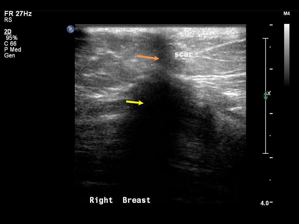 | 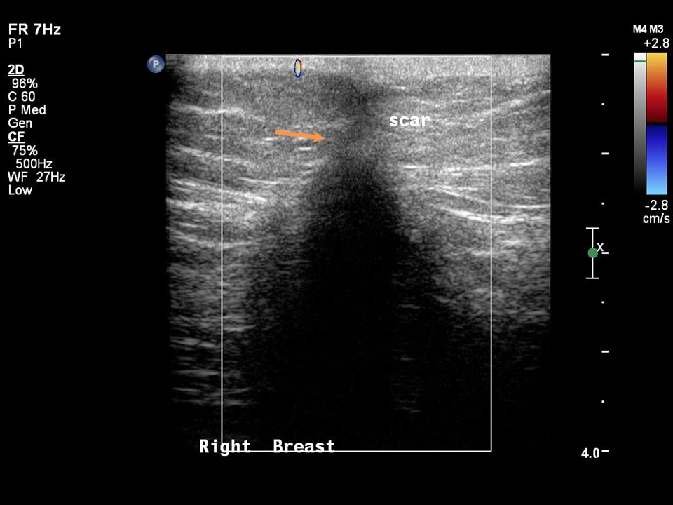 |
| Ultrasound features: 2018: Right breast, upper quadrants at 12 o'clock | |
| ‣ Mass | |
| • Location: | 2018: Right breast, upper quadrants at 12 o'clock |
| • Number: | None |
| • Size: | None |
| • Shape: | Irregular |
| • Orientation: | Not parallel |
| • Margins: | Angular |
| • Echo pattern: | Hypoechoic |
| • Posterior features: | Strong posterior shadowing |
| ‣ Calcifications: | Surgical scar macrocalcification |
| ‣ Associated features: | Architectural distortion |
| ‣ Special cases: | None |
BI-RADS:
BI-RADS Category (Right BCS, post operative breast): 2 (benign)Case summary:
| Postmenopausal woman who underwent breast-conserving surgery in 2003 for right breast carcinoma presented with firm nodularity along the scar tissue. Diagnosed as organized scar with typically benign calcifications at surgical site, BI-RADS 2 on imaging. |
Learning points:
|