


Home / Training / Manuals / Atlas of breast cancer early detection / Cases
Atlas of breast cancer early detection
Filter by language: English / Русский
Go back to the list of case studies
.png) Click on the pictures to magnify and display the legends
Click on the pictures to magnify and display the legends
| Case number: | 004 |
| Age: | 78 |
| Clinical presentation: | Postmenopausal woman with average risk of developing breast cancer presented with a large lump in the right breast. On examination, she had a large firm right breast lump. |
Mammography:
 |  |
| Breast composition: | ACR Category a (the breasts are almost entirely fatty) | Mammography features: |
| ‣ Location of the lesion: | Right breast, upper inner quadrant at 121 oclock, middle third |
| ‣ Mass: | |
| • Number: | 1 |
| • Size: | 3 cm |
| • Shape: | Oval |
| • Margins: | Circumscribed |
| • Density: | Equal |
| ‣ Calcifications: | |
| • Typically benign: | None |
| • Suspicious: | None |
| • Distribution: | None |
| ‣ Architectural distortion: | None |
| ‣ Asymmetry: | None |
| ‣ Intramammary node: | None |
| ‣ Skin lesion: | None |
| ‣ Solitary dilated duct: | None |
| ‣ Associated features: | None |
| Breast composition: | ACR category a (the breasts are almost entirely fatty) | Mammography features: |
| ‣ Location of the lesion: | Left breast, upper outer quadrant at 1 oclock, middle third |
| ‣ Mass: | |
| • Number: | 2 |
| • Size: | Largest 0.5 cm |
| • Shape: | Round |
| • Margins: | Circumscribed |
| • Density: | Equal |
| ‣ Calcifications: | |
| • Typically benign: | None |
| • Suspicious: | None |
| • Distribution: | None |
| ‣ Architectural distortion: | None |
| ‣ Asymmetry: | None |
| ‣ Intramammary node: | None |
| ‣ Skin lesion: | None |
| ‣ Solitary dilated duct: | None |
| ‣ Associated features: | None |
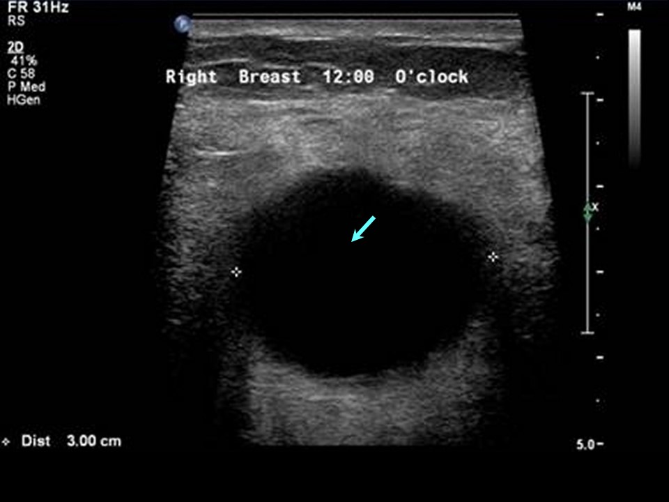
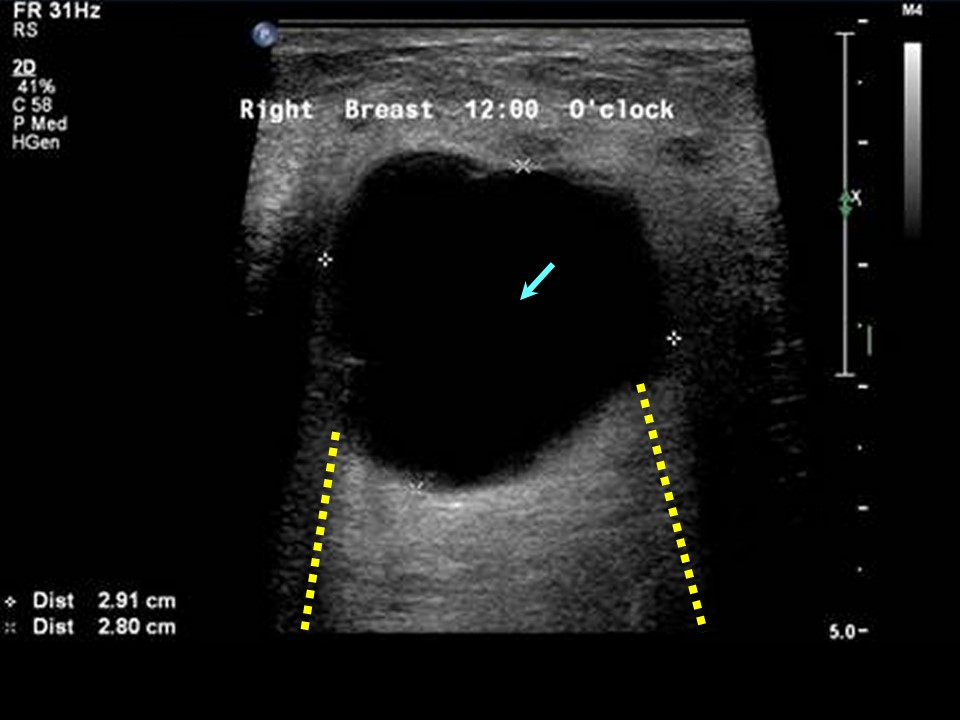
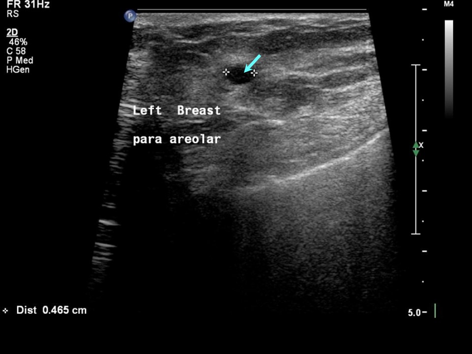
Ultrasound:
 |  |
 |
| Ultrasound features: Right breast, central portion of the breast at 12 oclock | |
| ‣ Mass | |
| • Location: | Right breast, central portion of the breast at 12 oclock |
| • Number: | Multiple |
| • Size: | Largest 3.0 cm |
| • Shape: | Round |
| • Orientation: | Parallel |
| • Margins: | Circumscribed |
| • Echo pattern: | Anechoic |
| • Posterior features: | Posterior shadowing |
| ‣ Calcifications: | None |
| ‣ Associated features: | None |
| ‣ Special cases: | Simple cyst |
| Ultrasound features: Left breast, central portion of the breast at 1 oclock | |
| ‣ Mass | |
| • Location: | Left breast, central portion of the breast at 1 oclock |
| • Number: | Multiple |
| • Size: | Largest 0.5 cm in greatest dimension |
| • Shape: | Oval |
| • Orientation: | Parallel |
| • Margins: | Circumscribed |
| • Echo pattern: | Anechoic |
| • Posterior features: | Posterior shadowing |
| ‣ Calcifications: | None |
| ‣ Associated features: | None |
| ‣ Special cases: | Simple cyst |
BI-RADS:
BI-RADS Category: 2 (benign)Further assessment:
Further assessment advised: Referral for cytologyCytology:
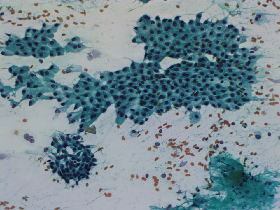 | 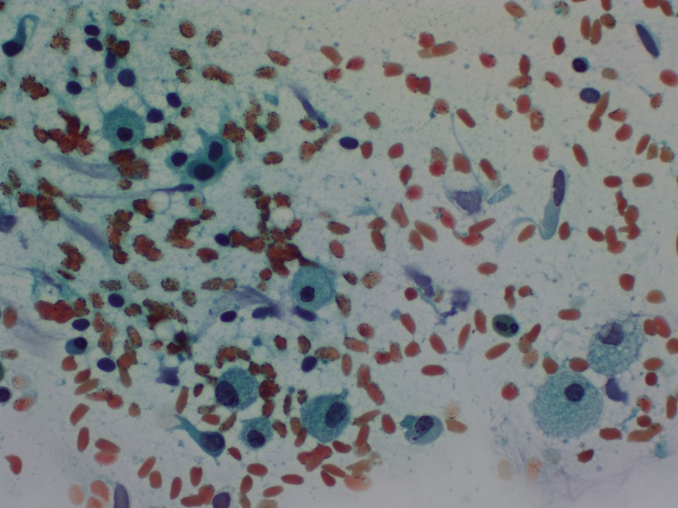 |
| Cytology features: | |
| ‣ Type of sample: | FNAC done on four occasions |
| ‣ Site of biopsy: | |
| • Laterality: | Right |
| • Quadrant: | Upper and central |
| • Localization technique: | Palpation |
| • Nature of aspirate: | 26 mL of yellow fluid aspirated each time |
| ‣ Cytological description: | Smears show tight clusters of benign ductal epithelial cells, some with apocrine metaplasia and many foamy histiocytes on a proteinaceous background |
| ‣ Reporting category: | Benign |
| ‣ Diagnosis: | Proliferative fibrocystic change |
| ‣ Comments: | None |
Histopathology:
Lump excision
 | 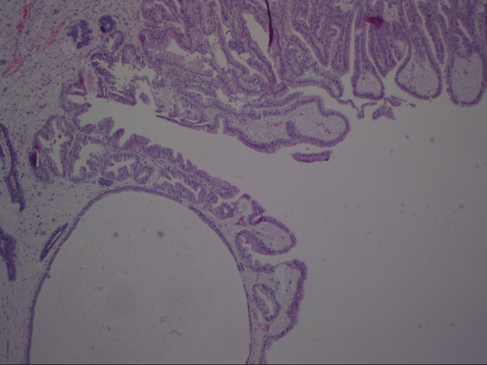 |
| Histopathology features: | |
| ‣ Specimen type: | Lump excision |
| ‣ Laterality: | Right |
| ‣ Macroscopy: | Irregular fibrofatty tissue bit (4.5 × 3.5 × 2.5 cm) with small piece of skin on one side (2.0 × 0.7 cm). Cut surface is yellowish, soft to firm, with a few whitish fibrous areas ranging in size from 0.3 cm to 1.0 cm and small cysts ranging in size from 0.3 cm to 0.5 cm in largest dimension |
| ‣ Histological type: | Many of the ducts are cystically dilated, some lined by flattened epithelium; a few show mild epitheliosis and papillary apocrine hyperplasia |
| ‣ Histological grade: | |
| ‣ Mitosis: | |
| ‣ Maximum invasive tumour size: | |
| ‣ Lymph node status: | |
| ‣ Peritumoural lymphovascular invasion: | |
| ‣ DCIS/EIC: | Not identified |
| ‣ Margins: | |
| ‣ Pathological stage: | |
| ‣ Biomarkers: | |
| ‣ Comments: | Negative for ADH/DCIS/malignancy |
Case summary:
| Postmenopausal woman presented with right breast lump. Diagnosed as multiple simple cysts in both breasts, BI-RADS 2 on imaging and as proliferative fibrocystic change on cytology and histopathology. |
Learning points:
|
Cysts in older women:
|