


Home / Training / Manuals / Atlas of breast cancer early detection / Cases
Atlas of breast cancer early detection
Filter by language: English / Русский
Go back to the list of case studies
.png) Click on the pictures to magnify and display the legends
Click on the pictures to magnify and display the legends
| Case number: | 002 |
| Age: | 49 |
| Clinical presentation: | Premenopausal woman with average risk of developing breast cancer presented with painful breast lumps in both breasts. On examination, she had multiple lumps with firm consistency in both breasts and a well-defined mobile lump, 5 × 3 cm. |
Mammography:
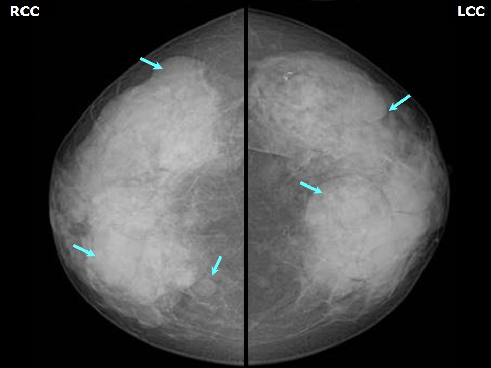 | 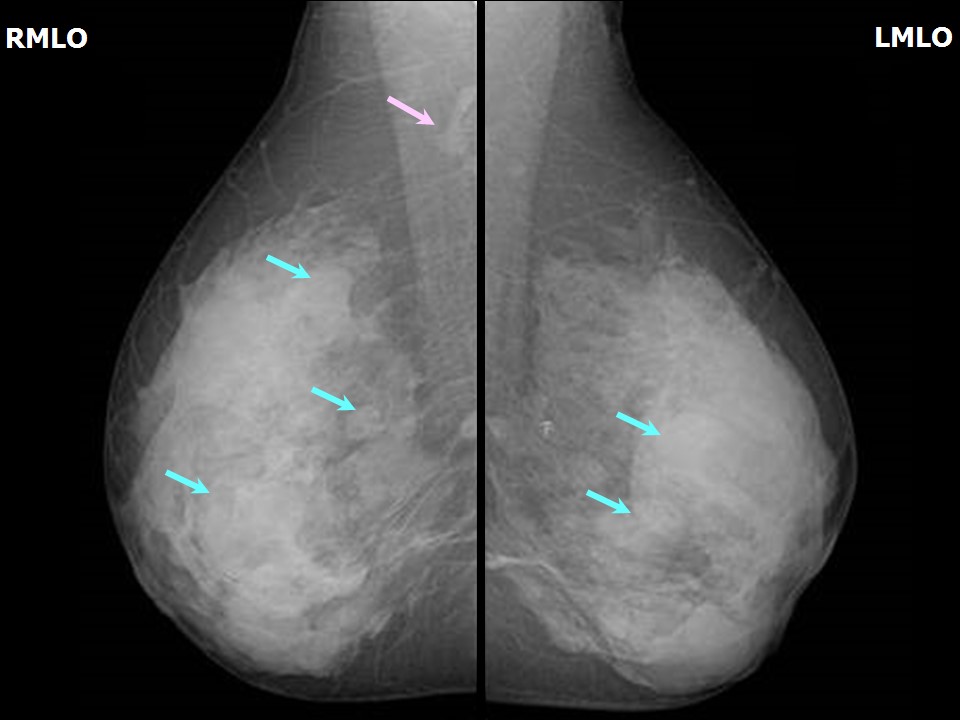 |
| Breast composition: | ACR category d (the breasts are extremely dense, which lowers the sensitivity of mammography) | Mammography features: |
| ‣ Location of the lesion: | Right breast, all quadrants, entire breast, all zones |
| ‣ Mass: | |
| • Number: | Multiple |
| • Size: | Largest 5.2 × 3.6 cm |
| • Shape: | Oval |
| • Margins: | Circumscribed with perilesional halo |
| • Density: | Equal |
| ‣ Calcifications: | |
| • Typically benign: | None |
| • Suspicious: | None |
| • Distribution: | None |
| ‣ Architectural distortion: | None |
| ‣ Asymmetry: | Global |
| ‣ Intramammary node: | None |
| ‣ Skin lesion: | None |
| ‣ Solitary dilated duct: | None |
| ‣ Associated features: | None |
| Breast composition: | ACR category d (the breasts are extremely dense, which lowers the sensitivity of mammography) | Mammography features: |
| ‣ Location of the lesion: | Left breast, all quadrants, entire breast, all zones |
| ‣ Mass: | |
| • Number: | Multiple |
| • Size: | Largest 6.2 × 5.3 cm |
| • Shape: | Oval |
| • Margins: | Circumscribed with perilesional halo |
| • Density: | Equal |
| ‣ Calcifications: | |
| • Typically benign: | Coarse |
| • Suspicious: | None |
| • Distribution: | None |
| ‣ Architectural distortion: | None |
| ‣ Asymmetry: | Global |
| ‣ Intramammary node: | None |
| ‣ Skin lesion: | None |
| ‣ Solitary dilated duct: | None |
| ‣ Associated features: | Calcification |
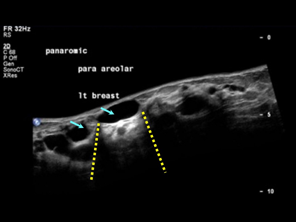
Ultrasound:
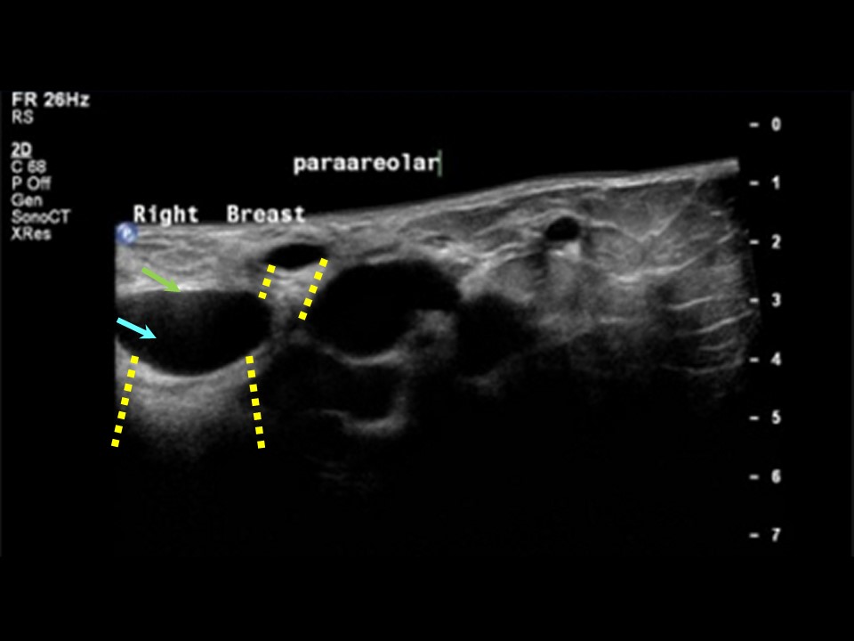 |  |
| Ultrasound features: Right breast, all quadrants | |
| ‣ Mass | |
| • Location: | Right breast, all quadrants |
| • Number: | Multiple |
| • Size: | Largest 4.5 × 3.0 cm |
| • Shape: | Round to oval |
| • Orientation: | Parallel |
| • Margins: | Circumscribed |
| • Echo pattern: | Anechoic |
| • Posterior features: | Posterior shadowing |
| ‣ Calcifications: | None |
| ‣ Associated features: | None |
| ‣ Special cases: | Simple cyst |
| Ultrasound features: Left breast, all quadrants | |
| ‣ Mass | |
| • Location: | Left breast, all quadrants |
| • Number: | Multiple |
| • Size: | Largest 5.2 × 4.2 cm |
| • Shape: | Round to oval |
| • Orientation: | Parallel |
| • Margins: | Circumscribed |
| • Echo pattern: | Anechoic |
| • Posterior features: | Posterior shadowing |
| ‣ Calcifications: | None |
| ‣ Associated features: | None |
| ‣ Special cases: | Simple cyst |
BI-RADS:
BI-RADS Category: 2 (benign)Further assessment:
Further assessment advised: Referral for cytologyCytology:
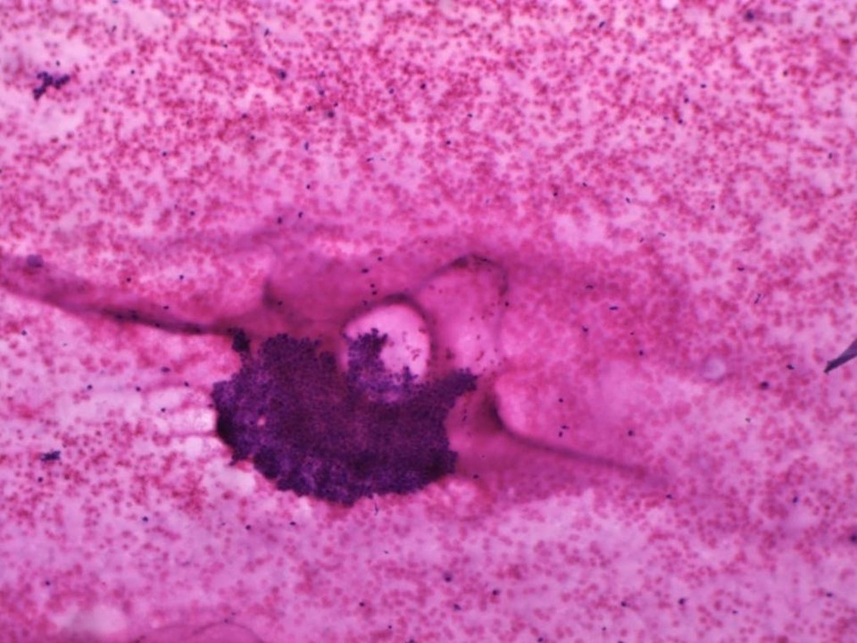 | 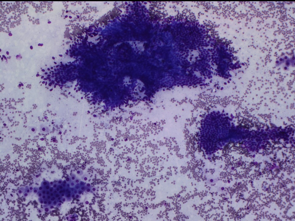 |
| Cytology features: | |
| ‣ Type of sample: | FNAC (cystic lesion) |
| ‣ Site of biopsy: | Bilateral breast lumps |
| • Laterality: | |
| • Quadrant: | Subareolar nodules in both breasts |
| • Localization technique: | Palpation |
| • Nature of aspirate: | 15 mL of thick yellowish fluid aspirated on multiple passes through the nodule in right breast; 12 mL of yellowish brown fluid aspirated on multiple passes through the nodule in left breast |
| ‣ Cytological description: | Smears from both sides show tight clusters of benign ductal epithelial cells without significant nuclear atypia; a few apocrine cells noted. A thick proteinaceous fluid with foamy histiocytes seen in the background |
| ‣ Reporting category: | Benign |
| ‣ Diagnosis: | Proliferative fibrocystic change |
| ‣ Comments: | None |
Case summary:
| Premenopausal woman presented with painful bilateral breast lumps. Diagnosed as multiple cysts, BI-RADS 2 on imaging and as proliferative fibrocystic change on cytology. |
Learning points:
|