


Home / Training / Manuals / Atlas of breast cancer early detection / Cases
Atlas of breast cancer early detection
Filter by language: English / Русский
Go back to the list of case studies
.png) Click on the pictures to magnify and display the legends
Click on the pictures to magnify and display the legends
| Case number: | 049 |
| Age: | 49 |
| Clinical presentation: | Premenopausal woman with average risk of developing breast cancer presented with a left breast lump. Examination revealed a hard 2 cm lump in the lower inner quadrant of the left breast. |
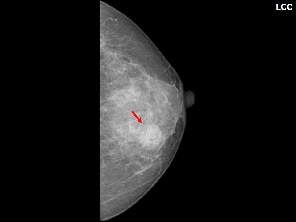
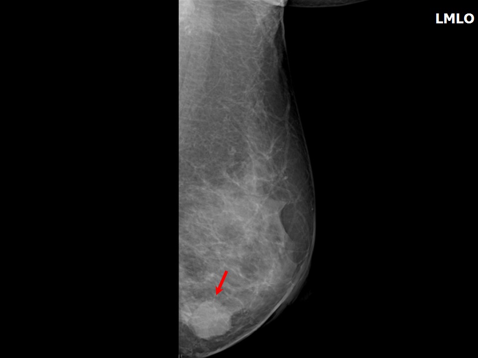
Mammography:
 |  |
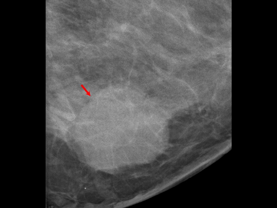 |
| Breast composition: | ACR category c (the breasts are heterogeneously dense, which may obscure small masses) | Mammography features: |
| ‣ Location of the lesion: | Left breast, lower inner quadrant at 78 oclock, middle third |
| ‣ Mass: | |
| • Number: | 1 |
| • Size: | 3.0 × 2.0 cm |
| • Shape: | Oval |
| • Margins: | Partly circumscribed and partly obscured |
| • Density: | Equal |
| ‣ Calcifications: | |
| • Typically benign: | None |
| • Suspicious: | None |
| • Distribution: | None |
| ‣ Architectural distortion: | None |
| ‣ Asymmetry: | None |
| ‣ Intramammary node: | None |
| ‣ Skin lesion: | None |
| ‣ Solitary dilated duct: | None |
| ‣ Associated features: | None |
Ultrasound:
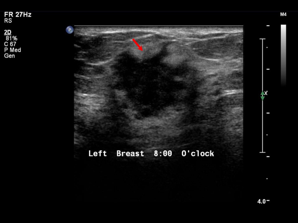 | 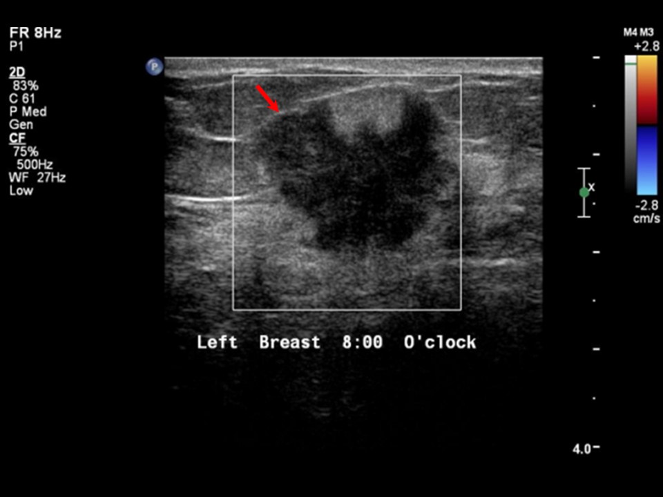 |
| Ultrasound features: Left breast, lower inner quadrant at 8 oclock | |
| ‣ Mass | |
| • Location: | Left breast, lower inner quadrant at 8 oclock |
| • Number: | 1 |
| • Size: | 1.9 cm |
| • Shape: | Round |
| • Orientation: | Parallel |
| • Margins: | indistinct |
| • Echo pattern: | Heteroechoic |
| • Posterior features: | No posterior features |
| ‣ Calcifications: | None |
| ‣ Associated features: | None |
| ‣ Special cases: | None |
BI-RADS:
BI-RADS Category: 5 (highly suggestive of malignancy)Further assessment:
Further assessment advised: Referral for cytologyCytology:
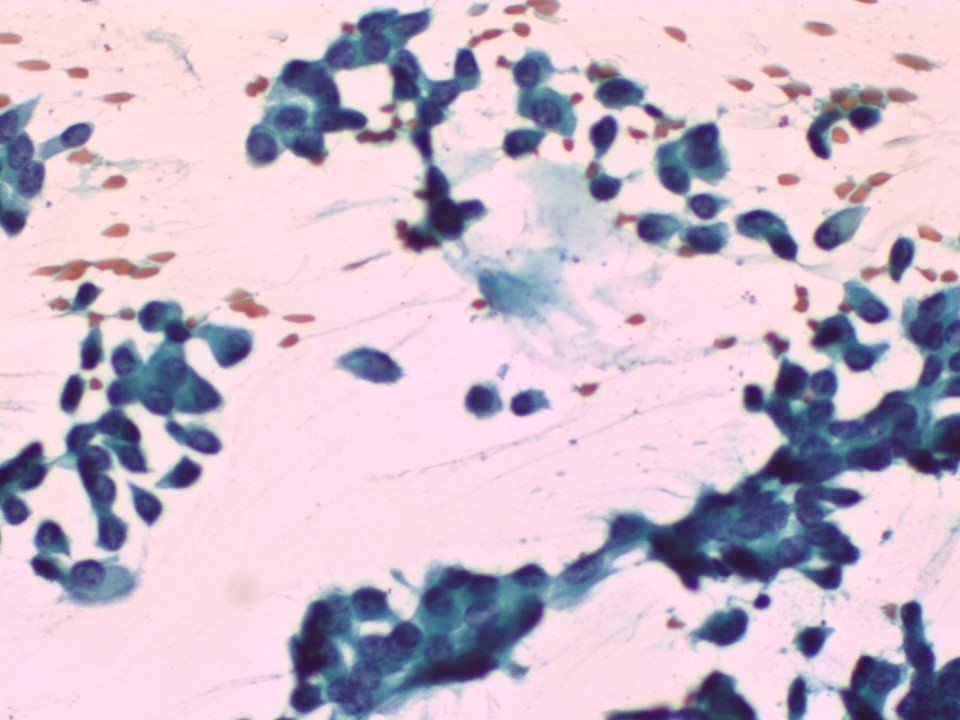 |
| Cytology features: | |
| ‣ Type of sample: | FNAC |
| ‣ Site of biopsy: | |
| • Laterality: | Left |
| • Quadrant: | Lower inner |
| • Localization technique: | Palpation |
| • Nature of aspirate: | Whitish |
| ‣ Cytological description: | Smear reveals loosely cohesive sheets and isolated malignant cells showing hyperchromatic pleomorphic nuclei with coarse chromatin. N:C ratio is high and there is a moderate amount of cytoplasm. Necrotic debris is seen in the background |
| ‣ Reporting category: | Malignant |
| ‣ Diagnosis: | Carcinoma |
| ‣ Comments: | None |
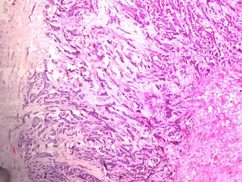
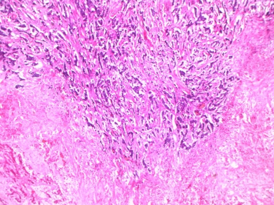
Histopathology:
Breast-conserving surgery
 |  |
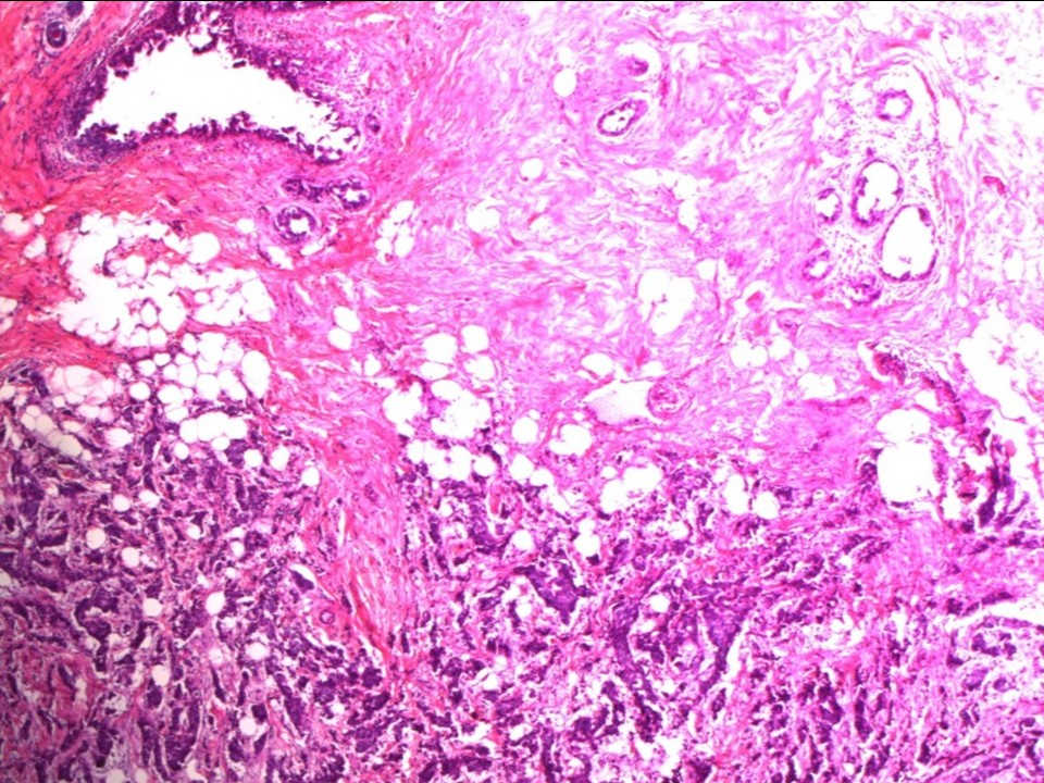 |
| Histopathology features: | |
| ‣ Specimen type: | Breast-conserving surgery |
| ‣ Laterality: | Left |
| ‣ Macroscopy: | Lumpectomy specimen (9.0 × 6.0 × 4.0 cm) oriented with long suture laterally and short suture superiorly. Skin flap (8.0 × 4.0 cm). Part of areola noted (2.0 × 1.0 cm). On serial sectioning, a greyish white tumour (3.0 × 2.3 × 2.2 cm) is identified. Areas of fibrosis noted. Tumour is located 0.6 cm from the skin, 1.8 cm from the posterior margin, 1.4 cm from the superior margin, 3.0 cm from the inferior margin, 1.3 cm from the medial margins, and 3.3 cm from the lateral margin. The remaining breast parenchyma is unremarkable |
| ‣ Histological type: | Invasive breast carcinoma |
| ‣ Histological grade: | Grade 2 (3 + 2 + 1 = 6) |
| ‣ Mitosis: | 6 |
| ‣ Maximum invasive tumour size: | 3 cm in greatest dimension |
| ‣ Lymph node status: | 0/13 |
| ‣ Peritumoural lymphovascular invasion: | Present |
| ‣ DCIS/EIC: | Clinging type DCIS of high nuclear grade; no necrosis |
| ‣ Margins: | Free of tumour |
| ‣ Pathological stage: | pT2N0 |
| ‣ Biomarkers: | |
| ‣ Comments: | The centre of the tumour shows extensive tumour necrosis. The adjacent breast parenchyma show extensive stromal fibrosis with hyalinization |
Case summary:
| Premenopausal woman presented with left breast lump. Diagnosed as left breast carcinoma, BI-RADS 5 on imaging, as left breast carcinoma on cytology, and as invasive breast carcinoma of no special type, pT2N0 on histopathology. |
Learning points:
|