


Home / Training / Manuals / Atlas of breast cancer early detection / Cases
Atlas of breast cancer early detection
Filter by language: English / Русский
Go back to the list of case studies
.png) Click on the pictures to magnify and display the legends
Click on the pictures to magnify and display the legends
| Case number: | 111 |
| Age: | 69 |
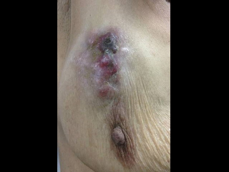
| Clinical presentation: | Postmenopausal woman with average risk of developing breast cancer presented with a painful lump in her right breast. On clinical examination, she was found to have a large hard lump in the upper quadrant of her right breast with ulceration of the overlying skin. |
 |
Mammography:
 |  |
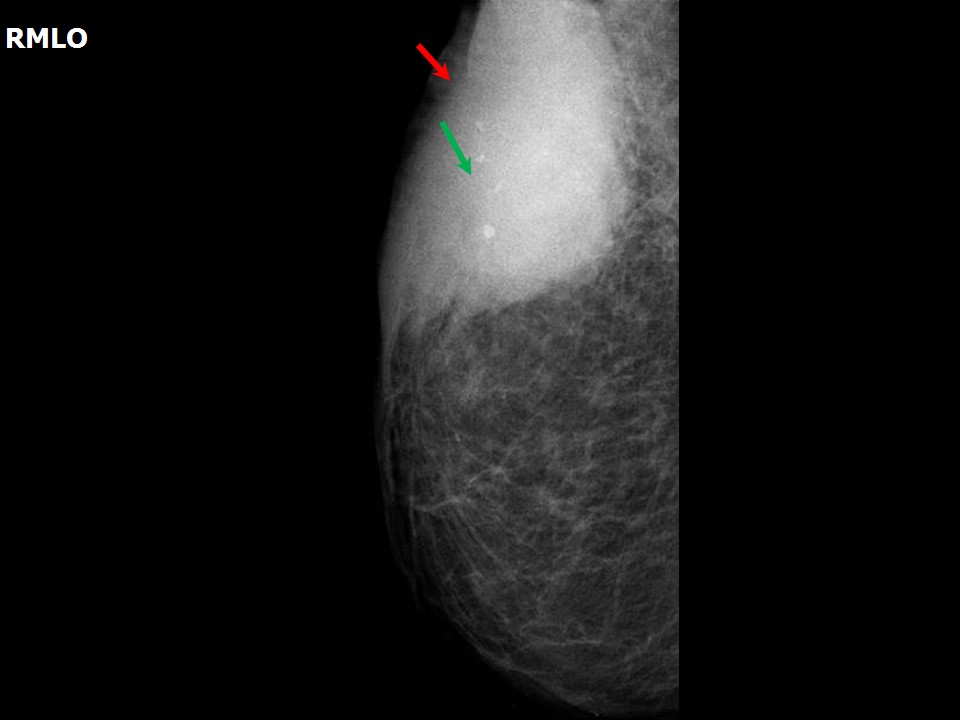 | 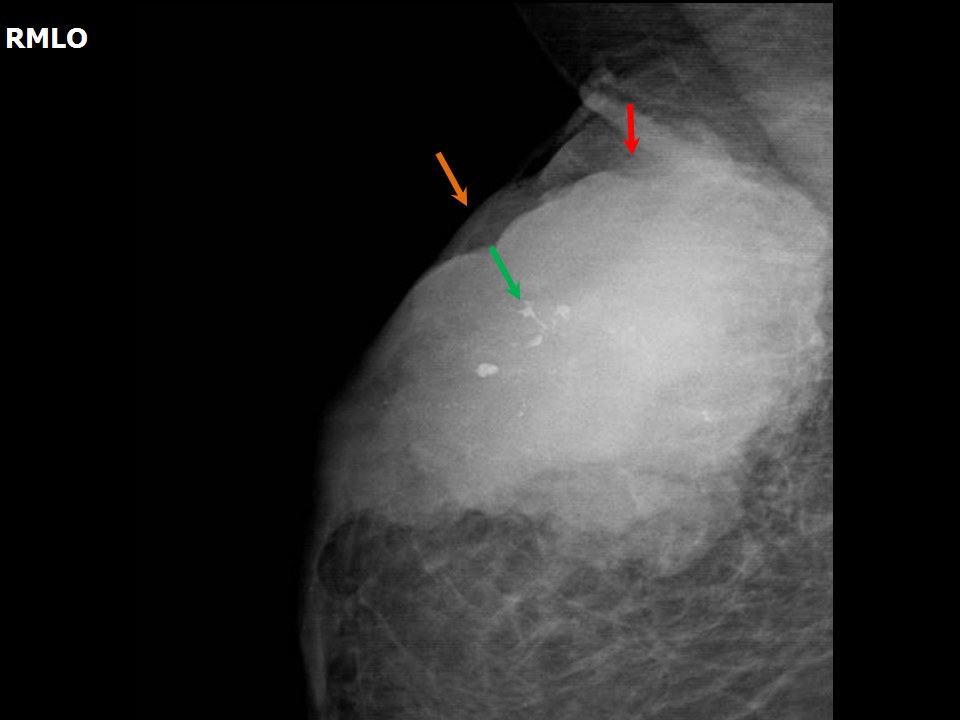 |
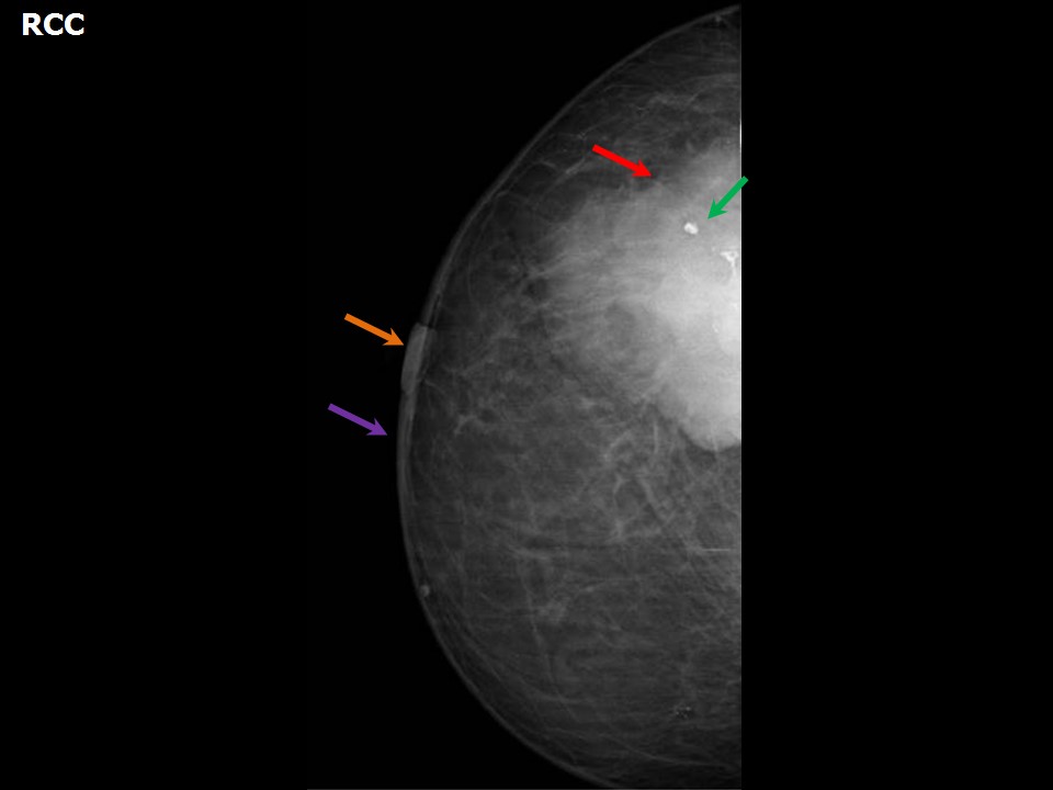
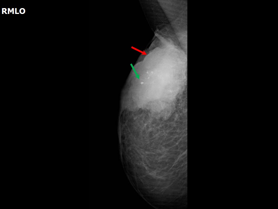
| Breast composition: | ACR category b (there are scattered areas of fibroglandular density) | Mammography features: |
| ‣ Location of the lesion: | Right breast, upper outer quadrant at 11 oclock, anterior, middle, and posterior thirds |
| ‣ Mass: | |
| • Number: | 1 |
| • Size: | 7.2 × 4.7 × 5.3 cm |
| • Shape: | Irregular |
| • Margins: | Microlobulated |
| • Density: | High |
| ‣ Calcifications: | |
| • Typically benign: | Coarse heterogeneous |
| • Suspicious: | None |
| • Distribution: | None |
| ‣ Architectural distortion: | None |
| ‣ Asymmetry: | None |
| ‣ Intramammary node: | Reactive |
| ‣ Skin lesion: | None |
| ‣ Solitary dilated duct: | None |
| ‣ Associated features: | Skin retraction, coarse calcification, and axillary adenopathy |
Ultrasound:
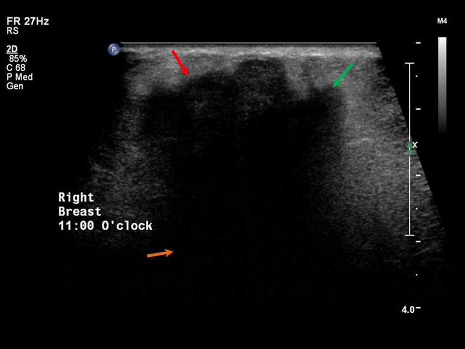 |
| Ultrasound features: Right breast, upper outer quadrant at 11 oclock | |
| ‣ Mass | |
| • Location: | Right breast, upper outer quadrant at 11 oclock |
| • Number: | 1 |
| • Size: | > 5.0 cm in greatest dimension |
| • Shape: | Irregular |
| • Orientation: | Not parallel |
| • Margins: | Angular |
| • Echo pattern: | Hypoechoic |
| • Posterior features: | Posterior shadowing |
| ‣ Calcifications: | Coarse calcifications in mass |
| ‣ Associated features: | Skin retraction and thickening, and metastatic axillary lymphadenopathy |
| ‣ Special cases: | None |
BI-RADS:
BI-RADS Category: 5 (highly suggestive of malignancy)Further assessment:
Further assessment advised: Referral for core biopsyHistopathology:
MRM
 |  |
 |  |
 |
| Histopathology features: | |
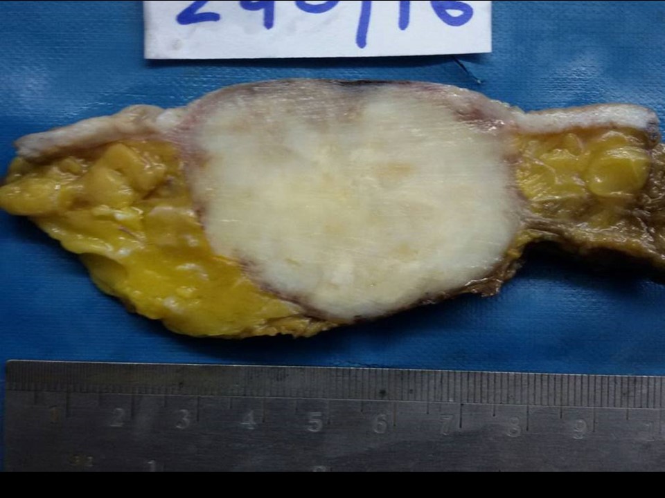
| ‣ Specimen type: | MRM |
| ‣ Laterality: | Right |
| ‣ Macroscopy: | On serial sectioning, a firm to hard, greyish white, well-circumscribed tumour is identified (4.5 × 4.0 × 3.5 cm) in the upper outer quadrant. Overlying skin shows an ulcerated indurated area (4.0 × 3.0 cm). The remaining breast tissue is unremarkable |
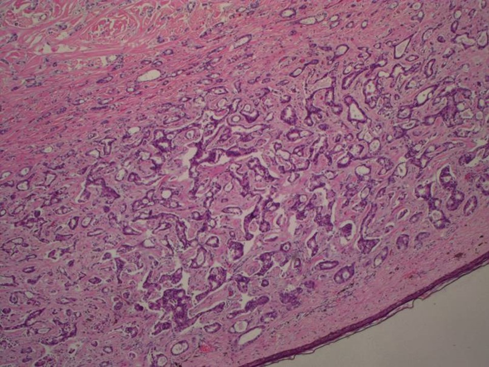
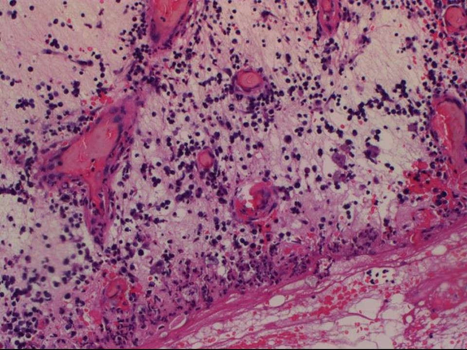
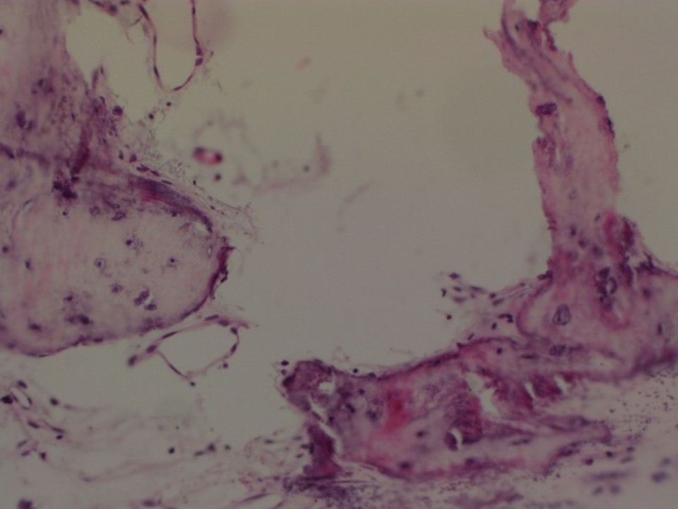
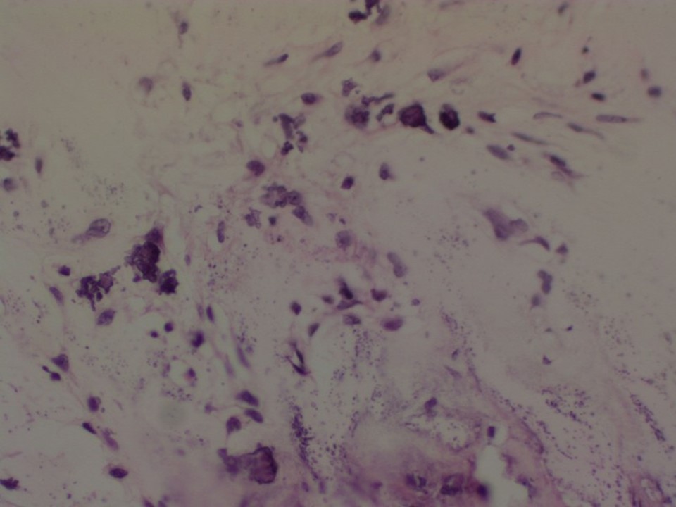
| ‣ Histological type: | Invasive carcinoma of no special type |
| ‣ Histological grade: | Grade 2 (2 + 2 + 1 = 5) |
| ‣ Mitosis: | 8 |
| ‣ Maximum invasive tumour size: | 4.5 cm in greatest dimension |
| ‣ Lymph node status: | 1/23 |
| ‣ Peritumoural lymphovascular invasion: | Not identified |
| ‣ DCIS/EIC: | Not identified |
| ‣ Margins: | Skin ulceration and oedema |
| ‣ Pathological stage: | pT4bN1 |
| ‣ Biomarkers: | |
| ‣ Comments: | Areas of calcification and ossification seen within the tumour |
Case summary:
| Postmenopausal woman presented with right breast lump with ulcerated skin. Diagnosed as right breast carcinoma with coarse calcifications within and overlying skin ulceration and thickening, BI-RADS 5 on imaging, as right breast carcinoma on cytology, and as invasive breast carcinoma of no special type, pT4bN1 on histopathology. |
Learning points:
|