Home / Training / Manuals / Atlas of breast cancer early detection / Learning

Atlas of breast cancer early detection

Filter by language: English / Русский

Breast imaging – Breast ultrasound – Ultrasound lexicon – Associated features – Vascularity


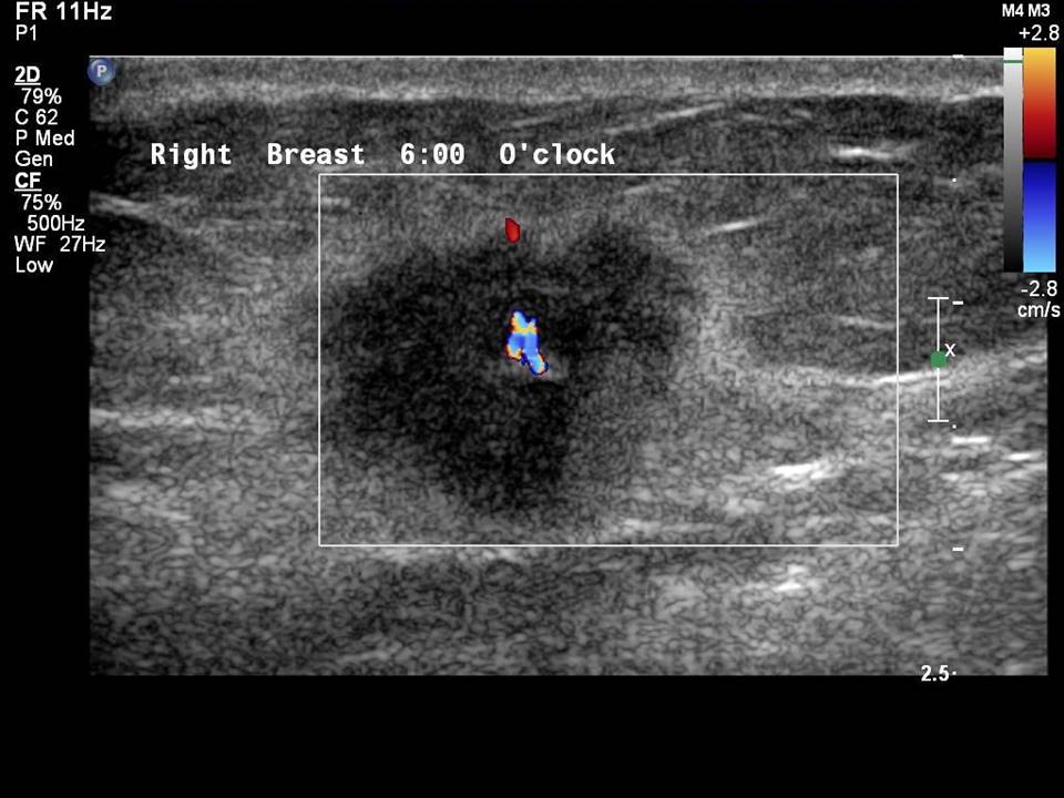
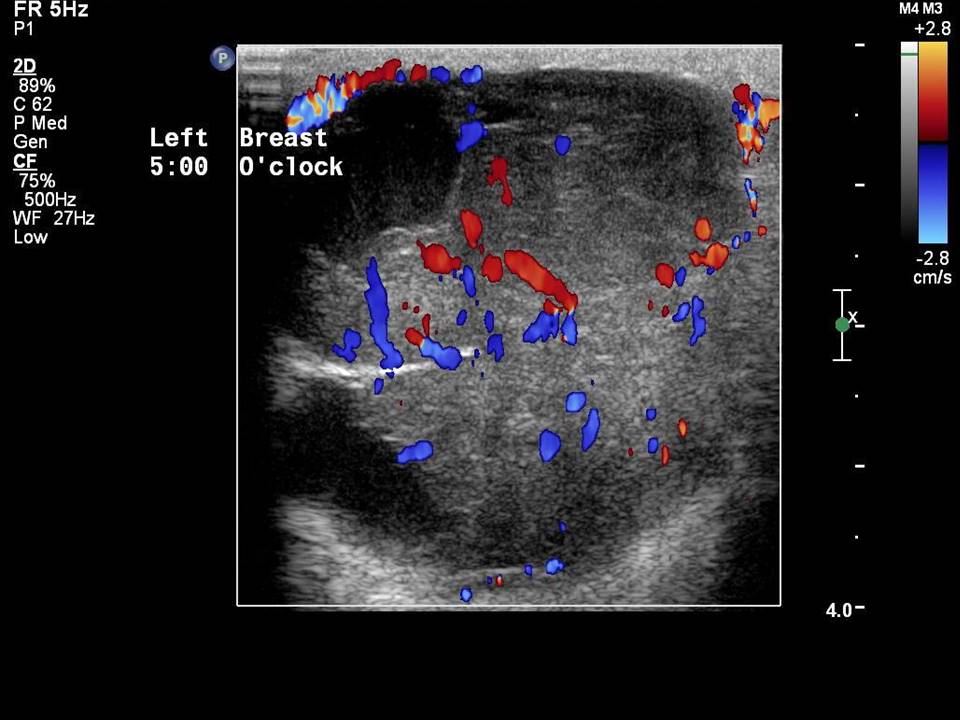
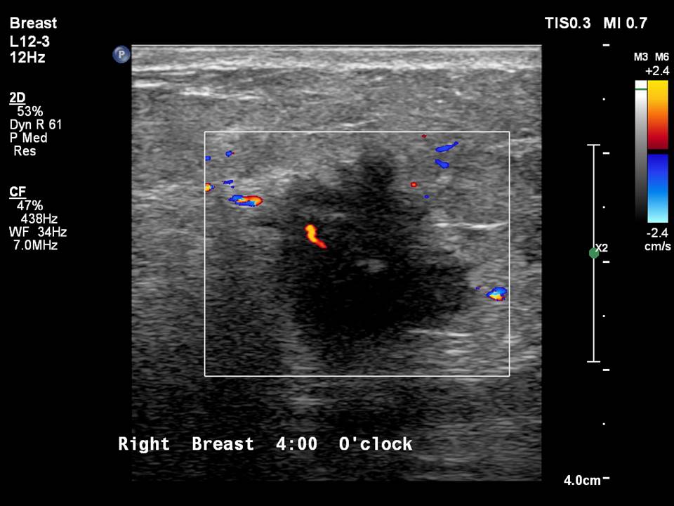
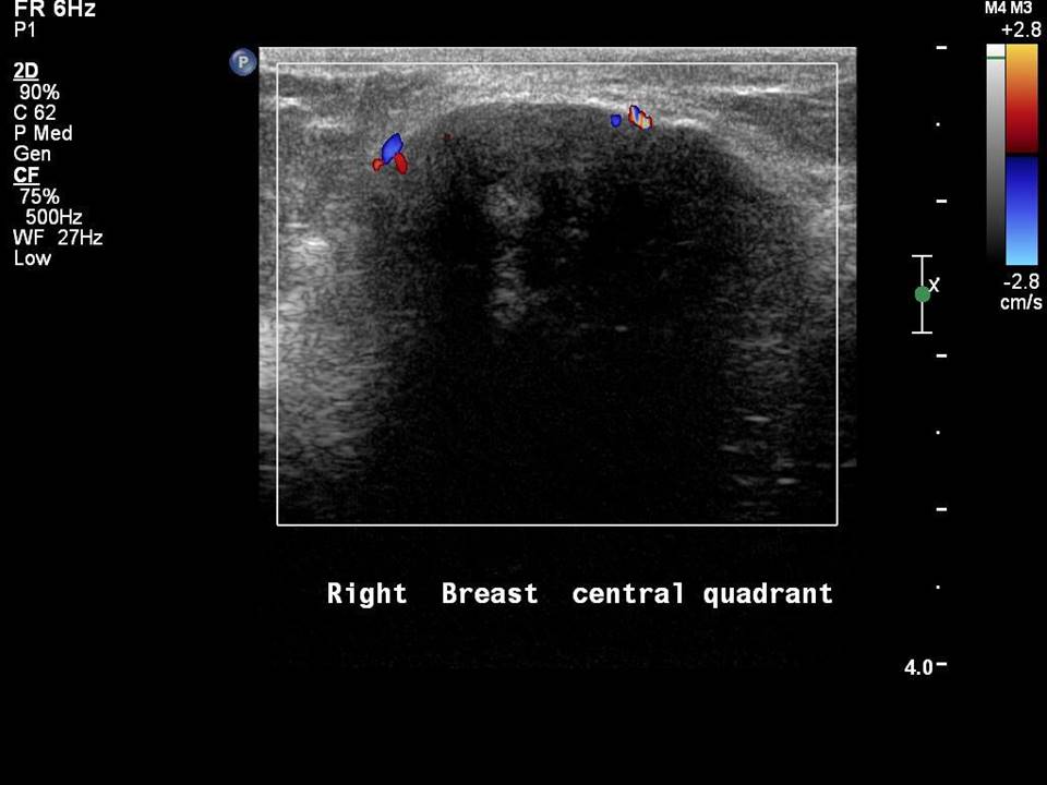
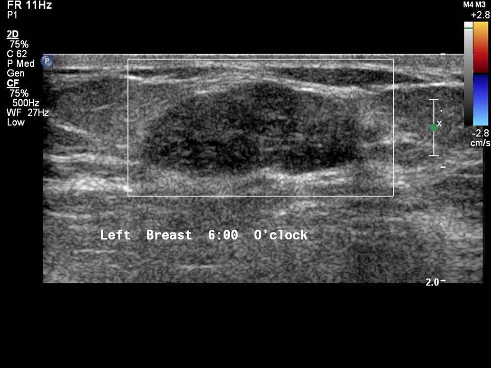
Vascularity in a mass or affected area of the breast can be detected by colour Doppler ultrasound. This complements the features of grey-scale B-mode ultrasound imaging. Colour Doppler ultrasound is used to determine internal vascularity, any vessels in the rim, and absent vascularity.

Internal vascularity
Internal vascularity describes the vessels seen within a mass lesion.


Vessels in rim
This term is used for vessels seen at the periphery of the mass lesion.


Absent vascularity
When no appreciable colour flow is seen in the region of the abnormality it is termed absent vascularity.


Vascularity in a lesion seen on Doppler ultrasound is caused by neoangiogenesis. It is not a specific feature that can be used to differentiate benign and malignant mass. Some features of colour Doppler ultrasound are useful for diagnosis.
  • Central or peripheral penetrating branching vessels in the lesion suggest neovascularization characteristic of malignant lesion .
  • Increased peripheral vascularity in the breast parenchyma helps to assess infective and inflammatory breast lesions .
  • Colour flow seen within the solid component of the complex cyst is a suspicious finding and biopsy is recommended .
  • Intraductal echoes can be caused by inspissated secretions or intraductal papillomas. Colour Doppler in the papilloma will differentiate the two, depending on the presence of vascularity .
  • It has been suggested that parallel arteries and veins in the lesion are a feature of a benign lesion.
  • A metastatic node is suggested on colour Doppler of axillary lymph nodes when there is loss of normal flow through the hilum. Non-hilar blood flow is demonstrated by peripheral vessels entering the thickened cortex of the metastatic node.
  • Poor vascularity or absent colour flow suggests a low-grade malignancy or a benign mass .


Click on the pictures to magnify and display the legends


Click on this icon to display a case study

25 avenue Tony Garnier CS 90627 69366, LYON CEDEX 07 France - Tel: +33 (0)4 72 73 84 85
© IARC 2024 - Terms of use  -  Privacy Policy.