


Home / Training / Manuals / Atlas of breast cancer early detection / Cases
Atlas of breast cancer early detection
Filter by language: English / Русский
Go back to the list of case studies
.png) Click on the pictures to magnify and display the legends
Click on the pictures to magnify and display the legends
| Case number: | 043 |
| Age: | 77 |
| Clinical presentation: | Postmenopausal woman with average risk of developing breast cancer presented with a large mass in the left breast first noticed 1 year ago. Examination revealed a large hard mass, 8 cm in diameter, fixed to the skin of the left breast. |
Mammography:
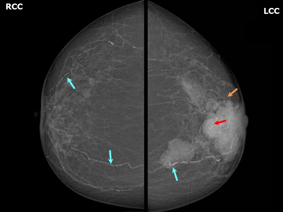 | 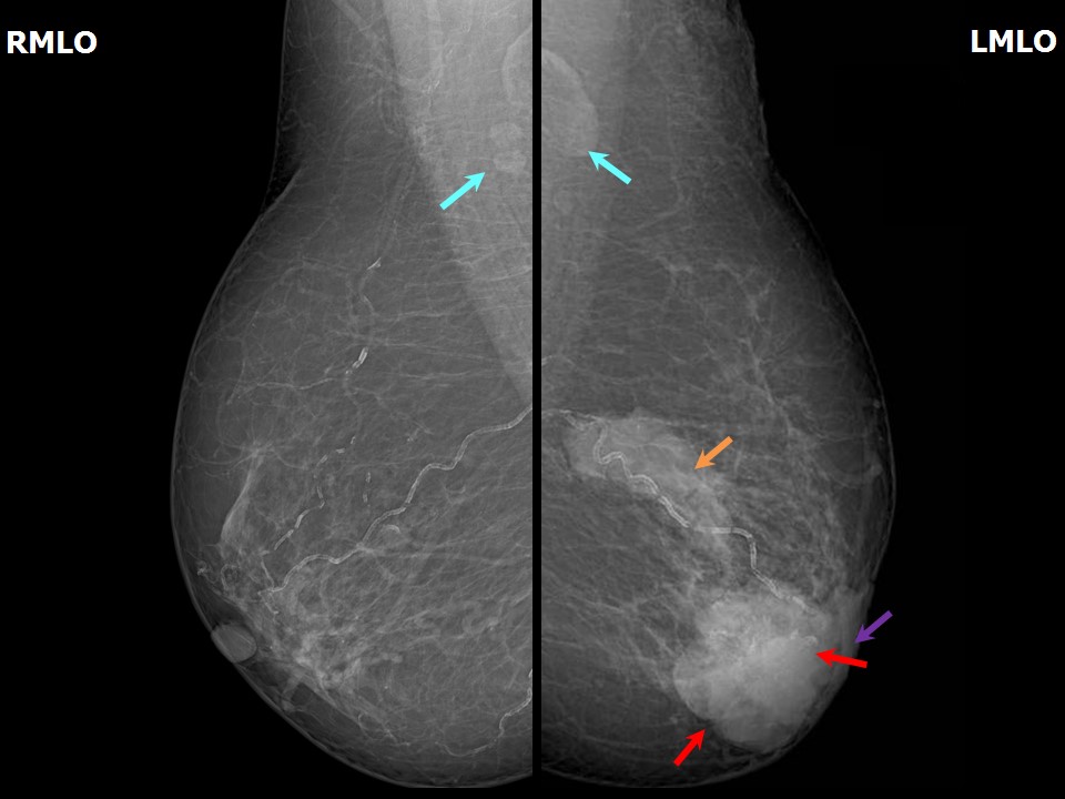 |
| Breast composition: | ACR category b (there are scattered areas of fibroglandular density) | Mammography features: |
| ‣ Location of the lesion: | Left breast, central portion of the breast, central zone, anterior third |
| ‣ Mass: | |
| • Number: | 1 |
| • Size: | 6.8 × 5.0 cm |
| • Shape: | Irregular |
| • Margins: | Indistinct |
| • Density: | High |
| ‣ Calcifications: | |
| • Typically benign: | Vascular calcification |
| • Suspicious: | Suspicious calcifications |
| • Distribution: | Regional |
| ‣ Architectural distortion: | None |
| ‣ Asymmetry: | None |
| ‣ Intramammary node: | None |
| ‣ Skin lesion: | None |
| ‣ Solitary dilated duct: | None |
| ‣ Associated features: | Coarse heterogeneous calcifications |
| Breast composition: | ACR category b (there are scattered areas of fibroglandular density) | Mammography features: |
| ‣ Location of the lesion: | Left breast, lower inner quadrant at 78 oclock, middle third |
| ‣ Mass: | |
| • Number: | 1 |
| • Size: | 3.0 × 2.0 cm |
| • Shape: | Irregular |
| • Margins: | Indistinct |
| • Density: | High |
| ‣ Calcifications: | |
| • Typically benign: | None |
| • Suspicious: | None |
| • Distribution: | None |
| ‣ Architectural distortion: | None |
| ‣ Asymmetry: | None |
| ‣ Intramammary node: | None |
| ‣ Skin lesion: | None |
| ‣ Solitary dilated duct: | None |
| ‣ Associated features: | Skin thickening |
| Breast composition: | ACR category b (there are scattered areas of fibroglandular density) | Mammography features: |
| ‣ Location of the lesion: | Left breast, upper outer quadrant at 1 oclock, middle third |
| ‣ Mass: | |
| • Number: | 1 |
| • Size: | 2.8 × 1.0 cm |
| • Shape: | Irregular |
| • Margins: | Indistinct |
| • Density: | High |
| ‣ Calcifications: | |
| • Typically benign: | None |
| • Suspicious: | None |
| • Distribution: | None |
| ‣ Architectural distortion: | None |
| ‣ Asymmetry: | None |
| ‣ Intramammary node: | None |
| ‣ Skin lesion: | None |
| ‣ Solitary dilated duct: | None |
| ‣ Associated features: | Skin thickening |
| Breast composition: | ACR category b (there are scattered areas of fibroglandular density) | Mammography features: |
| ‣ Location of the lesion: | Right breast, upper outer quadrant at 11 oclock, middle third |
| ‣ Mass: | |
| • Number: | 1 |
| • Size: | 0.7 cm in greatest dimension |
| • Shape: | Oval |
| • Margins: | Circumscribed |
| • Density: | Equal |
| ‣ Calcifications: | |
| • Typically benign: | Vascular calcification |
| • Suspicious: | None |
| • Distribution: | None |
| ‣ Architectural distortion: | None |
| ‣ Asymmetry: | None |
| ‣ Intramammary node: | None |
| ‣ Skin lesion: | None |
| ‣ Solitary dilated duct: | None |
| ‣ Associated features: | None |
Ultrasound:
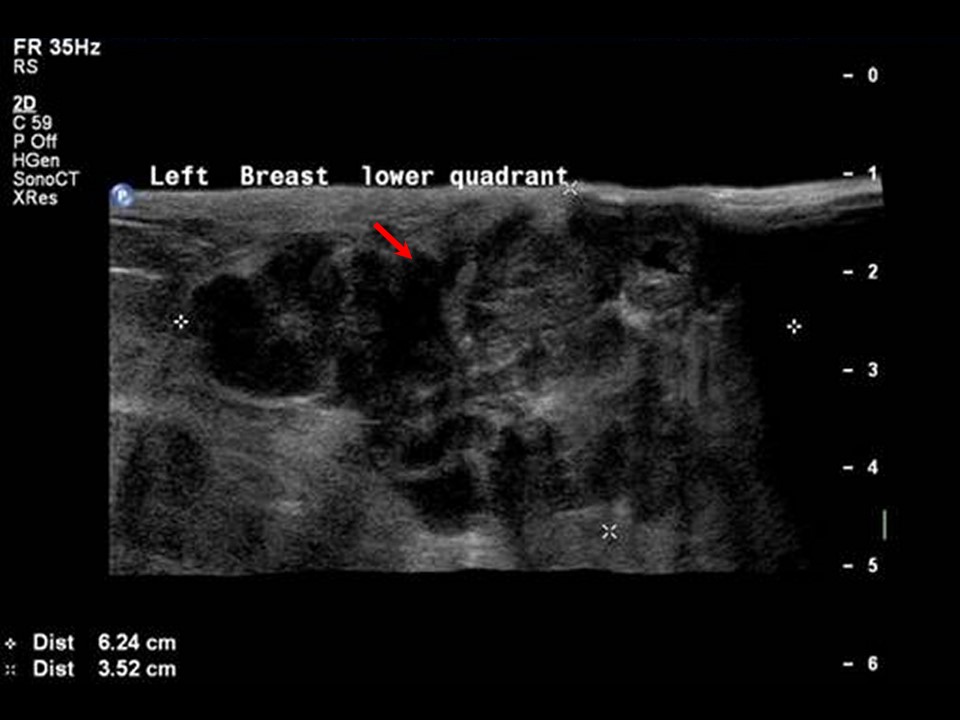 |
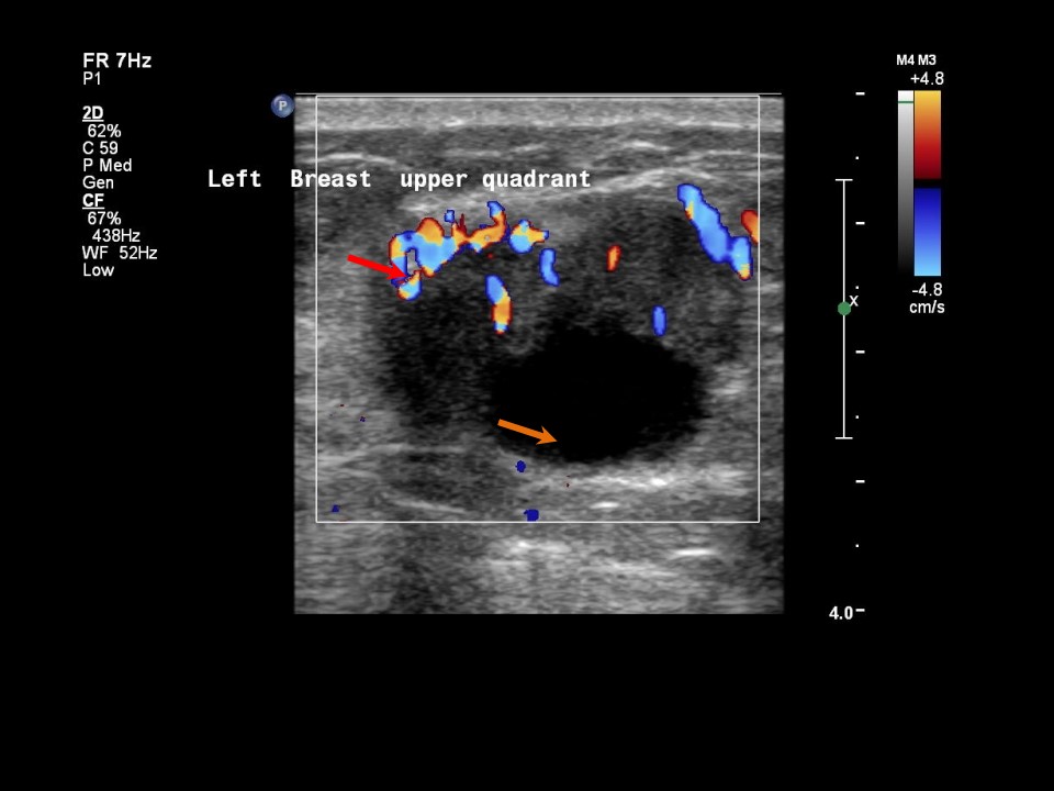
| Ultrasound features: Left breast, lower inner quadrant at 6-7 oclock | |
| ‣ Mass | |
| • Location: | Left breast, lower inner quadrant at 6-7 oclock |
| • Number: | 1 |
| • Size: | 6.5 × 3.5 cm |
| • Shape: | Irregular |
| • Orientation: | Not parallel |
| • Margins: | Angular |
| • Echo pattern: | Heteroechoic with necrotic areas |
| • Posterior features: | No posterior features |
| ‣ Calcifications: | None |
| ‣ Associated features: | Skin thickening and internal vascularity |
| ‣ Special cases: | None |
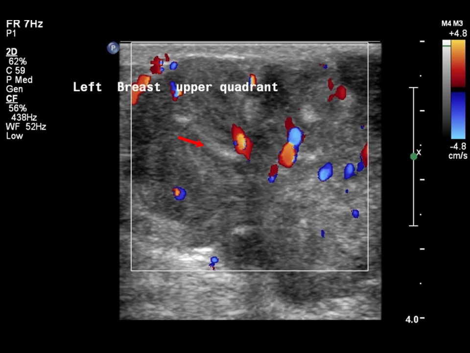 |  |
| Ultrasound features: Left breast, upper inner quadrant at 11 o'clock | |
| ‣ Mass | |
| • Location: | Left breast, upper inner quadrant at 11 o'clock |
| • Number: | 2 |
| • Size: | Largest 3.5 × 2.5 cm |
| • Shape: | Irregular |
| • Orientation: | Not parallel |
| • Margins: | Angular |
| • Echo pattern: | Heteroechoic with necrotic areas |
| • Posterior features: | No posterior features |
| ‣ Calcifications: | None |
| ‣ Associated features: | Internal vascularity |
| ‣ Special cases: | None |
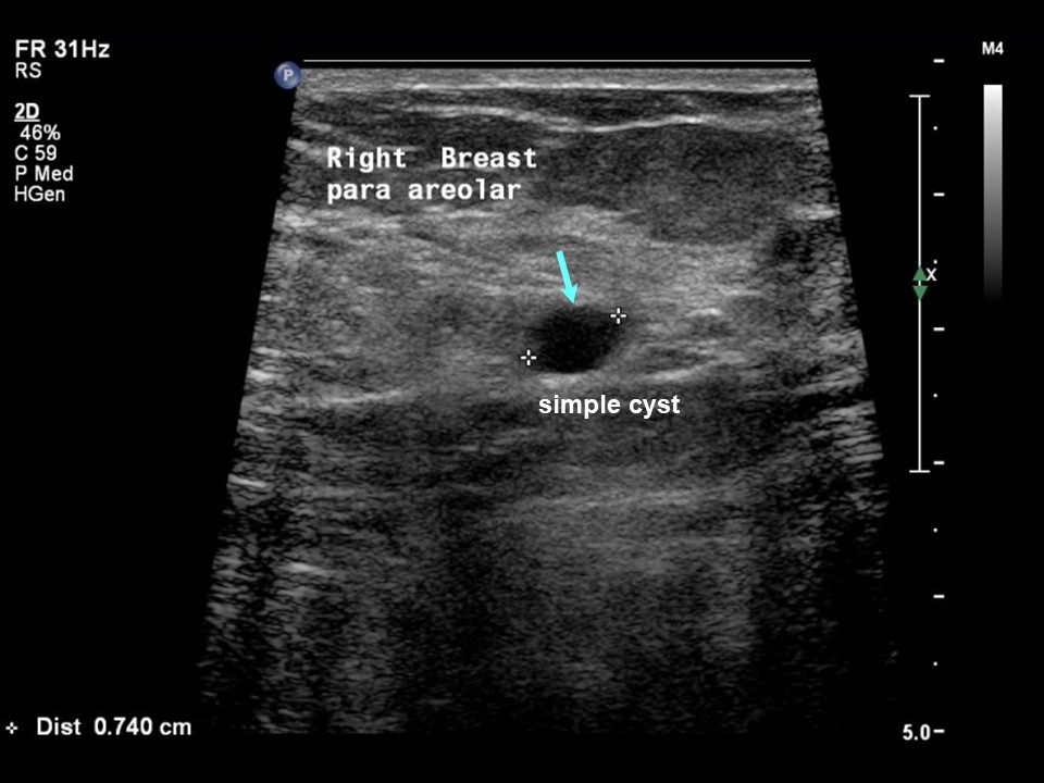 |
| Ultrasound features: Right breast, central portion of the breast | |
| ‣ Mass | |
| • Location: | Right breast, central portion of the breast |
| • Number: | 1 |
| • Size: | 0.7 × 0.6 cm |
| • Shape: | Oval |
| • Orientation: | Parallel |
| • Margins: | Circumscribed |
| • Echo pattern: | Anechoic |
| • Posterior features: | No posterior features |
| ‣ Calcifications: | None |
| ‣ Associated features: | None |
| ‣ Special cases: | Simple cyst |
BI-RADS:
BI-RADS Category: 5 (highly suggestive of malignancy)Further assessment:
Further assessment advised: Referral for cytology and for core biopsyCytology:
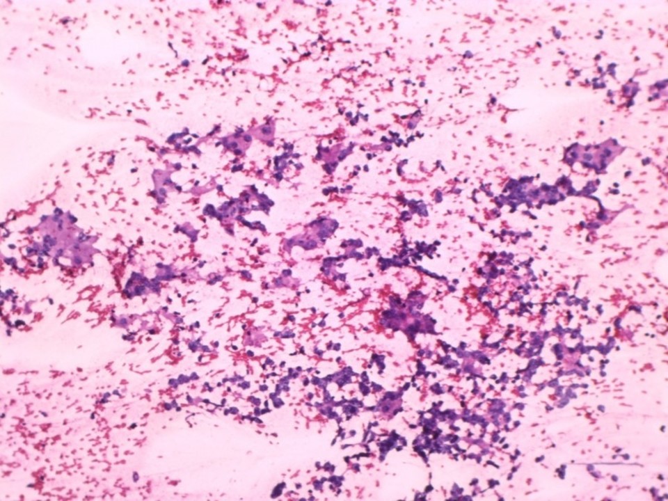 |
| Cytology features: | |
| ‣ Type of sample: | FNAC |
| ‣ Site of biopsy: | |
| • Laterality: | Left |
| • Quadrant: | |
| • Localization technique: | Palpation |
| • Nature of aspirate: | whitish |
| ‣ Cytological description: | Highly cellular smears reveal loosely cohesive sheets of ductal epithelial cells of breast showing anisonucleosis, coarse nuclear chromatin, and prominent nucleoli. A few multinucleated giant cells are noted |
| ‣ Reporting category: | Malignant |
| ‣ Diagnosis: | Carcinoma |
| ‣ Comments: | None |
Histopathology:
Incisional biopsy from left breast mass
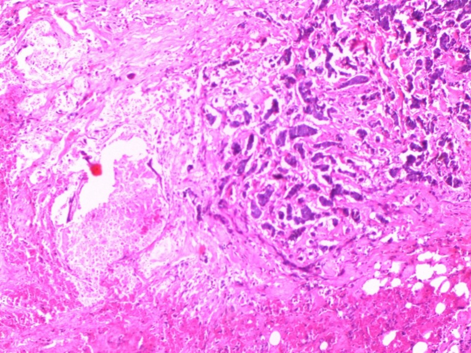 |
| Histopathology features: | |
| ‣ Specimen type: | Incisional biopsy from left breast mass |
| ‣ Laterality: | Left |
| ‣ Macroscopy: | Single tissue bit, partially covered with skin (1.4 × 1.0 × 0.7 cm). Skin surface appears unremarkable |
| ‣ Histological type: | Sections show fibrofatty tissue lined by skin. There is a focus of invasive breast carcinoma of no special type. Extensive tumour necrosis is seen. The tumour is 0.5 cm from the skin in the tissue submitted. There are also chronic inflammatory cells with eosinophils and neutrophils in the adipose tissue around the tumour |
| ‣ Histological grade: | Grade 2 (2 + 2 + 2 = 6) |
| ‣ Mitosis: | 12 |
| ‣ Maximum invasive tumour size: | |
| ‣ Lymph node status: | |
| ‣ Peritumoural lymphovascular invasion: | |
| ‣ DCIS/EIC: | |
| ‣ Margins: | |
| ‣ Pathological stage: | |
| ‣ Biomarkers: | |
| ‣ Comments: |
Case summary:
| Postmenopausal woman presented with large left breast lump fixed to the skin. Diagnosed as left breast carcinoma with coarse heterogeneous calcification within and overlying skin thickening, BI-RADS 5 on imaging, as left breast carcinoma on cytology, and as invasive breast carcinoma of no special type on incisional biopsy histopathology. |
Learning points:
|