


Home / Training / Manuals / Atlas of breast cancer early detection / Cases
Atlas of breast cancer early detection
Filter by language: English / Русский
Go back to the list of case studies
.png) Click on the pictures to magnify and display the legends
Click on the pictures to magnify and display the legends
| Case number: | 119 |
| Age: | 67 |
| Clinical presentation: | Postmenopausal woman with average risk of developing breast cancer presented with a left breast lump first noticed 3 months earlier. It had gradually increased in size in the last month. On examination, she was found to have a hard lump in the upper inner quadrant of the left breast. |
Mammography:
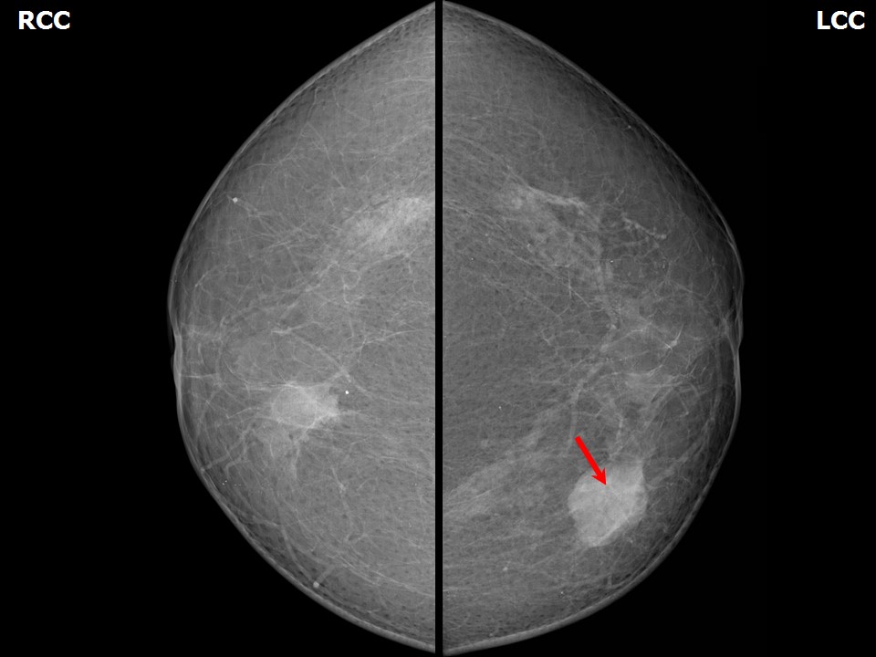 | 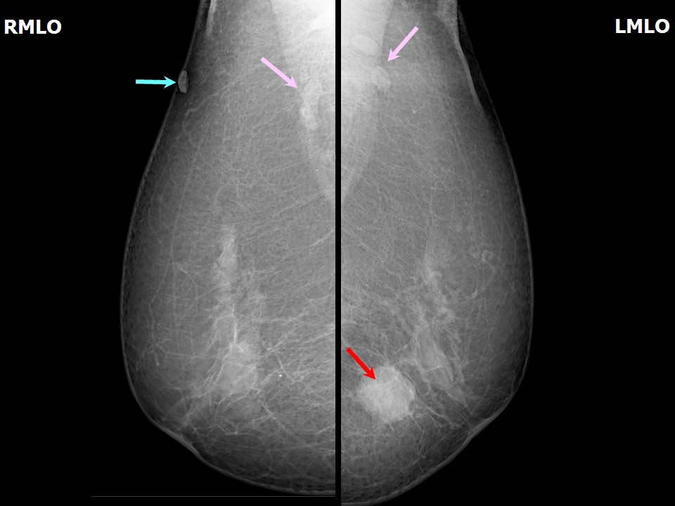 |
| Breast composition: | ACR category b (there are scattered areas of fibroglandular density) | Mammography features: |
| ‣ Location of the lesion: | Left breast, upper inner quadrant at 910 oclock, middle third |
| ‣ Mass: | |
| • Number: | 1 |
| • Size: | 3.3 × 2.7 cm |
| • Shape: | Oval |
| • Margins: | Indistinct |
| • Density: | High |
| ‣ Calcifications: | |
| • Typically benign: | None |
| • Suspicious: | None |
| • Distribution: | None |
| ‣ Architectural distortion: | None |
| ‣ Asymmetry: | None |
| ‣ Intramammary node: | None |
| ‣ Skin lesion: | None |
| ‣ Solitary dilated duct: | None |
| ‣ Associated features: | None |
| Breast composition: | ACR category b (there are scattered areas of fibroglandular density) | Mammography features: |
| ‣ Location of the lesion: | Right breast, lower inner quadrant at 5 oclock, middle third |
| ‣ Mass: | |
| • Number: | 1 |
| • Size: | 1.7 × 1.0 cm |
| • Shape: | Oval |
| • Margins: | Indistinct |
| • Density: | Equal |
| ‣ Calcifications: | |
| • Typically benign: | None |
| • Suspicious: | None |
| • Distribution: | None |
| ‣ Architectural distortion: | None |
| ‣ Asymmetry: | Focal, glandular tissue |
| ‣ Intramammary node: | None |
| ‣ Skin lesion: | None |
| ‣ Solitary dilated duct: | None |
| ‣ Associated features: | None |
| Breast composition: | ACR category b (there are scattered areas of fibroglandular density) | Mammography features: |
| ‣ Location of the lesion: | Right breast, upper outer quadrant at 12 oclock, axillary tail, skin lesion |
| ‣ Mass: | |
| • Number: | 1 |
| • Size: | 0.9 cm in greatest dimension |
| • Shape: | Oval |
| • Margins: | Circumscribed |
| • Density: | High |
| ‣ Calcifications: | |
| • Typically benign: | None |
| • Suspicious: | None |
| • Distribution: | None |
| ‣ Architectural distortion: | None |
| ‣ Asymmetry: | None |
| ‣ Intramammary node: | None |
| ‣ Skin lesion: | Present |
| ‣ Solitary dilated duct: | None |
| ‣ Associated features: | None |
Ultrasound:
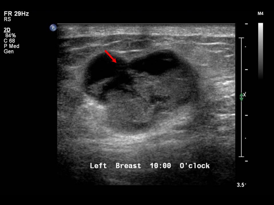 | 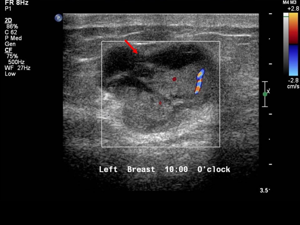 |
| Ultrasound features: Left breast, inner quadrants at 10 oclock | |
| ‣ Mass | |
| • Location: | Left breast, inner quadrants at 10 oclock |
| • Number: | 1 |
| • Size: | 2.5 × 2.1 cm |
| • Shape: | Irregular |
| • Orientation: | Parallel |
| • Margins: | Indistinct |
| • Echo pattern: | Complex cystic and solid |
| • Posterior features: | No posterior features |
| ‣ Calcifications: | None |
| ‣ Associated features: | Internal vascularity in intracysticsolid echogenic region |
| ‣ Special cases: | Mass in or on skin |
BI-RADS:
BI-RADS Category: 5 (highly suggestive of malignancy)Further assessment:
Further assessment advised: Referral for cytologyCytology:
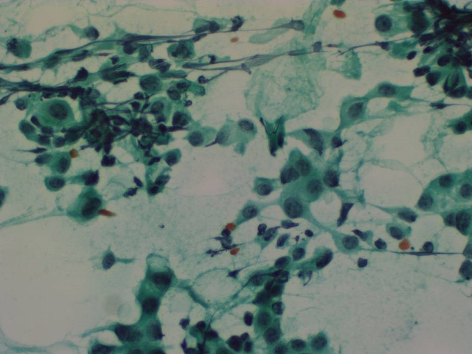 |
| Cytology features: | |
| ‣ Type of sample: | FNAC |
| ‣ Site of biopsy: | |
| • Laterality: | Left |
| • Quadrant: | Upper inner |
| • Localization technique: | Palpation |
| • Nature of aspirate: | 2 mL of blood-stained fluid |
| ‣ Cytological description: | Cellular smears reveal loosely cohesive sheets of malignant cells as well as scattered single isolated malignant cells on a haemorrhagic background. Individual cells reveal mild to moderate anisonucleosis, roughened chromatin, and occasional mitosis. A few cohesive sheets of benign ductal cells and myoepithelial cells are also seen |
| ‣ Reporting category: | Malignant |
| ‣ Diagnosis: | Carcinoma |
| ‣ Comments: | None |
Histopathology:
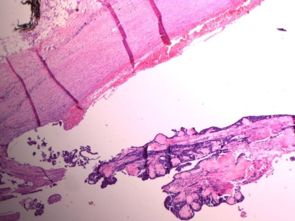
Breast-conserving surgery
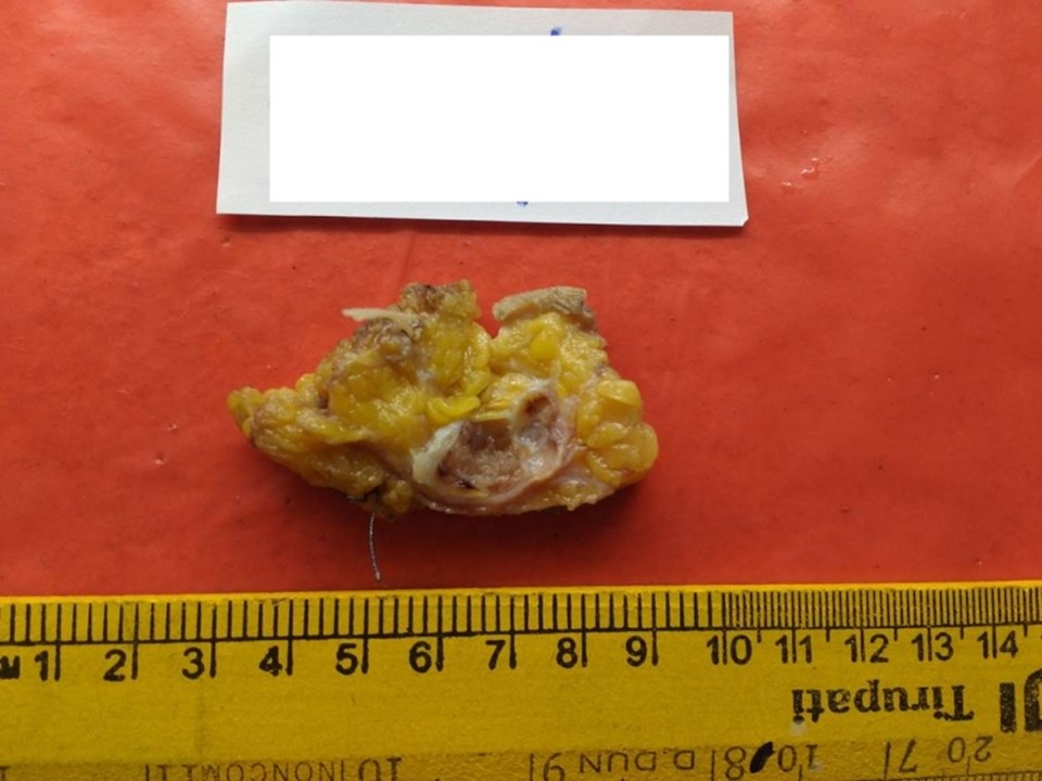 |  |
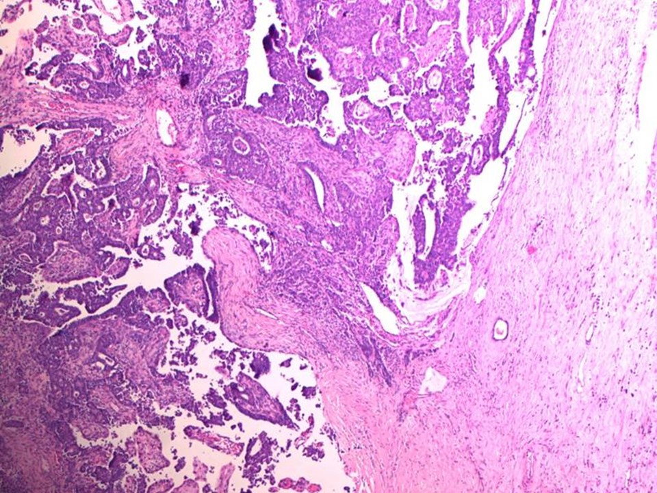 |
| Histopathology features: | |
| ‣ Specimen type: | Breast-conserving surgery |
| ‣ Laterality: | Left |
| ‣ Macroscopy: | Specimen (8.0 × 6.0 × 3.0 cm) with skin flap (5.0 × 3.5 cm). On serial sectioning, a cystic cavity (2.2 × 1.8 × 2.0 cm) containing a friable mass with papillary excrescences is identified. It is located 1.7 cm from the skin (anterior margin), 0.5 cm from the posterior margin, 2.5 cm from the superior margin, 0.2 cm from the inferior margin, 1.0 cm from the medial margin, and 2.5 cm from the lateral margin. The remaining breast parenchyma shows a firm whitish area (2.0 × 1.5 cm) medial to the tumour |
| ‣ Histological type: | Encapsulated papillary carcinoma with adjacent foci of invasive carcinoma of no special type |
| ‣ Histological grade: | 1 + 1 + 1 = 3 |
| ‣ Mitosis: | 3 |
| ‣ Maximum invasive tumour size: | Microinvasion 0.1 cm in greatest dimension |
| ‣ Lymph node status: | 0/21 |
| ‣ Peritumoural lymphovascular invasion: | Absent |
| ‣ DCIS/EIC: | No associated DCIS in the surrounding breast tissue |
| ‣ Margins: | Free of tumour |
| ‣ Pathological stage: | pT1miN0 |
| ‣ Biomarkers: | |
| ‣ Comments: | The firm whitish area medial to the tumour shows only fibrosis |
Case summary:
| Postmenopausal woman presented with lump in the left breast. Diagnosed as complex cystic and solid mass lesion in the left breast, BI-RADS 5 on imaging, as breast carcinoma on cytology, and as encapsulated papillary carcinoma with adjacent foci of microinvasive breast carcinoma of no special type, pT1miN0 on histopathology. |
Learning points:
|