|
Breast imaging – Mammography interpretation – Mammography lexicon – Special cases |   |
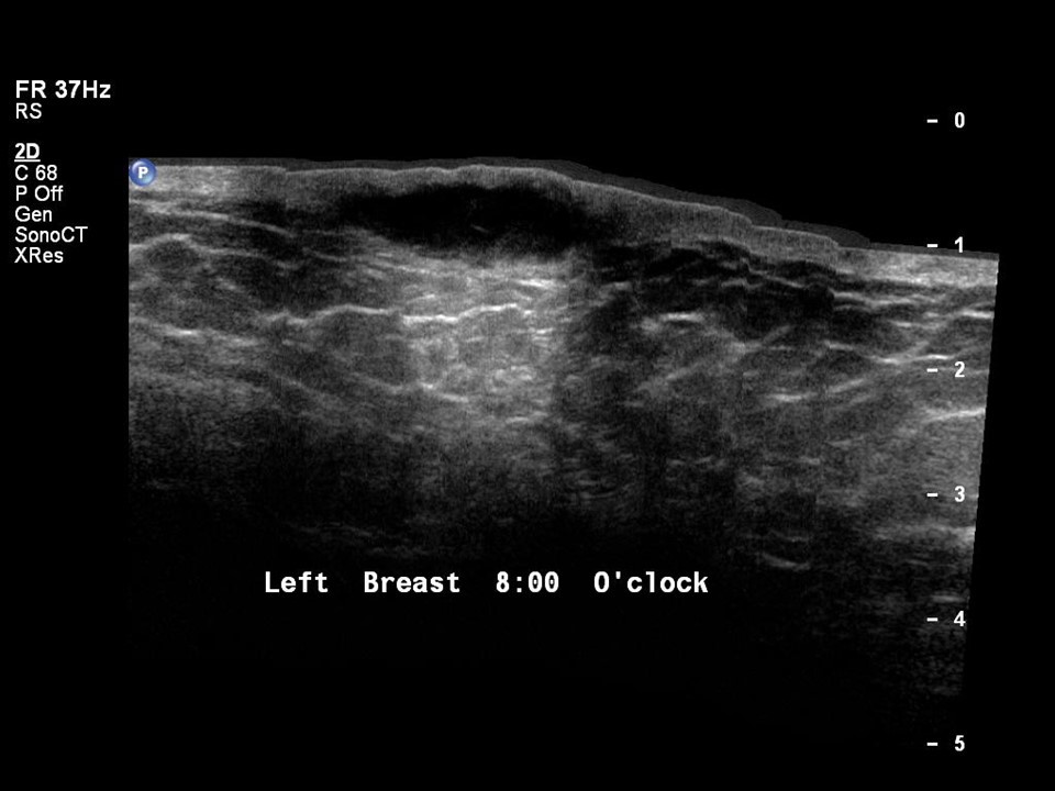
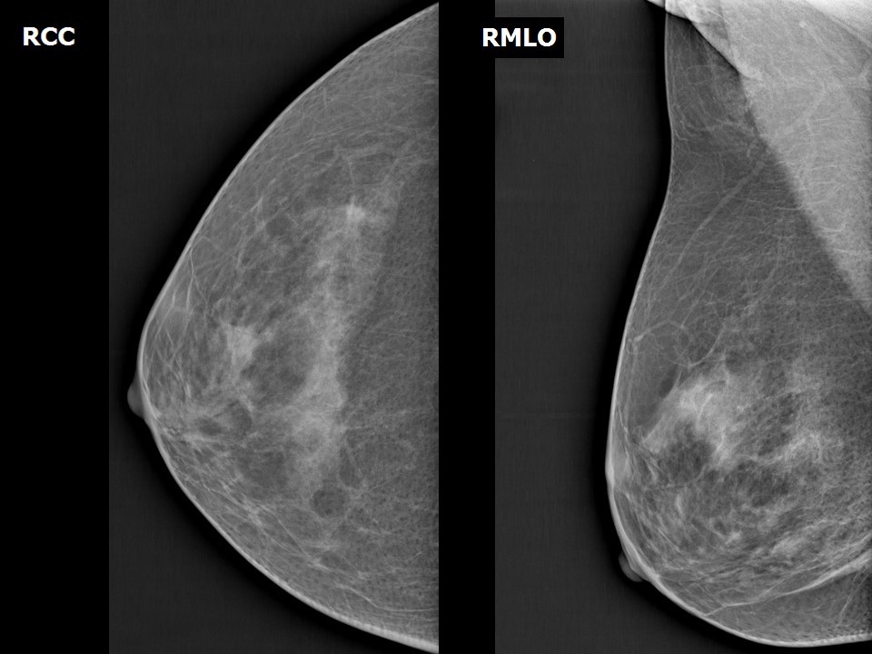
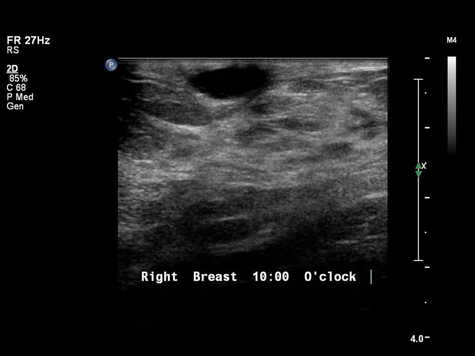
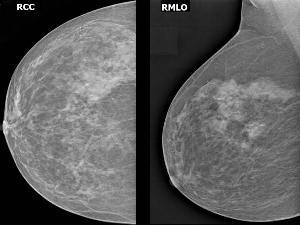
Findings with unique diagnostic features on mammography include intramammary lymph node, skin lesion, and solitary dilated duct.
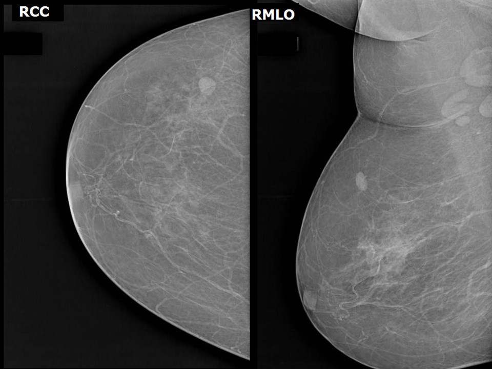
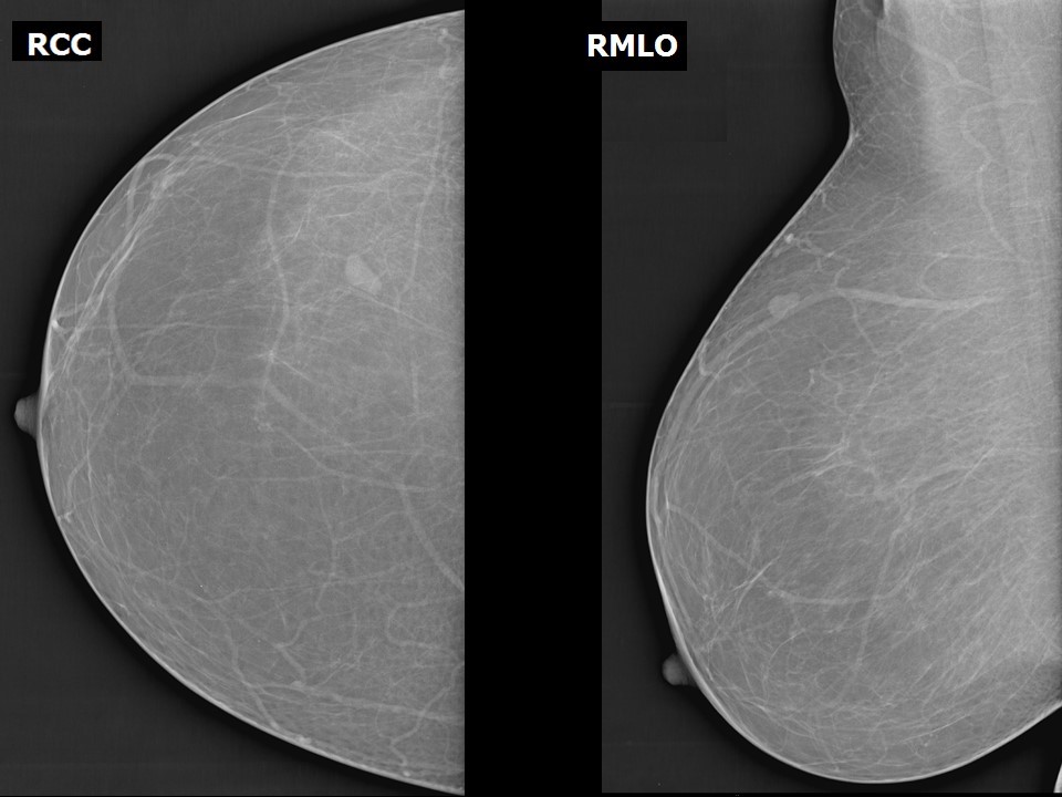
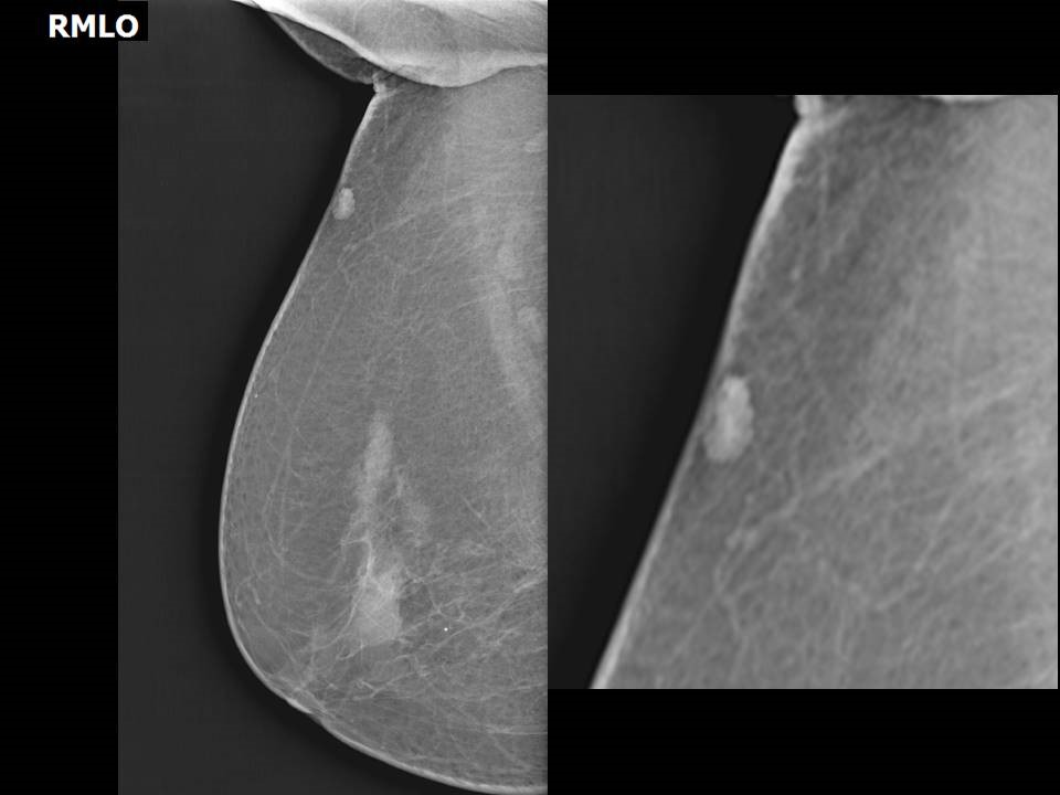
Intramammary lymph nodes 
- Intramammary lymph nodes are seen in the vast majority of mammograms in the outer quadrant of the breast, but can occur in other parts of the breast.
- They are a common visible recognizable finding in approximately 5% of mammograms.
- They have circumscribed margins, are usually < 1 cm across, and have a lucent central hilum.
- The appearance is characteristic and does not require further investigation.
- Identifying and marking out the presence of intramammary nodes is necessary to rule out a new developing asymmetry on mammography and to avoid unnecessary biopsies.
Screening mammogram: intramammary node seen
Stable intramammary node
Skin lesion
The radiologist needs to ascertain whether a lesion is intramammary or on the skin. Before the breast is positioned and the mammogram obtained, a radiopaque marker is placed on the skin lesion to confirm the mammographic location. Air trapped in the interstices distinguishes a skin lesion from an intramammary mass.
Mole on the skin
Keratinous cyst
Nevus
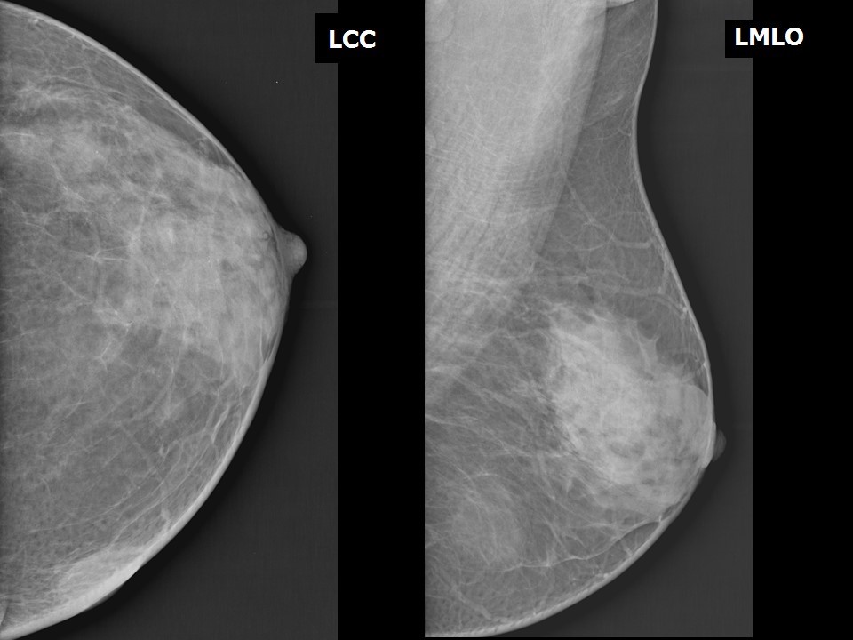
Solitary dilated duct
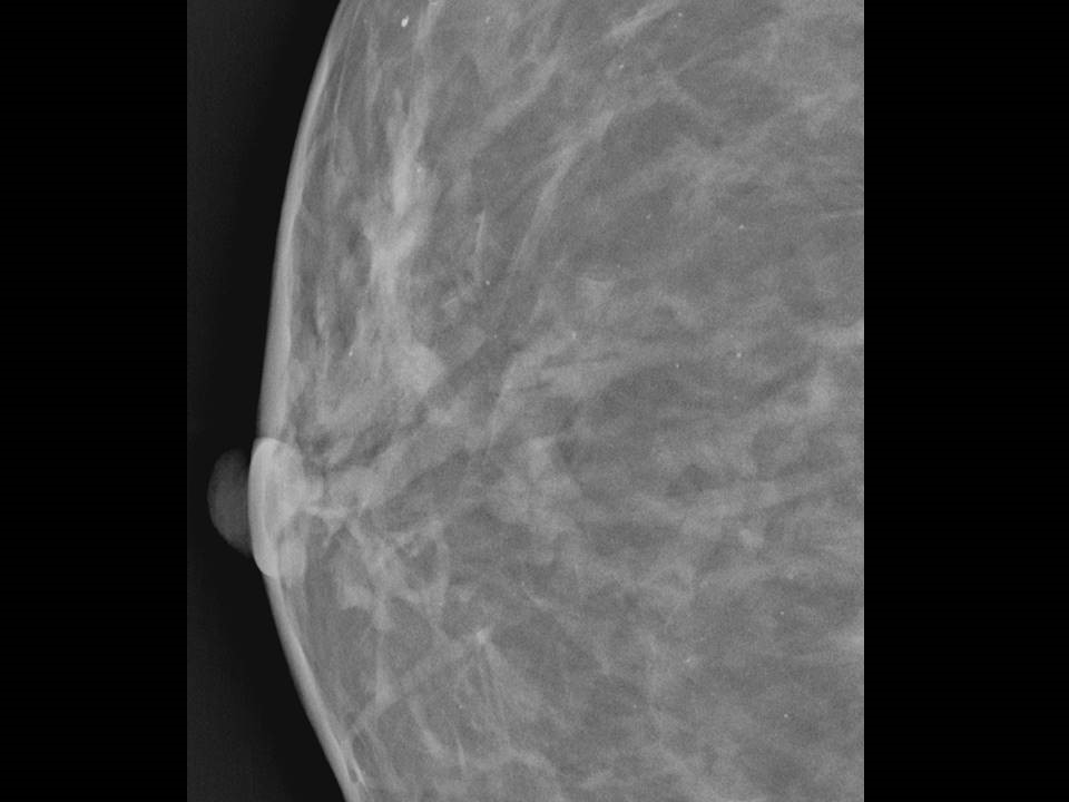
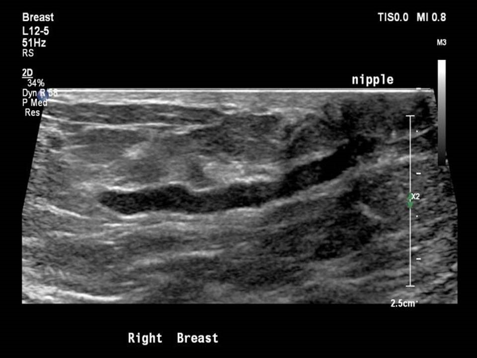
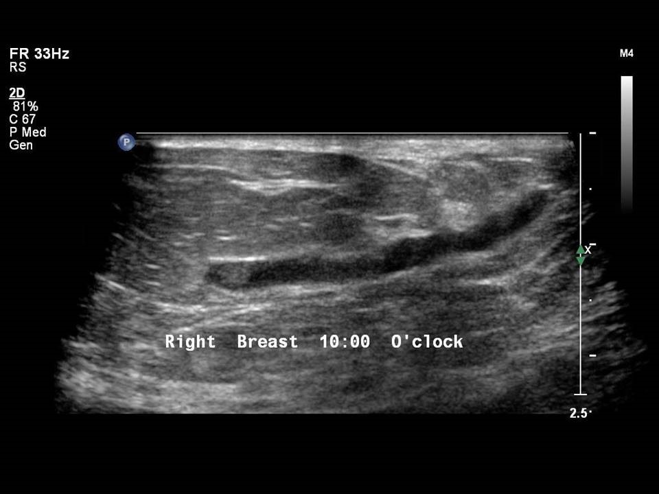
- A single dilated channel in the subareolar region is seen on mammography as a tubular opacity of equal density posterior to the nipple shadow in the subareolar region. If a woman presents with nipple discharge or changes in the nipple–areolar complex a detailed assessment of the dilated duct on ultrasound is performed to rule out an intraductal solid mass
 . Stability of the dilated duct on serial follow-up mammograms will decrease the likelihood of an intraductal mass . Stability of the dilated duct on serial follow-up mammograms will decrease the likelihood of an intraductal mass 
|
.png)
Click on the pictures to magnify and display the legends

Click on this icon to display a case study
25 avenue Tony Garnier CS 90627 69366, LYON CEDEX 07 France - Tel: +33 (0)4 72 73 84 85 © IARC 2026 - Terms of use - Privacy Policy.
| .png)


