


Home / Training / Manuals / Atlas of breast cancer early detection / Cases
Atlas of breast cancer early detection
Filter by language: English / Русский
Go back to the list of case studies
.png) Click on the pictures to magnify and display the legends
Click on the pictures to magnify and display the legends
BI-RADS Category (MRI 2014): 3 (probably benign)
BI-RADS Category (MRI 2017 and Mammography 2018): 2 (benign)
| Case number: | 106 |
| Age: | 49 |
| Clinical presentation: | Premenopausal woman presented with increased risk of developing breast cancer because of family history of endometrial and breast cancer. She presented for follow-up screening. Examination did not reveal any lumps in the breasts or axillae. |
Mammography:
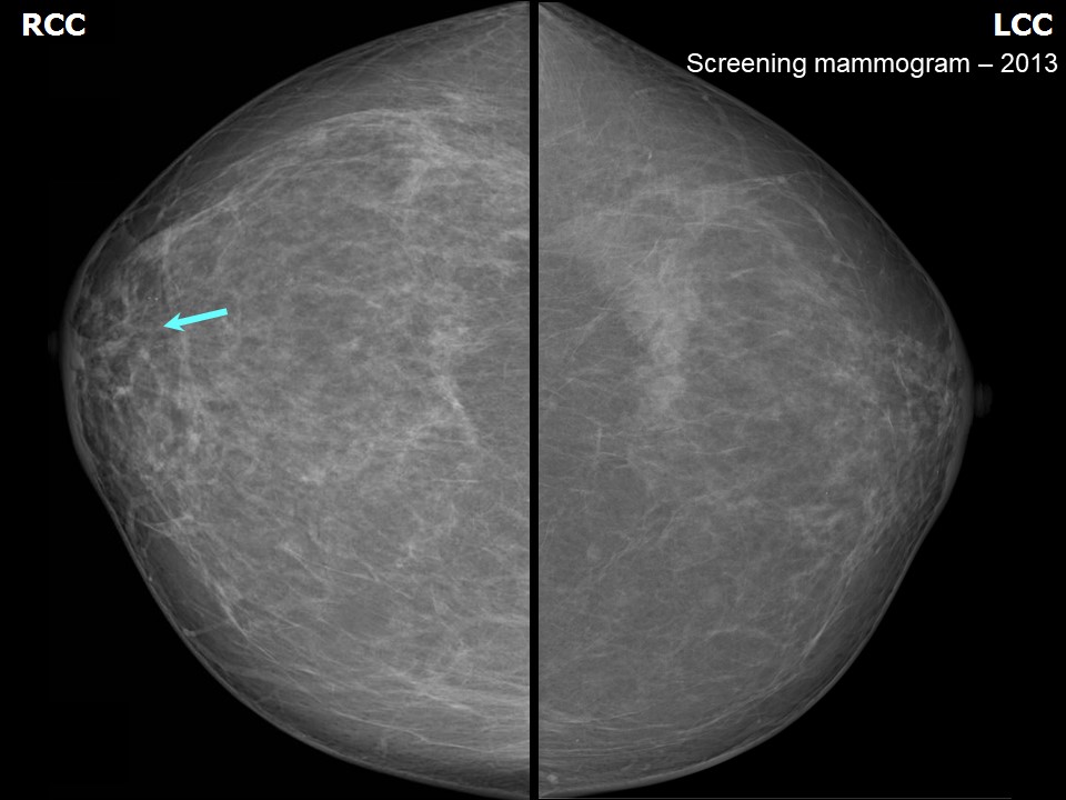 | 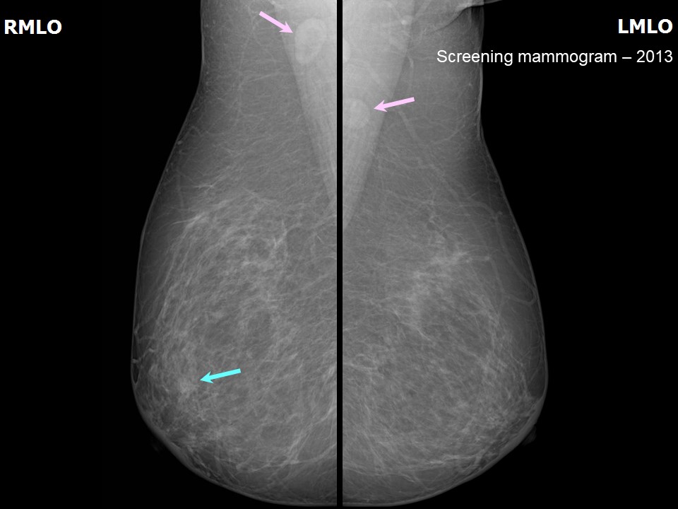 |
| Breast composition: | ACR category a (the breasts are almost entirely fatty) | Mammography features: |
| ‣ Location of the lesion: | 2013: Right breast, central portion of the breast, central zone, anterior third |
| ‣ Mass: | |
| • Number: | 1 |
| • Size: | None |
| • Shape: | Irregular |
| • Margins: | Indistinct |
| • Density: | Equal |
| ‣ Calcifications: | |
| • Typically benign: | None |
| • Suspicious: | None |
| • Distribution: | None |
| ‣ Architectural distortion: | None |
| ‣ Asymmetry: | None |
| ‣ Intramammary node: | None |
| ‣ Skin lesion: | None |
| ‣ Solitary dilated duct: | Present |
| ‣ Associated features: | None |
 |  |
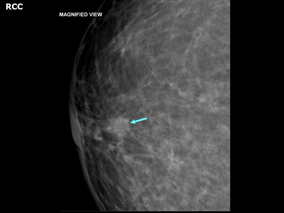 |  |
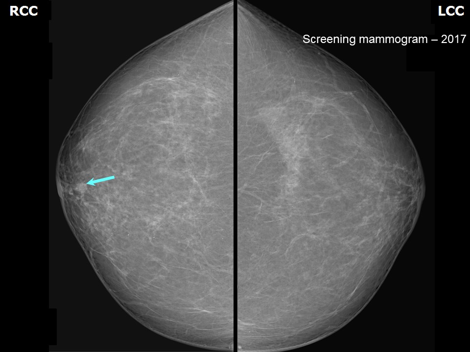
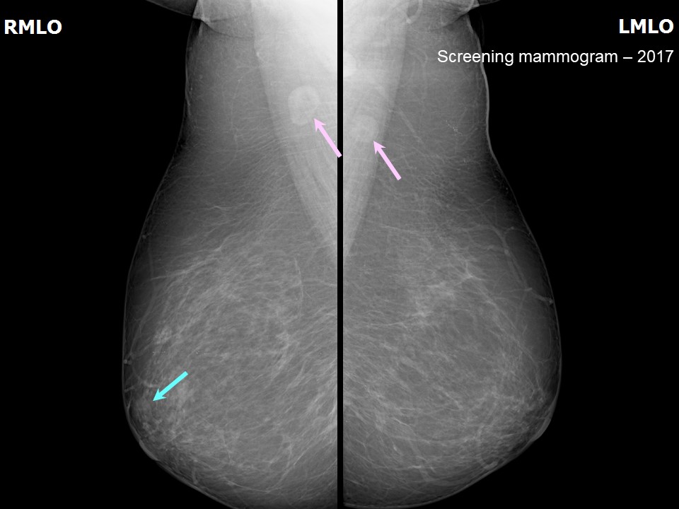
| Breast composition: | ACR category b (there are scattered areas of fibroglandular density) | Mammography features: |
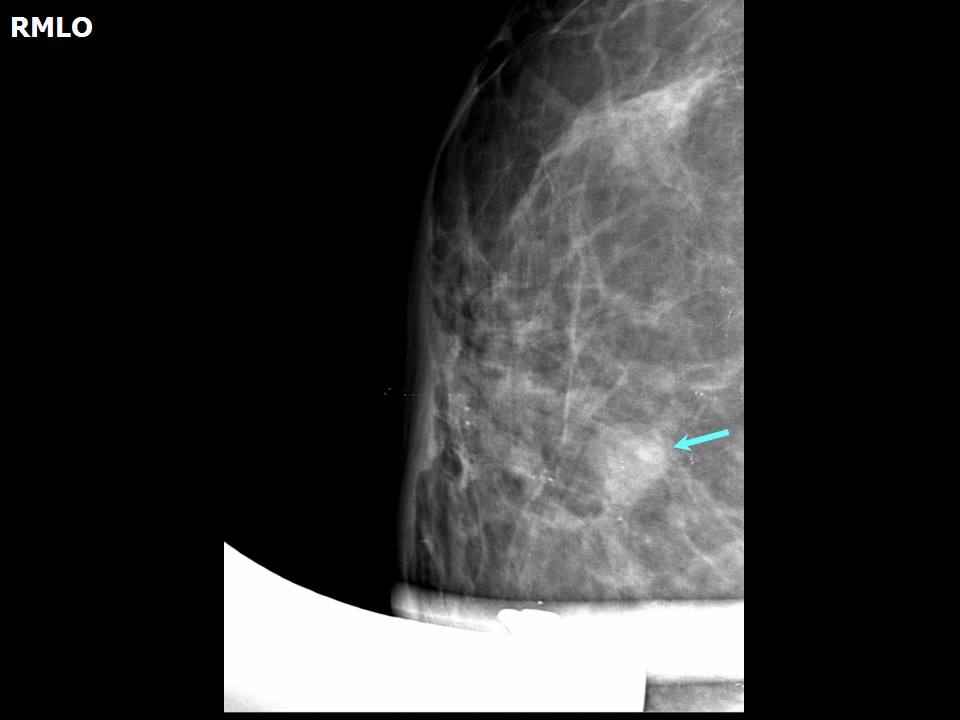
| ‣ Location of the lesion: | 2017: Right breast, central portion of the breast, central zone, anterior third |
| ‣ Mass: | |
| • Number: | 1 |
| • Size: | 0.6 cm |
| • Shape: | Round |
| • Margins: | Circumscribed |
| • Density: | Equal |
| ‣ Calcifications: | |
| • Typically benign: | None |
| • Suspicious: | None |
| • Distribution: | None |
| ‣ Architectural distortion: | None |
| ‣ Asymmetry: | None |
| ‣ Intramammary node: | None |
| ‣ Skin lesion: | None |
| ‣ Solitary dilated duct: | Present |
| ‣ Associated features: | None |
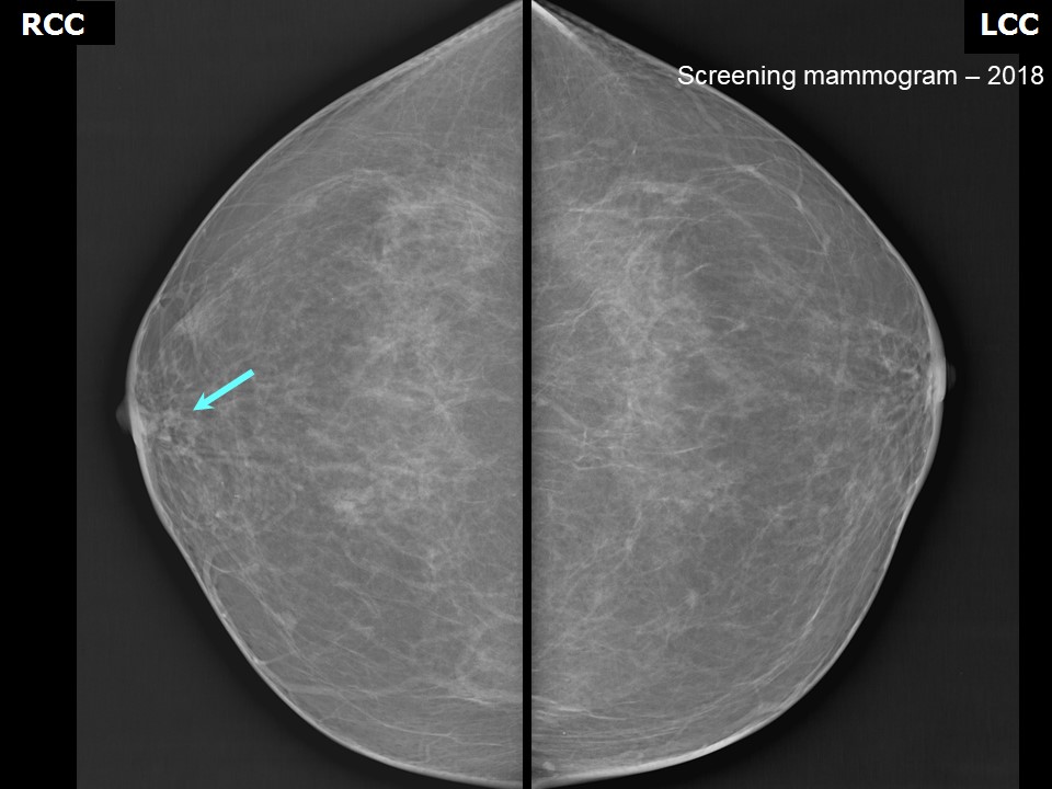 | 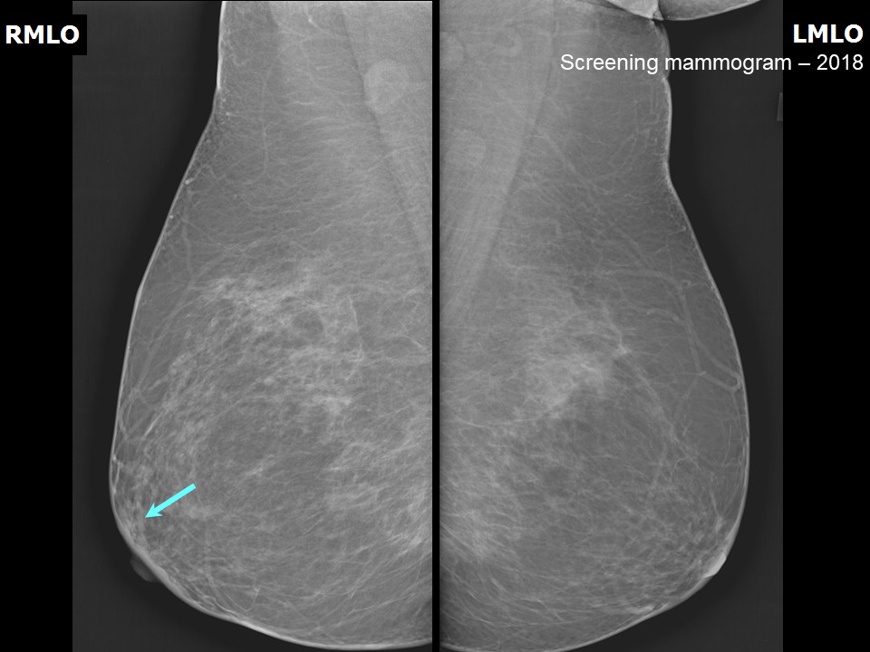 |
| Breast composition: | ACR category b (there are scattered areas of fibroglandular density) | Mammography features: |
| ‣ Location of the lesion: | 2018: Right breast, central portion of the breast, central zone, anterior third |
| ‣ Mass: | |
| • Number: | 0 |
| • Size: | None |
| • Shape: | None |
| • Margins: | None |
| • Density: | None |
| ‣ Calcifications: | |
| • Typically benign: | None |
| • Suspicious: | None |
| • Distribution: | None |
| ‣ Architectural distortion: | None |
| ‣ Asymmetry: | None |
| ‣ Intramammary node: | None |
| ‣ Skin lesion: | None |
| ‣ Solitary dilated duct: | Present |
| ‣ Associated features: | None |
Ultrasound:
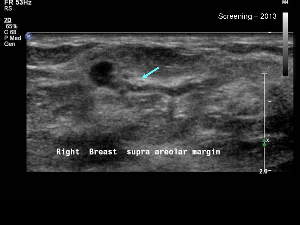 |
| Ultrasound features: 2013: Right breast, upper outer quadrant at 10 o'clock | |
| ‣ Mass | |
| • Location: | 2013: Right breast, upper outer quadrant at 10 o'clock |
| • Number: | 1 |
| • Size: | None |
| • Shape: | None |
| • Orientation: | Parallel |
| • Margins: | Circumscribed |
| • Echo pattern: | Hypoechoic |
| • Posterior features: | No posterior features |
| ‣ Calcifications: | None |
| ‣ Associated features: | Solitary dilated duct with bulbous terminal end and internal echoes |
| ‣ Special cases: | Solitary dilated duct |
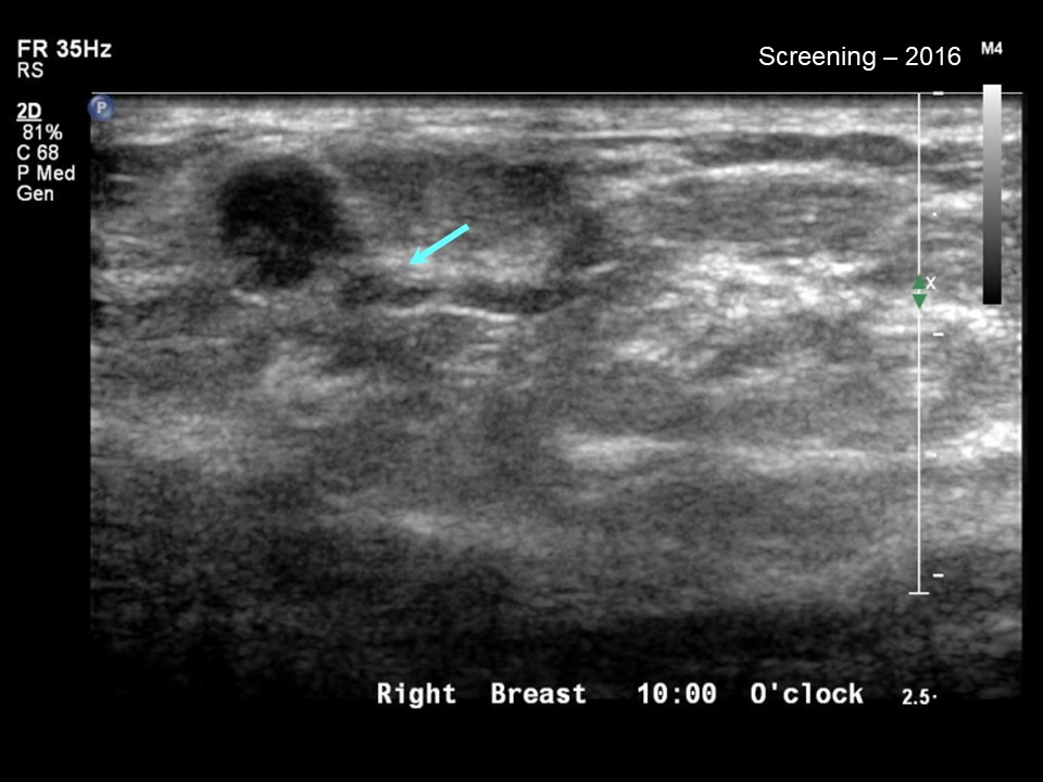 |
| Ultrasound features: 2016: Right breast, upper outer quadrant at 10 o'clock | |
| ‣ Mass | |
| • Location: | 2016: Right breast, upper outer quadrant at 10 o'clock |
| • Number: | 1 |
| • Size: | None |
| • Shape: | None |
| • Orientation: | Parallel |
| • Margins: | Circumscribed |
| • Echo pattern: | Hypoechoic |
| • Posterior features: | No posterior features |
| ‣ Calcifications: | None |
| ‣ Associated features: | Stable solitary dilated duct with bulbous terminal end and internal echoes |
| ‣ Special cases: | Solitary dilated duct |
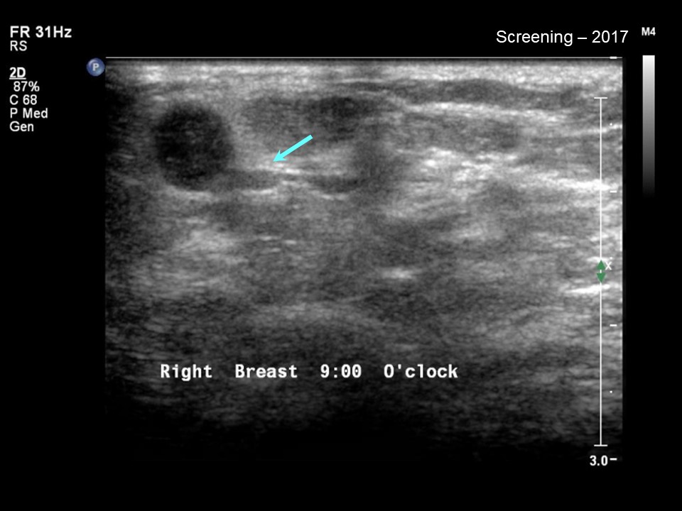 |
| Ultrasound features: 2017: Right breast, outer quadrants at 9 o'clock | |
| ‣ Mass | |
| • Location: | 2017: Right breast, outer quadrants at 9 o'clock |
| • Number: | 1 |
| • Size: | None |
| • Shape: | None |
| • Orientation: | Parallel |
| • Margins: | Circumscribed |
| • Echo pattern: | Hypoechoic |
| • Posterior features: | No posterior features |
| ‣ Calcifications: | None |
| ‣ Associated features: | Stable solitary dilated duct with bulbous terminal end and internal echoes |
| ‣ Special cases: | Solitary dilated duct |
BI-RADS:
BI-RADS Category (2013): 3 (probably benign)BI-RADS Category (MRI 2014): 3 (probably benign)
BI-RADS Category (MRI 2017 and Mammography 2018): 2 (benign)
Further assessment:
Further assessment advised: Further imaging with breast MRIMRI:
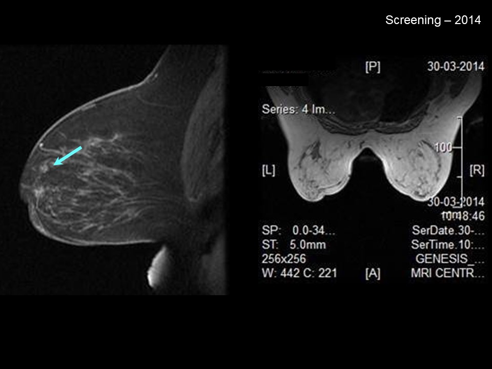 |
| MRI features: | ||
| ‣ MRI features: | MRI 2014: Amount of fibroglandular tissue: ACR category a (the breasts are almost entirely fatty). Background parenchymal enhancement: Minimal (< 25%), symmetrical | |
| ‣ Location: | Right breast, upper quadrant | |
| ‣ Focus: | No | |
| ‣ Mass: | ||
| • Shape: | Irregular | |
| • Margin: | Indistinct | |
| • Internal enhancement: | Heterogeneous | |
| • Kinetic curve: | No | |
| ‣ Non-mass enhancement: | ||
| • Distribution: | No | |
| • Internal enhancement: | No | |
| ‣ Non-enhancing findings: | No | |
| ‣ Associated features: | No | |
| ‣ Axillary nodes: | No | |
Cytology:
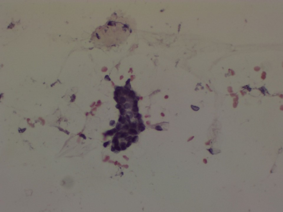 |
| Cytology features: | |
| ‣ Type of sample: | FNAC |
| ‣ Site of biopsy: | |
| • Laterality: | Right |
| • Quadrant: | Non-palpable lesion seen on ultrasound at 9 oclock |
| • Localization technique: | Ultrasound-guided |
| • Nature of aspirate: | Fatty material |
| ‣ Cytological description: | Smear reveals blood, a single cluster of benign ductal cells, and a few fragments of adipose tissue |
| ‣ Reporting category: | Insufficient material |
| ‣ Diagnosis: | Insufficient. Core biopsy advised |
| ‣ Comments: | None |
Case summary:
| Premenopausal woman with increased risk of developing breast cancer presented for routine screening, diagnosed as solitary dilated duct in right breast of suspicious morphology on initial assessment, BI-RADS category 3 on imaging. Regular follow-up for more than 5 years in view of family risk in many relatives, diagnosed as stable unchanged solitary dilated duct in right breast, BI-RADS category 2 on imaging. |
Learning points:
|