English
Français
Home
Research projects
Breast cancer
Cancer control
Cervical cancer
Colorectal cancer
Oral cancer
Training
Manuals
eLearning courses
Digital learning series
Video tutorials
Other useful screening videos
Audio presentations
Quick clinical reference charts
Online library
SCR publications
Scientific papers
Collaborators
About the group
Visitors and news
Menu
Home
Research projects
Cervical cancer
Breast cancer
Oral cancer
HPV vaccine
Training materials
Manuals
eLearning courses
Digital learning series
Video tutorials
Other useful screening videos
Audio presentations
Quick clinical reference charts
Online Library
SCR publications
Scientific papers
Collaborators
About the group
Visitors and news
Foreword
Introduction
Breast anatomy
Introduction
Gross anatomy
Breast tissue
Microscopic anatomy
Vascular and lymphatic supply
Development of the breast
Changes in the breast with ageing
Clinical breast examination (CBE)
Breast cancer screening versus early diagnosis
Performing a breast examination
Practical tips when performing breast examinations
Common abnormalities
Interpretation: normal/negative cases
Interpretation: abnormal/positive cases
Documentation
Pitfalls and limitations
Breast imaging
Introduction
Evolution of breast imaging
Techniques
Principles of mammography
Mammography technique
Mammography unit
Basic functioning
Mammography procedure
Scheduling a woman for mammography
Obtaining mammographic images
Procedural steps
Right MLO view
Right CC view
Left MLO view
Left CC view
Technical adequacy of acquired mammographic views
Mediolateral oblique (MLO) view
Craniocaudal (CC) view
Additional mammographic views
Introduction
Spot compression magnification view
90 degrees lateral views
Exaggerated craniocaudal view
Cleavage view
Axillary tail view
Lumpectomy specimen mammography
Pitfalls
Incorrect MLO positioning
Inadequate breast compression
Screen artefacts on mammographic image
Overstretched RCC and RMLO views
Opacities from body parts causing artefacts in the examination field
Breast tissue inadequately pulled onto the image receptor
Image processing
Mammography interpretation
Introduction
Getting organized
Understanding the normal mammogram and the physiological variations
Normal mammogram
Physiological variations
Breast variations with age
Interpreting the abnormal mammogram
Mammography lexicon
Mammography lexicon
Breast composition
Mass
Calcifications
Calcifications
Typically benign calcifications
Suspicious morphology
Distribution of calcifications
Change over time
Artefacts
Architectural distortion
Asymmetry
Special cases
Associated features
Breast ultrasound
Introduction
Indications
Equipment: Basic components and functioning
Technique
Getting ready
Procedural steps
Benefits
Limitations
Learning breast ultrasound
Normal anatomy
Breast changes in special stages of life
Breast changes in special stages of life (2)
Interpreting the abnormalities
Ultrasound lexicon
Ultrasound lexicon
Breast tissue composition
Breast masses
Breast masses
Shape
Orientation
Margin
Echo pattern
Posterior features
Calcifications
Associated features
Associated features
Architectural distortion
Oedema
Duct changes
Skin changes
Vascularity
Special cases
Special cases
Cysts
Mass in skin
Foreign body
Lymph nodes – Intramammary
Lymph nodes – Axillary
Postsurgical fluid collection
Fat necrosis
Ultrasound-guided interventional procedures
Advantages
Fine-needle aspiration cytology (FNAC)
Core needle biopsy of the breast
Reporting and final assessment categories
Standard reporting of breast imaging
Report template
Final assessment categories
BI-RADS 0
BI-RADS 1
BI-RADS 2
BI-RADS 3
BI-RADS 4
BI-RADS 5
BI-RADS 6
Breast pathology
Introduction
Cytopathology of the breast
Procedure for palpable breast masses
FNAC procedure
Equipment
Getting ready
Locating the palpable breast lump
Aspiration
Smear preparation technique
Fixation of the smears
Staining procedure
Additional points to remember in breast FNAC procedure
Reporting terminology
Histopathology of the breast
Specimen types
Processing of tissue for histopathology
Specimen transport
Specimen grossing
Grossing of excision biopsy specimens
Localization of lesion with mammography
Histological techniques
Histopathology reporting format
Description and significance of the components of the report
Histological grade
Histological subtype
Tumour size
Margins
Features of DCIS
Lymphovascular invasion
Immunohistochemistry for biomarkers
Pathological staging
Case studies
BI-RADS 1
ACR category a
ACR category b
ACR category c
ACR category d
BI-RADS 2
Cystic
Simple cyst
Complicated cyst
Solid
Fibroadenoma
Phyllodes
Adenosis
Fat-containing solid
Lipoma
Hamartoma
Galactocele
Calcified benign mass
Involuting fibroadenoma
Calcifications – typically benign
Skin
Vascular
Coarse or popcorn-like
Large rod-like
Round
Rim
Dystrophic
Milk of calcium
Suture
Diffusely scattered calcifications bilaterally
Miscellaneous
Trauma
Infection
Inflammatory
Granulomatous mastitis
Intramammary node
Fibrocystic changes
Accessory breast
Fat necrosis
Oil cyst
BI-RADS 3
BI-RADS 3
BI-RADS 4
4A-Low suspicion for malignancy
4B-Moderate suspicion for malignancy
4C-High suspicion for malignancy
BI-RADS 5
IBC-Microcalcifications
IBC-Skin thickening
IBC-Skin retraction
IBC-Nipple retraction
IBC-Architectural distortion
IBC-Spiculated margin
IBC-Indistinct margin
IBC-Obscured margin
IBC-Microlobulated margin
IBC-Metaplastic carcinoma
IBC-Nipple erosion
IBC–Occult primary in breast with nodal metastases
IBC with metastatic intramammary node
IBC with coarse heterogeneous calcifications
IBC with Paget disease
IBC with neuroendocrine differentiation
IBC in neurofibromatosis
Encapsulated papillary carcinoma
Multifocal
Multicentric
Invasive cribriform carcinoma
Infiltrating lobular carcinoma
Inflammatory carcinoma
Malignant phyllodes
Medullary carcinoma
Mucinous carcinoma
Bilateral synchronous carcinoma
Calcification of suspicious morphology
BI-RADS 6
BI-RADS 6
Miscellaneous cases
Post-operative breast
Developing asymmetries
Screen-positive
TRAM flap
Metastatic breast
Quiz
Acknowledgements
Authors
Suggested citation
Copyright
Home
/
Training
/
Manuals
/
Atlas of breast cancer early detection
/ Quiz
Atlas of breast cancer early detection
Filter by language:
English
/
Русский
Quiz
Case number:
151
Age:
56
Clinical presentation:
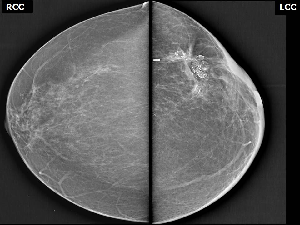
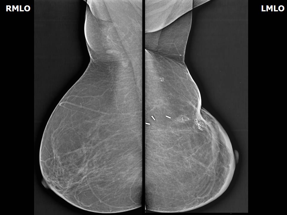
Postmenopausal woman with increased risk of developing breast cancer presented for postoperative surveillance. She had undergone left breast-conserving surgery for invasive carcinoma and also has a family history of breast carcinoma.
Mammography:
Mammography assessment:
Mammography features:
Breast composition:
----
ACR category a (the breasts are almost entirely fatty)
ACR category b (there are scattered areas of fibroglandular density)
ACR category c (the breasts are heterogeneously dense, which may obscure small masses)
ACR category d (the breasts are extremely dense, which lowers the sensitivity of mammography)
‣ Location of the lesion:
• Laterality:
----
None
Bilateral breasts
Left breast
Right breast
Bilateral axillae
Left axilla
Right axilla
• Quadrant:
----
None
All quadrants
Central portion of the breast
Lower inner quadrant
Lower outer quadrant
Upper inner quadrant
Upper outer quadrant
Inner quadrants
Lower quadrants
Outer quadrants
Upper quadrants
Lower and outer quandrants
All quadrants (left), lower quadrants (right)
• O'clock:
01:
02:
03:
04:
05:
06:
07:
08:
09:
10:
11:
12:
Central zone:
• Zone:
----
None
Anterior third
All zones
Anterior and middle thirds
Middle third
Middle and posterior thirds
Posterior third
‣ Mass:
• Number:
----
0
1
2
3
?????????????
• Size:
----
None
Less than 1 cm
More than 1 cm
• Shape:
----
None
Focal asymmetry
Indistinct
Irregular
Linear dilated duct
Oval
Round
Round to oval
• Margins:
----
None
Angular
Circumscribed
Spiculated
Indistinct
Partly circumscribed and partly obscured
Microlobulated
Obscured
Partly circumscribed and partly indistinct
• Density:
----
None
Calcified mass
Equal
Equal with dense calcifications
Equal with dense macrocalcification
Equal with fat-containing areas
Fat-containing
High
‣ Calcifications:
• Typically benign:
⁃ Skin:
----
None
Present
⁃ Vascular:
----
None
Present
⁃ Coarse or “popcorn-like”:
----
None
Present
⁃ Large rod-like:
----
None
Present
⁃ Round:
----
None
Present
⁃ Rim:
----
None
Present
⁃ Dystrophic:
----
None
Present
⁃ Milk of calcium:
----
None
Present
⁃ Suture:
----
None
Present
• Suspicious:
⁃ Amorphous:
----
None
Present
⁃ Coarse heterogeneous:
----
None
Present
⁃ Fine pleomorphic:
----
None
Present
⁃ Fine linear or fine-linear branching:
----
None
Present
• Distribution:
⁃ Diffuse or scattered:
----
None
Present
⁃ Regional:
----
None
Present
⁃ Grouped or clustered:
----
None
Present
⁃ Linear:
----
None
Present
⁃ Segmental:
----
None
Present
‣ Architectural distortion:
----
None
Present
‣ Asymmetry:
----
None
Present
Present, Developing asymmetry
Present, Focal
Present, Global
‣ Intramammary node:
----
None
Present
Present, enlarged
Present, reactive
‣ Skin lesion:
----
None
Present
‣ Solitary dilated duct:
----
None
Present
‣ Associated features:
• Skin retraction:
----
None
Present
• Post-operative skin retraction:
----
None
Present
• Skin thickening:
----
None
Present
• Nipple retraction:
----
None
Present
• Trabecular thickening:
----
None
Present
• Architectural distortion:
----
None
Present
• Calcifications:
----
None
Present
• Axillary lymphadenopathy:
----
None
Present
Case summary:
BI-RADS:
----
None
1 (negative)
2 (benign)
3 (probably benign)
4 (suspicious)
4A (low level of suspicion for malignancy)
4B (moderate suspicion of malignancy)
4C (high suspicion for malignancy)
5 (highly suggestive of malignancy)
6 (known biopsy-proven malignancy)
Further assessment advised:
----
None
Referral for cytology
Referral for cytology and for core biopsy
Referral for cytology, for core biopsy and further imaging with breast MRI
Referral for cytology and for excision biopsy
Referral for core biopsy
Referral for core biopsy and further imaging with breast MRI
Referral for excision biopsy
Further imaging with breast MRI