


Home / Training / Manuals / Atlas of breast cancer early detection / Cases
Atlas of breast cancer early detection
Filter by language: English / Русский
Go back to the list of case studies
.png) Click on the pictures to magnify and display the legends
Click on the pictures to magnify and display the legends
| Case number: | 121 |
| Age: | 52 |
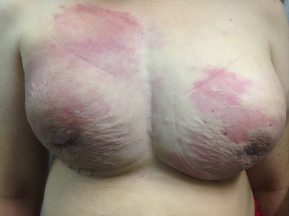
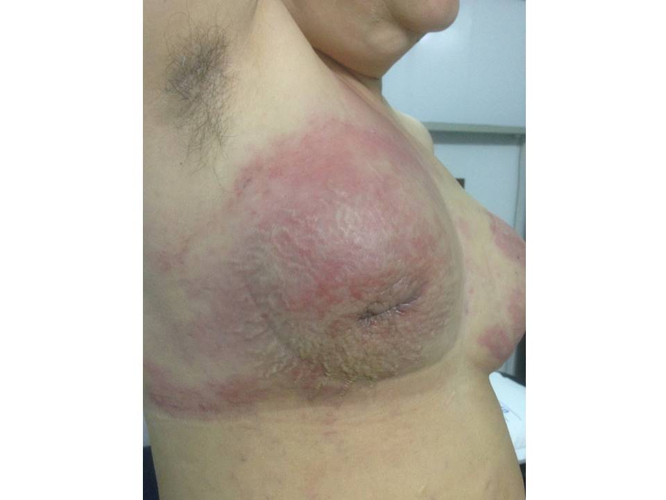
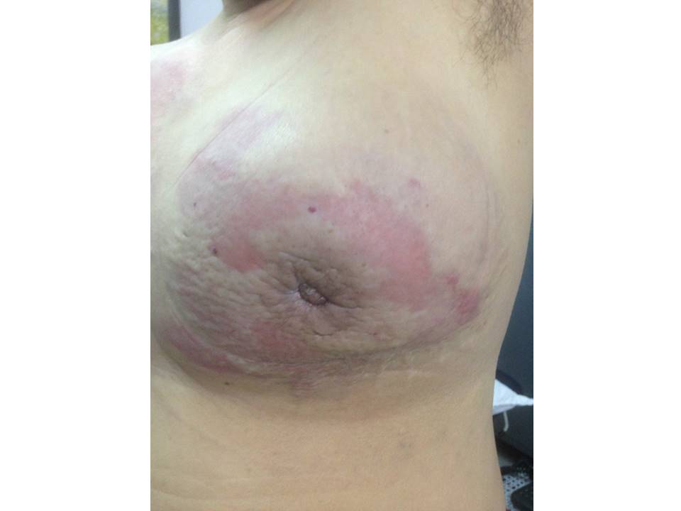
| Clinical presentation: | Postmenopausal woman presented with bilateral breast enlargement and redness of skin over both breasts. Examination revealed large hard lumps and inflammatory changes on the overlying skin in both breasts. |
 |  |
 |
Mammography:
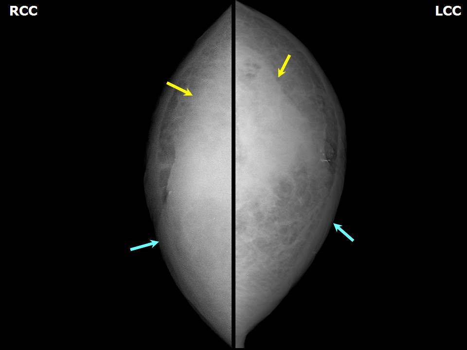 | 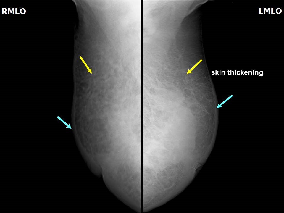 |
| Breast composition: | ACR category d (the breasts are extremely dense, which lowers the sensitivity of mammography) | Mammography features: |
| ‣ Location of the lesion: | Bilateral breasts extremely high density limits evaluation |
| ‣ Mass: | |
| • Number: | 0 |
| • Size: | Not seen |
| • Shape: | None |
| • Margins: | None |
| • Density: | None |
| ‣ Calcifications: | |
| • Typically benign: | None |
| • Suspicious: | None |
| • Distribution: | None |
| ‣ Architectural distortion: | Present |
| ‣ Asymmetry: | None |
| ‣ Intramammary node: | None |
| ‣ Skin lesion: | None |
| ‣ Solitary dilated duct: | None |
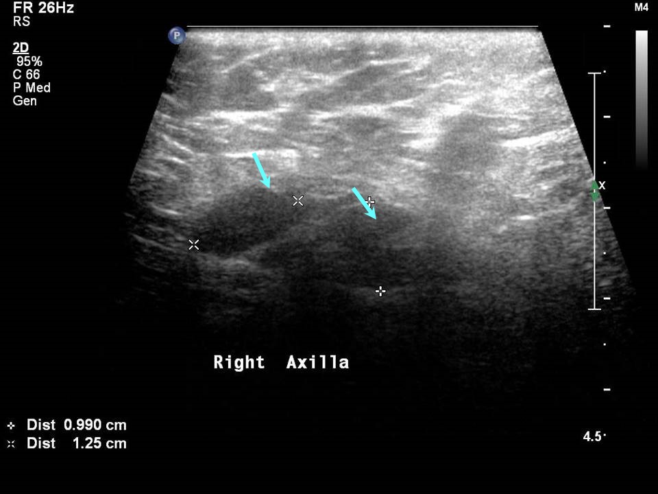
| ‣ Associated features: | Skin thickening, trabecular thickening, architectural distortion, axillary lymphadenopathy, and enlarged nodes with loss of fatty hilum in both axillae |
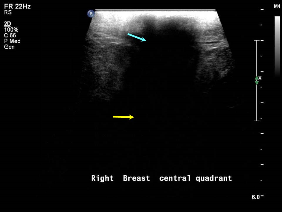
Ultrasound:
 |  |
 |
| Ultrasound features: Right breast, central portion of the breast | |
| ‣ Mass | |
| • Location: | Right breast, central portion of the breast |
| • Number: | 1 |
| • Size: | 3.5 cm in greatest dimension |
| • Shape: | Irregular |
| • Orientation: | Not parallel |
| • Margins: | Angular |
| • Echo pattern: | Hypoechoic |
| • Posterior features: | Strong posterior shadowing |
| ‣ Calcifications: | None |
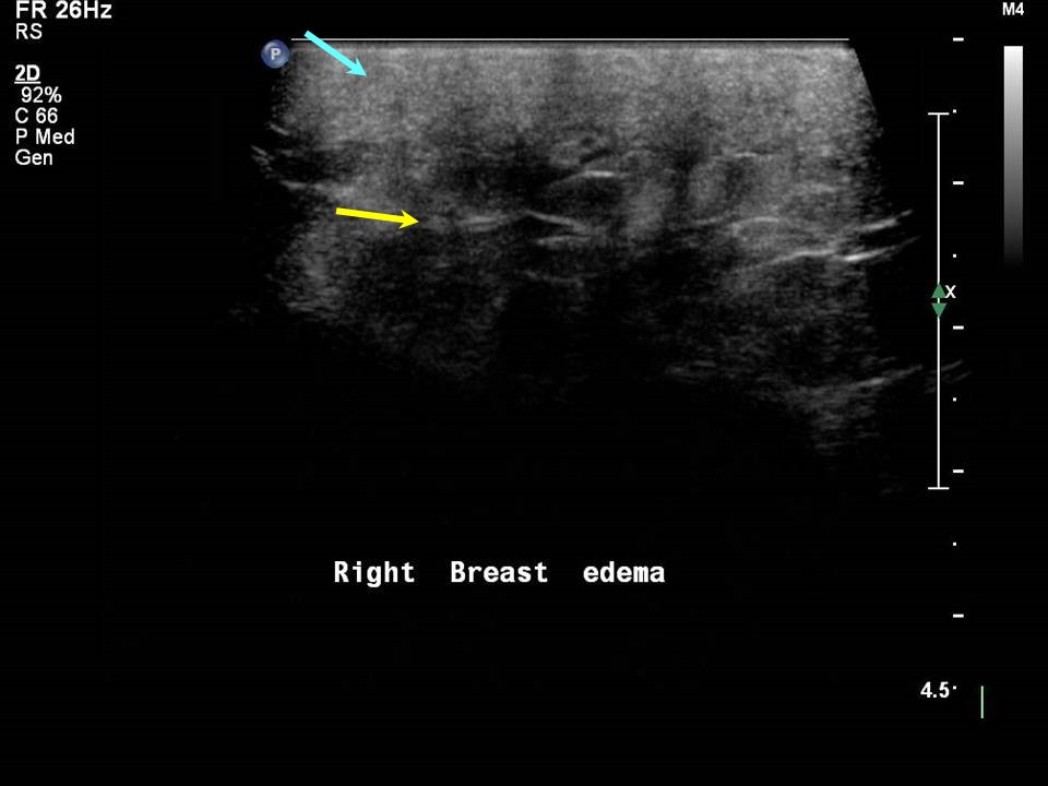
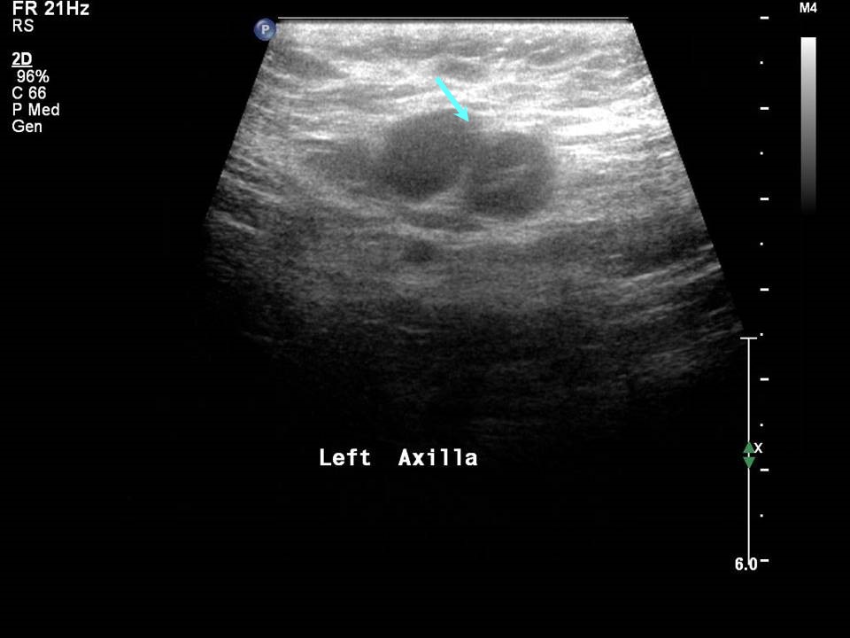
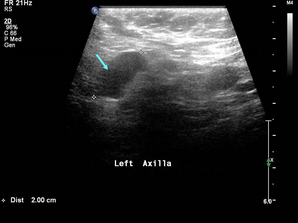
| ‣ Associated features: | Skin thickening, oedema, architectural distortion, and axillary lymphadenopathy |
| ‣ Special cases: | None |
 |  |
 |  |
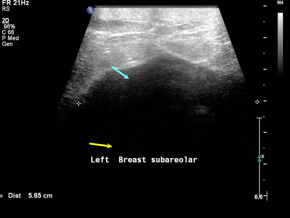
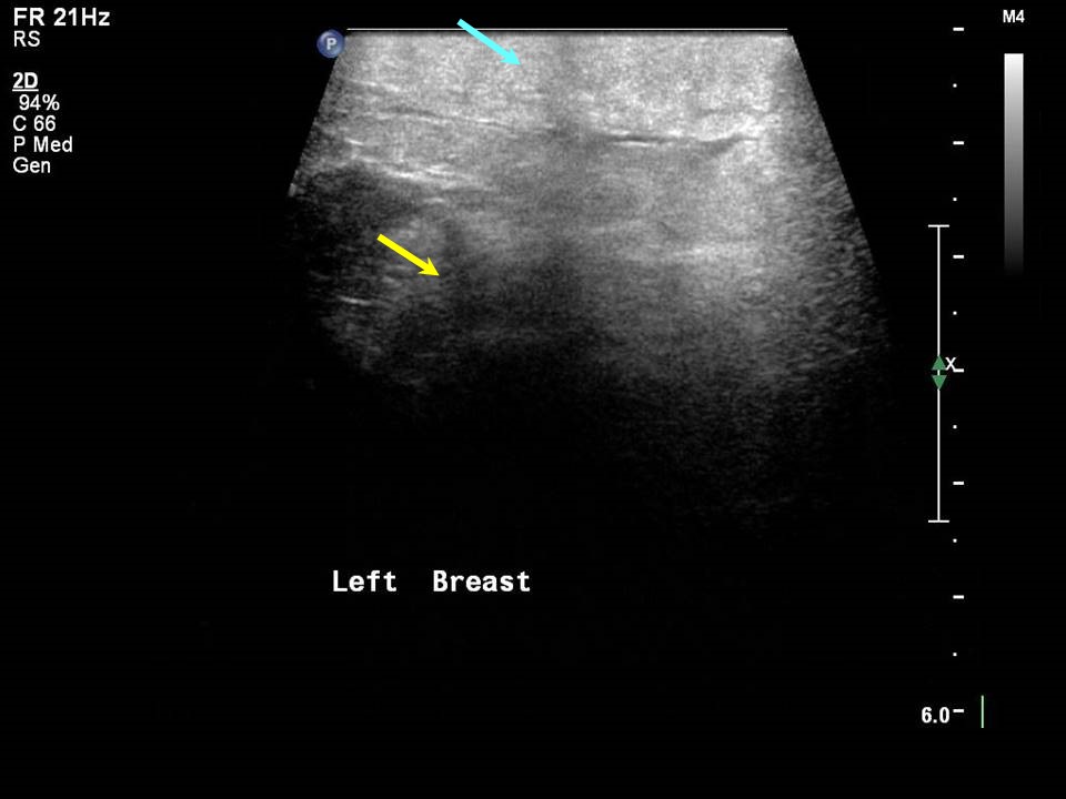
| Ultrasound features: Left breast, central portion of the breast | |
| ‣ Mass | |
| • Location: | Left breast, central portion of the breast |
| • Number: | 1 |
| • Size: | 5.6 cm in greatest dimension |
| • Shape: | Irregular |
| • Orientation: | Not parallel |
| • Margins: | Indistinct |
| • Echo pattern: | Hypoechoic |
| • Posterior features: | Strong posterior shadowing |
| ‣ Calcifications: | None |
| ‣ Associated features: | Skin thickening, oedema, architectural distortion, and axillary lymphadenopathy |
| ‣ Special cases: | None |
BI-RADS:
BI-RADS Category (bilateral): 5 (highly suggestive of malignancy)Further assessment:
Further assessment advised: Referral for cytology and for core biopsyCytology:
 |
| Cytology features: | |
| ‣ Type of sample: | FNAC |
| ‣ Site of biopsy: | |
| • Laterality: | Right |
| • Quadrant: | Subareolar |
| • Localization technique: | Palpation |
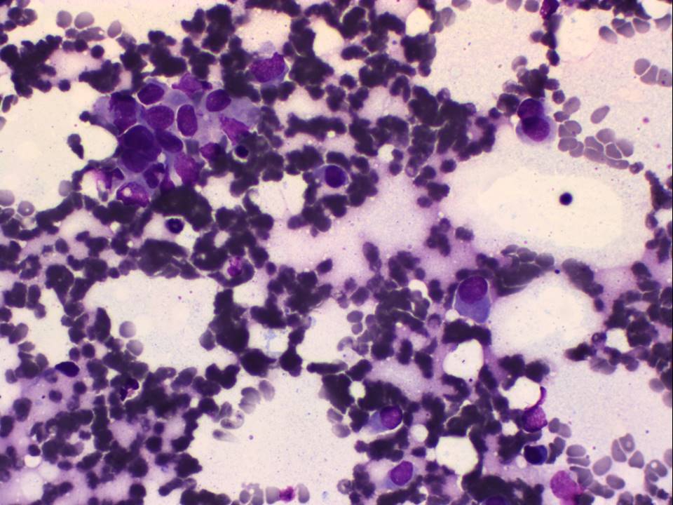
| • Nature of aspirate: | Haemorrhagic |
| ‣ Cytological description: | Smear shows mainly haemorrhage and adipose tissue and a single loosely cohesive group of a few ductal epithelial cells, which are highly suspicious for malignancy |
| ‣ Reporting category: | Suspicious, probably in situ or invasive carcinoma |
| ‣ Diagnosis: | Suspicious for malignancy |
| ‣ Comments: |
 |
| Cytology features: | |
| ‣ Type of sample: | FNAC |
| ‣ Site of biopsy: | |
| • Laterality: | Left |
| • Quadrant: | Subareolar |
| • Localization technique: | Palpation |
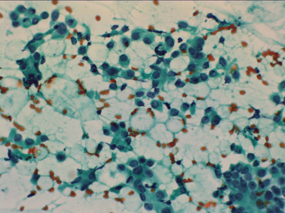
| • Nature of aspirate: | Haemorrhagic |
| ‣ Cytological description: | Smears are haemorrhagic with adipose tissue fragments. A few loosely cohesive cells showing an increased N:C ratio with prominent nucleoli and a moderate amount of cytoplasm are seen |
| ‣ Reporting category: | Malignant |
| ‣ Diagnosis: | Breast carcinoma high grade |
| ‣ Comments: |
Histopathology:
Left breast incision biopsy
| Histopathology features: | |
| ‣ Specimen type: | Left breast incision biopsy |
| ‣ Laterality: | Left |
| ‣ Macroscopy: | Single tissue bit (1.8 × 1.0 cm), submitted entirely |
| ‣ Histological type: | Invasive breast carcinoma of no special type |
| ‣ Histological grade: | Grade 3 (3 + 3 + 2 = 8) |
| ‣ Mitosis: | |
| ‣ Maximum invasive tumour size: | |
| ‣ Lymph node status: | |
| ‣ Peritumoural lymphovascular invasion: | |
| ‣ DCIS/EIC: | |
| ‣ Margins: | |
| ‣ Pathological stage: | |
| ‣ Biomarkers: | ER negative, PR negative, and HER2 positive (score 3+) |
| ‣ Comments: |
Right breast skin biopsy
| Histopathology features: | |
| ‣ Specimen type: | Right breast skin biopsy |
| ‣ Laterality: | Right |
| ‣ Macroscopy: | Skin-covered tissue bit (1.0 × 0.8 cm), submitted entirely |
| ‣ Histological type: | Invasive breast carcinoma of no special type involving the dermis of the skin. Epidermis is free of tumour |
| ‣ Histological grade: | Grade 3 (3 + 3 + 2 = 8) |
| ‣ Mitosis: | |
| ‣ Maximum invasive tumour size: | |
| ‣ Lymph node status: | |
| ‣ Peritumoural lymphovascular invasion: | |
| ‣ DCIS/EIC: | |
| ‣ Margins: | |
| ‣ Pathological stage: | |
| ‣ Biomarkers: | ER negative, PR negative, and HER2 positive (score 3+) |
| ‣ Comments: |
Right breast incision biopsy
| Histopathology features: | |
| ‣ Specimen type: | Right breast incision biopsy |
| ‣ Laterality: | Right |
| ‣ Macroscopy: | Single tissue bit (1.0 × 0.7 cm), submitted entirely |
| ‣ Histological type: | Invasive breast carcinoma of no special type |
| ‣ Histological grade: | Grade 3 (3 + 3 + 2 = 8) |
| ‣ Mitosis: | |
| ‣ Maximum invasive tumour size: | |
| ‣ Lymph node status: | |
| ‣ Peritumoural lymphovascular invasion: | |
| ‣ DCIS/EIC: | |
| ‣ Margins: | |
| ‣ Pathological stage: | |
| ‣ Biomarkers: | ER negative, PR negative, and HER2 positive (score 3+) |
| ‣ Comments: |
Case summary:
| Postmenopausal women presented with bilateral breast enlargement and redness of skin over both breasts. Examination revealed large hard lumps and inflammatory changes on the overlying skin on both breasts. Diagnosed as bilateral breast carcinoma with breast oedema, skin thickening, axillary metastatic lymph nodes, BI-RADS 5 bilateral synchronous on imaging, as bilateral breast carcinoma on cytology, and as invasive breast carcinoma of no special type on incisional biopsies bilaterally. |
Learning points:
|