Home / Training / Manuals / Atlas of breast cancer early detection / Learning

Atlas of breast cancer early detection

Clinical breast examination (CBE) – Performing a breast examination


Clinical breast examination (CBE), also known as physical examination of the breast (PBE), is the systematic clinical examination of both the breasts, axillae and supraclavicular region to detect any abnormalities. Women with symptoms suggestive of breast cancer need to be appropriately evaluated with CBE by a trained primary care provider (e.g., a nurse or a clinician). Depending on the outcomes of CBE, diagnostic imaging (mammography and/or ultrasound of the breast) and FNAC or core biopsy are advised. A combination of CBE, diagnostic imaging and tissue diagnosis is referred to as “triple assessment”, which is also a key step in breast cancer screening pathway as all screen-positive women need to undergo these procedures.

In addition to being a component of triple assessment, CBE is also recognized as a breast cancer screening test that is simple and feasible in limited resource settings. It can be provided by trained nurses or health workers and is less expensive compared with mammography-based screening programmes for populations.

The steps of CBE are the same whether the procedure is performed in the context of screening or as a diagnostic evaluation.

CBE involves the following steps:
  1. Inspection of the breasts to identify physical signs of breast cancer.
  2. Palpation of the breasts and the axillary regions using the finger pads to physically examine all areas of breast tissue and the axillae to identify any lumps or enlarged lymph nodes.


Getting ready
  1. Ensure adequate privacy.
  2. Greet the woman respectfully and with kindness.
  3. Tell the woman why you are going to examine her breasts and explain how you are going to do the examination.
  4. Take a medical history and fill in the requisite forms.
  5. Check for any breast complaints. If she has presented because of concerns about a lump, ask about its location.
  6. Ask the woman to undress from her waist up. Have her sit on the examining table with her arms at her sides.
  7. Wash your hands thoroughly and dry them. If necessary, put on examination gloves on both hands.


It is better to avoid CBE during and just before menstruation as the breasts may be tender.


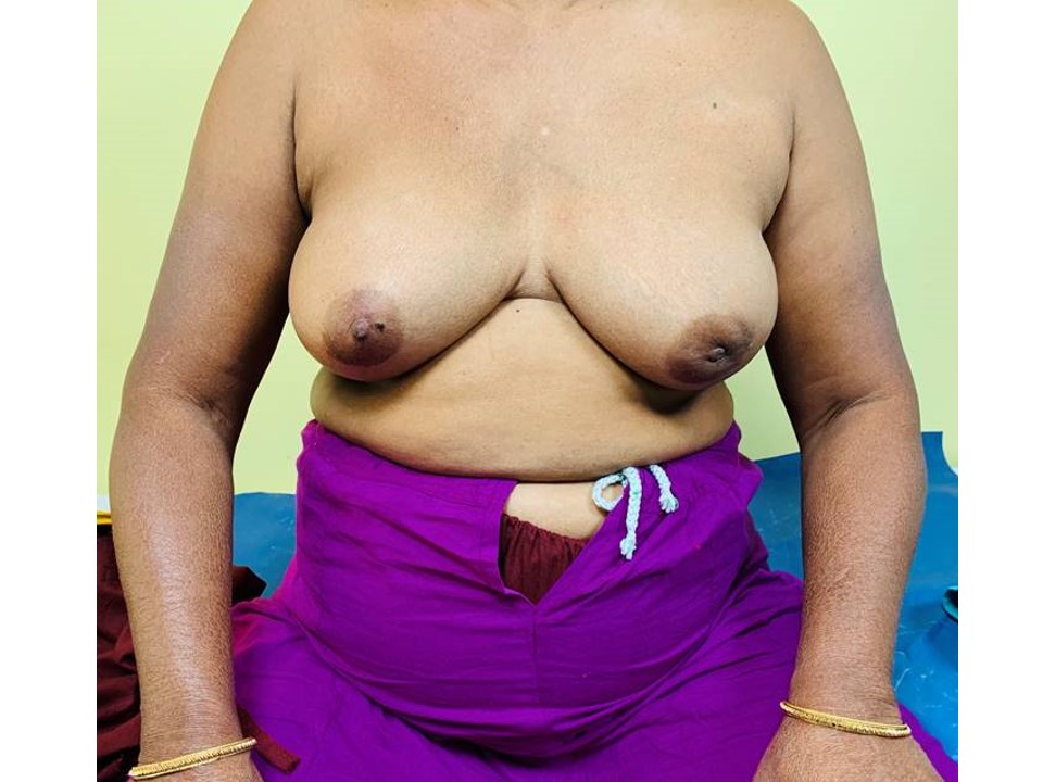
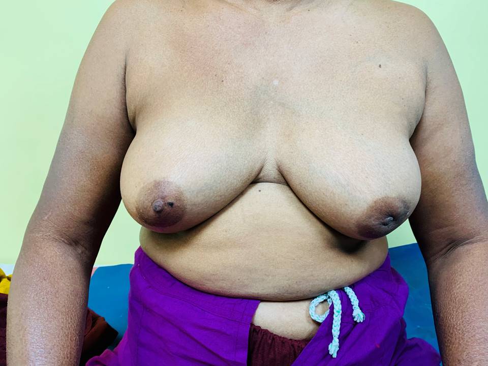
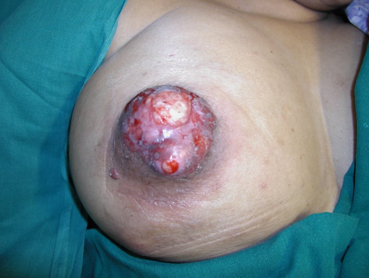
Steps of inspection
1. Inspect both the breasts with the woman sitting comfortably with both arms resting on her thighs.


2. Repeat inspection with the woman pressing her hands into her hips so that the pectoralis muscles are contracted.


3. Ask the woman to place her hands behind her head and lean forward a little. Repeat the inspection.


Look for the following abnormalities while you inspect the breasts:
Symmetry
  • Is there any asymmetry of the size and shape of the breasts?
  • Do the breasts hang evenly?
Skin changes
  • Is there any discoloration or puckering of the skin over the breasts?
  • Is there any localized thickening of the skin? (the thickened skin may have the look of an orange peel with dimpling also known as peau d’orange appearance)
  • Are there any visible scars or sinuses?
  • If there is any puckering of the skin, does it get accentuated when the woman presses her hands into her hips?
  • If there is any skin dimpling or puckering or breast asymmetry, does it get exaggerated as the women raises her arms?
Nipple changes
  • What is the position of the nipples and is there any disparity in the level of the two nipples?
  • Are there any ulcerations or excoriations of the nipple and the areola?
  • Are either of the nipples retracted (pulled inside)?
  • Is there any spontaneous, unilateral, blood-stained discharge from either of the nipples?
Swelling/ulcer
  • Is there any swelling visible in the breast with or without accompanying redness of the overlying skin?
  • Is there any obvious ulcer or swelling visible on the breast?
  • If there is a visible swelling or ulcer, does it move when the pectoralis muscles contract as the women presses her hands into her hips?


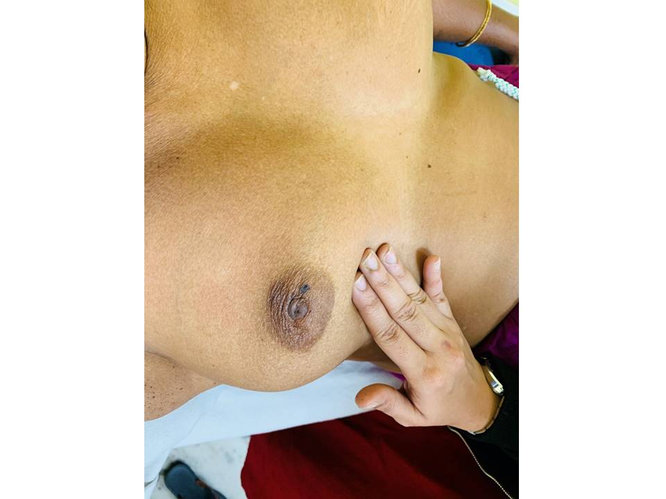
Steps of palpation of the breasts and axillae
Step 1. Palpate the breasts in the lying down position
  1. Ask the woman to lie on her back on the examination table.
  2. Stand on the right side of the patient.
  3. If the woman has breast symptoms, examine the normal breast first.
  4. To examine the left breast first, place a pillow under the patient’s left shoulder and ask her to place her left arm over her head.
  5. Palpate the entire breast using one of the techniques (linear or dial of clock) described below, with the pads of the three middle fingers. Note any lumps or tenderness.
  6. The finger pads of the three middle fingers should be used to palpate the breast in a circular motion.
  7. Palpation pressure: Apply steady pressure, pushing down to the level of the chest wall.
    • Apply enough pressure to palpate to three levels of depth.
    • Apply light pressure for superficial breast tissue.
    • Apply medium pressure for the intermediate layer.
    • Apply deep pressure for tissue close to the chest wall.
  8. Note if there is any discharge from the nipple during the examination.
  9. Change the position of the pillow and ask the woman to place her right hand behind her head to repeat the procedure on the right breast.


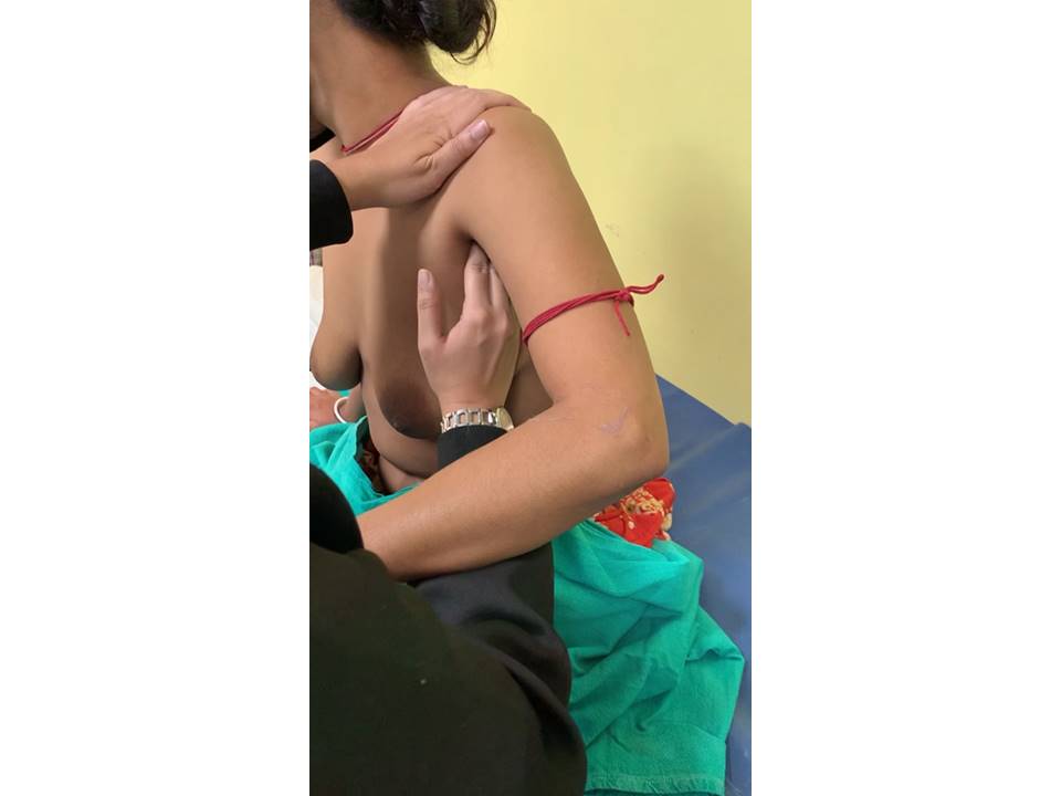
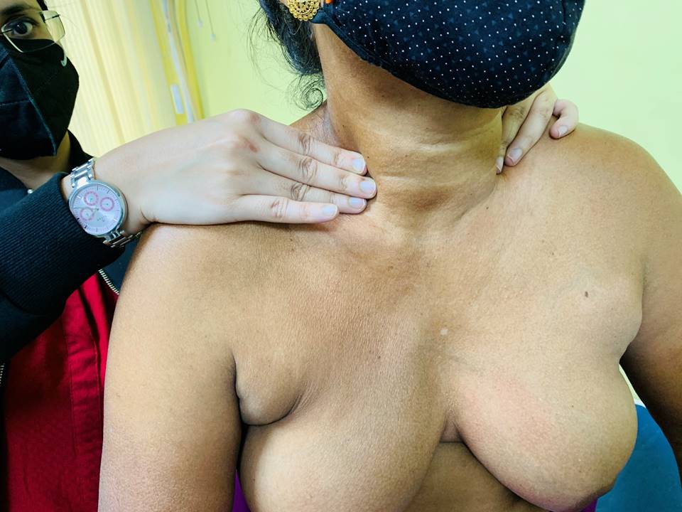
Step 2. Palpate the breasts in the sitting position
  • 1. Ask the woman to sit on the examination table facing you.
  • 2. Palpate the axilla of the breast using the following technique:
    • To examine the left breast and axilla, keep your left arm on the woman's left shoulder to prevent the shoulder from moving up. Allow the woman to rest her left arm on your forearm so as to relax the pectoralis muscles ridge. Then your right hand examines the left axilla of the woman while her arm and pectoralis muscles are placed in a comfortable position.
    • Palpate the lymph nodes by rolling the tissue against the lateral border of the pectoralis major muscle and lateral chest wall.
    • Insert the examining fingers gently towards the neck to palpate the apex of the axilla.


  • 3. Repeat these steps for the right side and use your left hand to examine the right axilla.
  • 4. Examine the supraclavicular areas of both sides to complete the examination.


  • 5. After completing the examination, tell the patient that the examination is now finished and that she can get dressed.
  • 6. Explain the abnormal findings on examination, if any, to the woman. Describe any further course of action that needs to be taken.
  • 7. Reassure her if there are no abnormal findings and show her how she can check the breasts herself for any lump or skin changes or nipple changes from time to time.


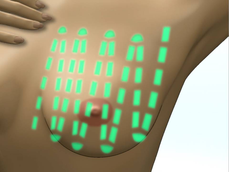
  • Linear method of breast palpation


    Starting from the sternal border, palpate the breast by rolling the tissue against the underlying chest wall in small circles. Cover the breast tissue in multiple strips, starting from under the clavicle down to the inframammary region and move upwards again, placing the fingers slightly laterally overlapping the previous area. The breast tissue is thus covered in multiple strips.


    Dial of clock method of breast palpation

    The breast is visualized as the face of a clock and divided into areas accordingly. The 12 o’clock region is at the top centrally and the 6 o’clock region is at the bottom centre. The breast tissue is rolled in small circles starting from the nipple and radiating outwards. Start at 12 o’clock and continue clockwise or anticlockwise to return back to 12 o’clock. The entire breast tissue is thus systematically covered.


    Palpation of axilla


    Look for the following abnormalities while you palpate the breasts and the axilla:

    • Confirm the findings of inspection.
    • Is there any rise of local skin temperature?
    • Is there tenderness in any part of the breast or axilla?
    • Is there a lump in the breast? If yes, look for: location, size, shape, consistency, mobility, fixity to breast tissue and/or skin and/or chest wall, overlying skin changes.
    • Is there any irregularity/nodularity in the breast that is different from the opposite breast?
    • Are there any palpable lymph nodes in the axillary and supraclavicular areas? What is the feel of the lymph nodes? Are they hard or tender? Are they fixed to each other and/or deeper tissues?


    Readers can download the step-by-step CBE examination checklist, which acts as a ready reckoner in the clinic. The checklist is also useful during a training programme to assess the trainee’s skill.


    Click on the pictures to magnify and display the legends


    Click on this icon to display a case study

    IARC, 150 Cours Albert Thomas, 69372 Lyon CEDEX 08, France - Tel: +33 (0)4 72 73 84 85 - Fax: +33 (0)4 72 73 85 75
    © IARC 2024 - All Rights Reserved.