


Home / Training / Manuals / Atlas of breast cancer early detection / Cases
Atlas of breast cancer early detection
Filter by language: English / Русский
Go back to the list of case studies
.png) Click on the pictures to magnify and display the legends
Click on the pictures to magnify and display the legends
| Case number: | 176 |
| Age: | 61 |
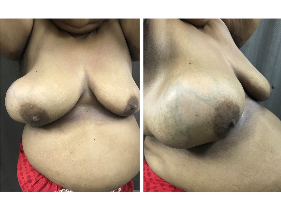
| Clinical presentation: | Postmenopausal woman with average risk of developing breast cancer presented with a right breast lump first noticed more than 10 months ago. It has increased significantly in size since then. |
 |
Mammography:
 |  |
 |
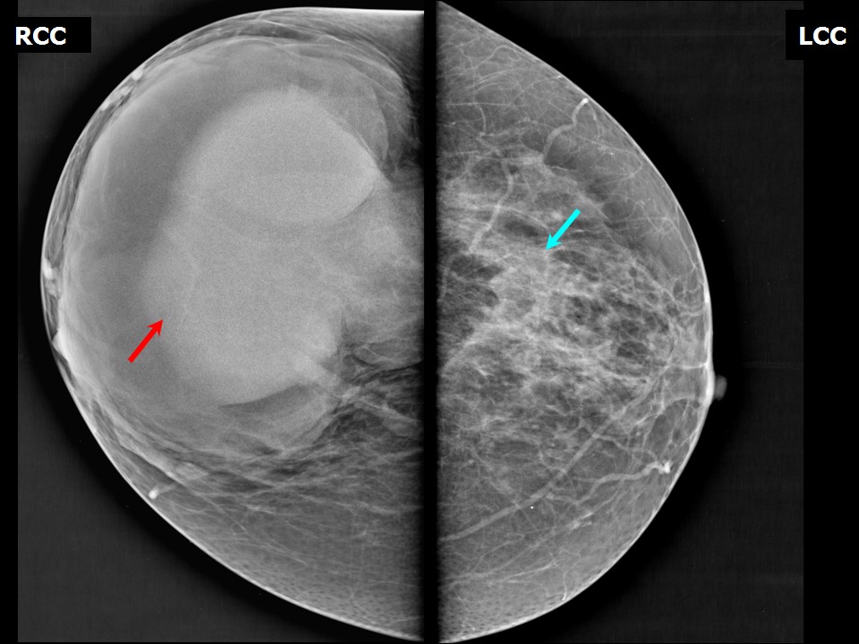
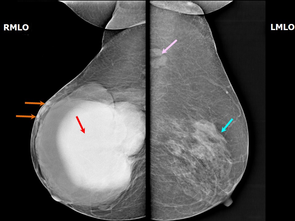
| Breast composition: | ACR category b (there are scattered areas of fibroglandular density) | Mammography features: |
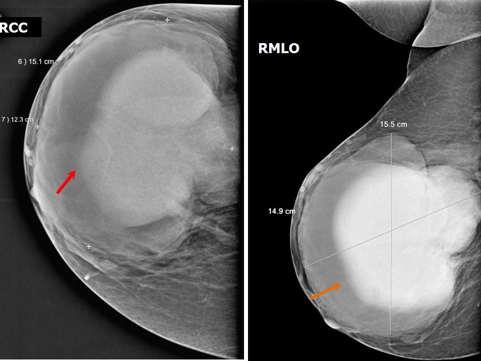
| ‣ Location of the lesion: | Right breast, lower and outer quadrants at 512 oclock, anterior, middle, and posterior thirds |
| ‣ Mass: | |
| • Number: | 1 |
| • Size: | 15.5 × 15.0 × 12.0 cm |
| • Shape: | Oval |
| • Margins: | Circumscribed |
| • Density: | Equal |
| ‣ Calcifications: | |
| • Typically benign: | None |
| • Suspicious: | None |
| • Distribution: | None |
| ‣ Architectural distortion: | None |
| ‣ Asymmetry: | None |
| ‣ Intramammary node: | None |
| ‣ Skin lesion: | None |
| ‣ Solitary dilated duct: | None |
| ‣ Associated features: | None |
Ultrasound:
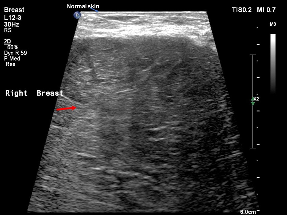 | 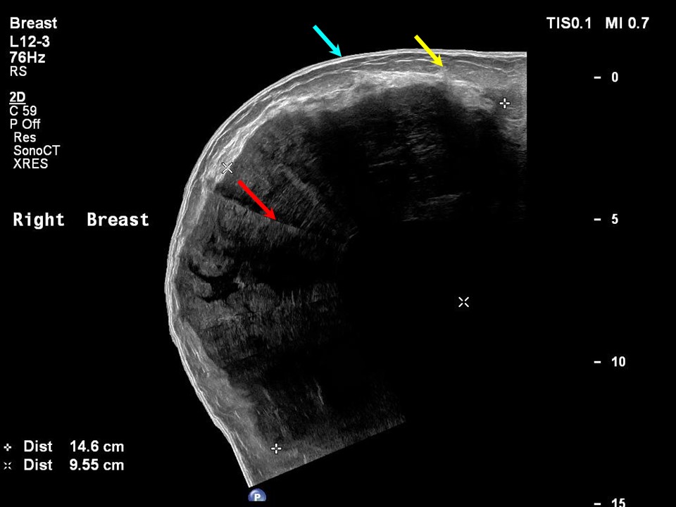 |
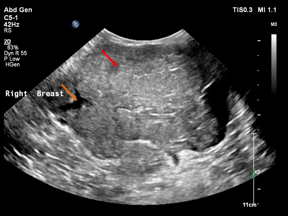 | 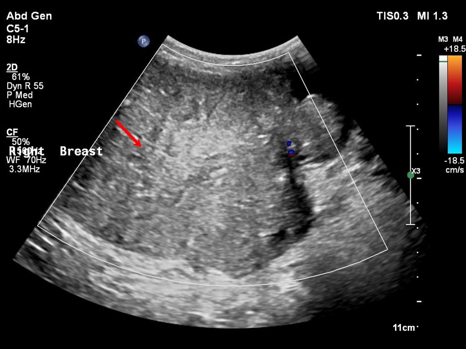 |
| Ultrasound features: Right breast, outer quadrants at 5-12 o'clock | |
| ‣ Mass | |
| • Location: | Right breast, outer quadrants at 5-12 o'clock |
| • Number: | 1 |
| • Size: | 15.0 cm in greatest dimension |
| • Shape: | Oval |
| • Orientation: | Parallel |
| • Margins: | Circumscribed |
| • Echo pattern: | Hypoechoic |
| • Posterior features: | No posterior features |
| ‣ Calcifications: | None |
| ‣ Associated features: | None |
| ‣ Special cases: | None |
BI-RADS:
BI-RADS Category: 4A (low level of suspicion for malignancy)Further assessment:
Further assessment advised: Referral for cytologyCytology:
 |  |
| Cytology features: | |
| ‣ Type of sample: | FNAC |
| ‣ Site of biopsy: | |
| • Laterality: | Right |
| • Quadrant: | Large lump involving all quadrants |
| • Localization technique: | Palpation |
| • Nature of aspirate: | Whitish aspirate admixed with blood |
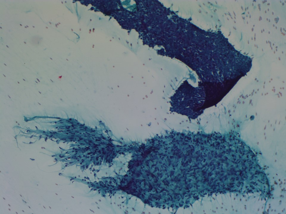
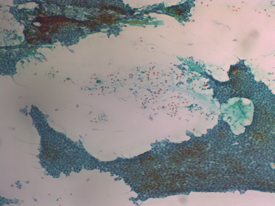
| ‣ Cytological description: | Smears are very cellular. Many flat cohesive sheets of benign ductal epithelial cells, a few bare nuclei, and cellular stromal fragments are seen. The background shows proteinaceous fluid material. Some of the smears also show many acute inflammatory cells. Numerous foamy histiocytes seen in some smears |
| ‣ Reporting category: | Benign |
| ‣ Diagnosis: | Benign proliferative breast lesion with fibrocystic changes. Phyllodes tumour should be considered because of the cellular stromal fragments |
| ‣ Comments: | None |
Histopathology:
.
 |  |
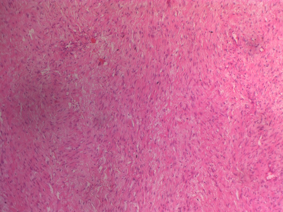 | 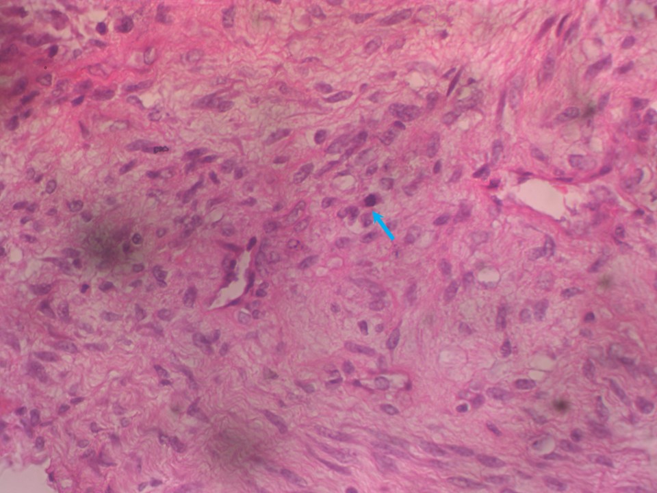 |
| Histopathology features: | |
| ‣ Specimen type: | . |
| ‣ Laterality: | Right |
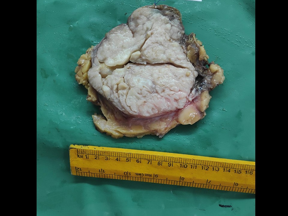
| ‣ Macroscopy: | Specimen (18.0 × 16.0 × 11.0 cm) with overlying skin flap (15.0 × 14.0 cm). The nipple and areola are present and unremarkable. On serial sectioning, a well-circumscribed, encapsulated, grey white, solid, lobulated mass (14.5 × 12.0 × 11.5 cm) is present with numerous clefts involving almost the entire specimen. The tumour is located 1.8 cm below the skin and abutting the base (posterior margin). It is located 0.9 cm from the medial margin, 1.2 cm from the lateral margin, 1.0 cm from the superior margin, and 1.5 cm from the inferior margin |
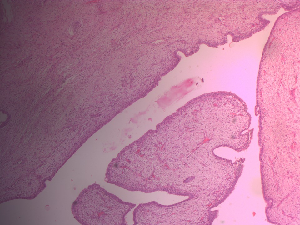
| ‣ Histological type: | Sections reveal a lesion showing proliferation of both epithelial and stromal elements. The stroma appears hypercellular. This stromal hypercellularity is more pronounced around the epithelial components. There is also proliferation of the epithelial components and they are stretched into clefts around the stromal fronds. There are many areas of apocrine metaplasia and UDH. The stromal cells show 56 mitoses per 10 hpf. Impression: Borderline phyllodes tumour |
| ‣ Histological grade: | Borderline phyllodes tumour |
| ‣ Mitosis: | 6 |
| ‣ Maximum invasive tumour size: | 14.5 cm in greatest dimension |
| ‣ Lymph node status: | Not received |
| ‣ Peritumoural lymphovascular invasion: | |
| ‣ DCIS/EIC: | Absent |
| ‣ Margins: | Free of tumour |
| ‣ Pathological stage: | |
| ‣ Biomarkers: | |
| ‣ Comments: |
Case summary:
| Postmenopausal woman presented with right breast lump with recent rapid increase in size. Diagnosed as large circumscribed mass occupying almost entire right breast, likely phyllodes, BI-RADS 4A on imaging, as benign proliferative lesion on cytology, and as phyllodes tumour on histopathology. |
Learning points:
|