


Home / Training / Manuals / Atlas of breast cancer early detection / Cases
Atlas of breast cancer early detection
Filter by language: English / Русский
Go back to the list of case studies
.png) Click on the pictures to magnify and display the legends
Click on the pictures to magnify and display the legends
BI-RADS Category (2012): 2 (benign)
BI-RADS Category (2017): 2 (benign)
| Case number: | 162 |
| Age: | 43 |
| Clinical presentation: | Premenopausal woman with high risk of breast carcinoma because of family history (first-degree relative with bilateral breast carcinoma) presented with a right breast lump. CBE revealed a firm lump in the upper outer quadrant of the right breast. |
Mammography:
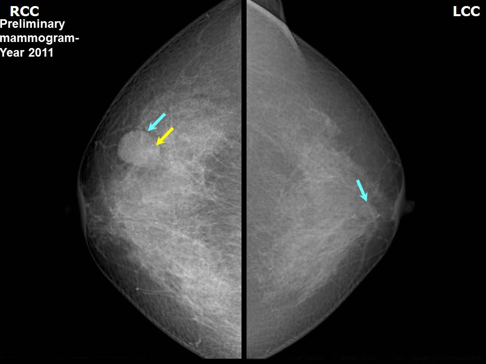 | 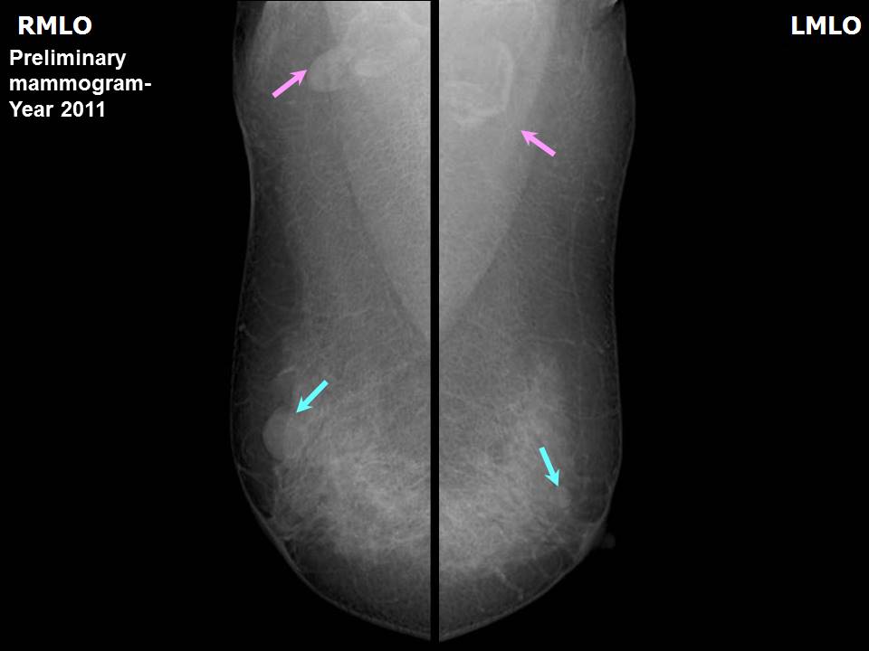 |
| Breast composition: | ACR category b (there are scattered areas of fibroglandular density) | Mammography features: |
| ‣ Location of the lesion: | 2011: Right breast, upper outer quadrant at 11 oclock, anterior third |
| ‣ Mass: | |
| • Number: | 1 |
| • Size: | 1.9 × 1.5 cm |
| • Shape: | Oval |
| • Margins: | Circumscribed |
| • Density: | Equal |
| ‣ Calcifications: | |
| • Typically benign: | None |
| • Suspicious: | None |
| • Distribution: | None |
| ‣ Architectural distortion: | None |
| ‣ Asymmetry: | None |
| ‣ Intramammary node: | None |
| ‣ Skin lesion: | None |
| ‣ Solitary dilated duct: | None |
| ‣ Associated features: | None |
| Mammography features: | |
| ‣ Location of the lesion: | 2011: Left breast, upper inner quadrant at 11 oclock, anterior third |
| ‣ Mass: | |
| • Number: | 1 |
| • Size: | 1.0 cm in greatest dimension |
| • Shape: | Oval |
| • Margins: | Circumscribed |
| • Density: | Equal |
| ‣ Calcifications: | |
| • Typically benign: | None |
| • Suspicious: | None |
| • Distribution: | None |
| ‣ Architectural distortion: | None |
| ‣ Asymmetry: | None |
| ‣ Intramammary node: | None |
| ‣ Skin lesion: | None |
| ‣ Solitary dilated duct: | None |
| ‣ Associated features: | None |
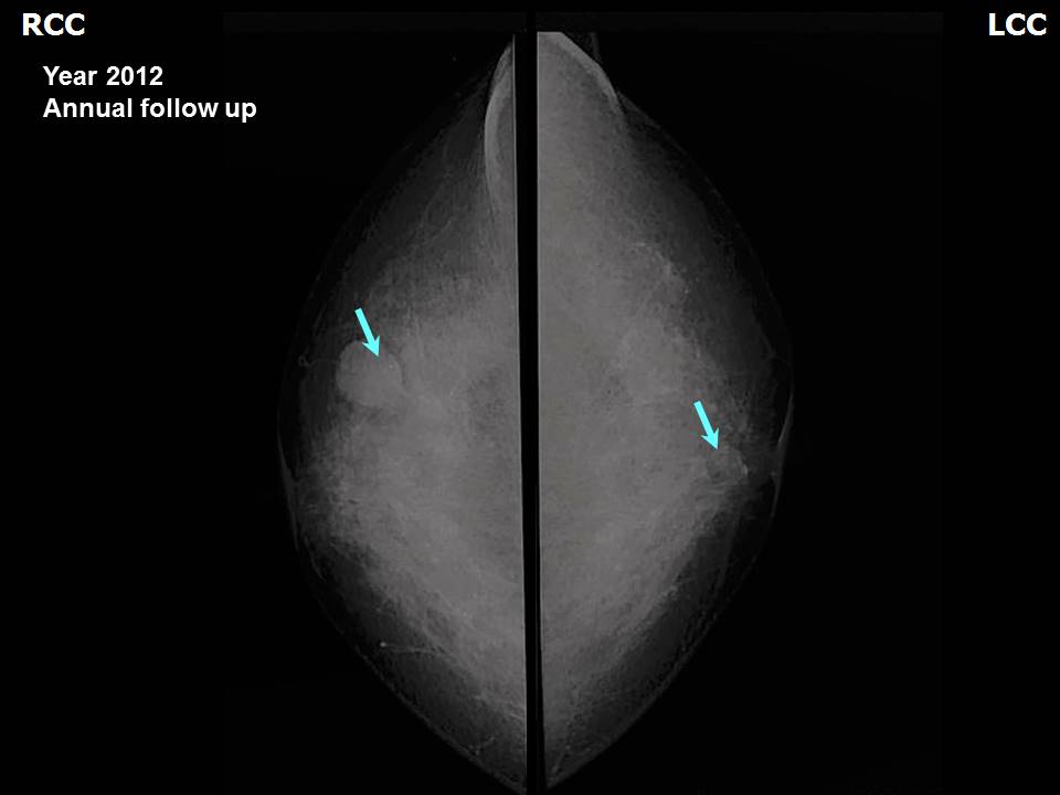 | 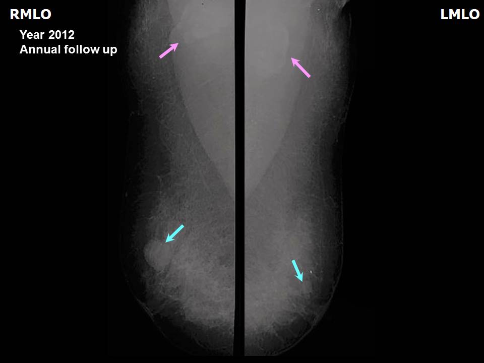 |
| Breast composition: | ACR category b (there are scattered areas of fibroglandular density) | Mammography features: |
| ‣ Location of the lesion: | 2012: Right breast, upper outer quadrant at 11 oclock, anterior third |
| ‣ Mass: | |
| • Number: | 1 |
| • Size: | 1.9 × 1.5 cm |
| • Shape: | Oval |
| • Margins: | Circumscribed |
| • Density: | Equal |
| ‣ Calcifications: | |
| • Typically benign: | None |
| • Suspicious: | None |
| • Distribution: | None |
| ‣ Architectural distortion: | None |
| ‣ Asymmetry: | None |
| ‣ Intramammary node: | None |
| ‣ Skin lesion: | None |
| ‣ Solitary dilated duct: | None |
| ‣ Associated features: | None |
| Mammography features: | |
| ‣ Location of the lesion: | 2012: Left breast, upper inner quadrant at 11 oclock, anterior third |
| ‣ Mass: | |
| • Number: | 1 |
| • Size: | 1.0 cm in greatest dimension |
| • Shape: | Oval |
| • Margins: | Circumscribed |
| • Density: | Equal |
| ‣ Calcifications: | |
| • Typically benign: | None |
| • Suspicious: | None |
| • Distribution: | None |
| ‣ Architectural distortion: | None |
| ‣ Asymmetry: | None |
| ‣ Intramammary node: | None |
| ‣ Skin lesion: | None |
| ‣ Solitary dilated duct: | None |
| ‣ Associated features: | None |
 |  |
 |
| Breast composition: | ACR category b (there are scattered areas of fibroglandular density) | Mammography features: |
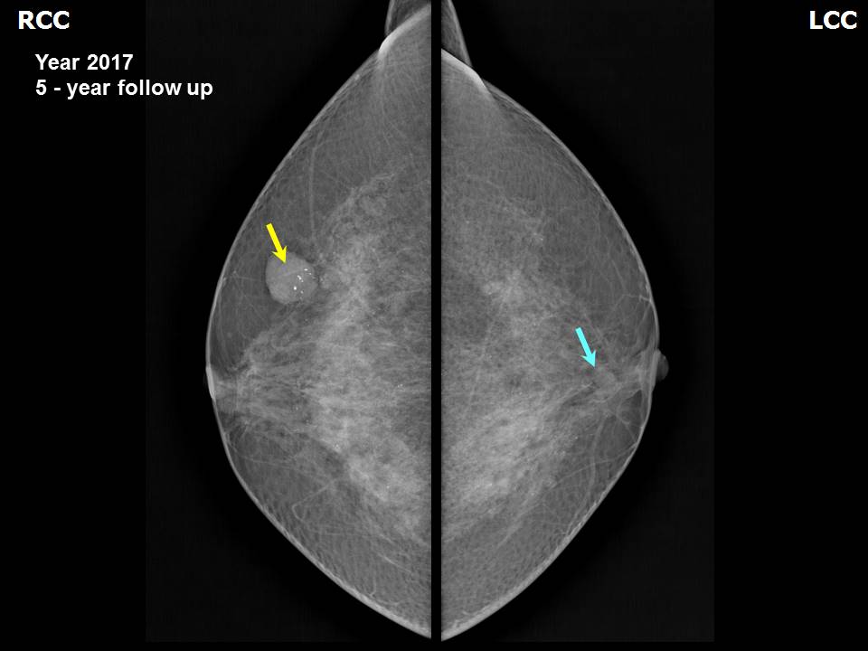
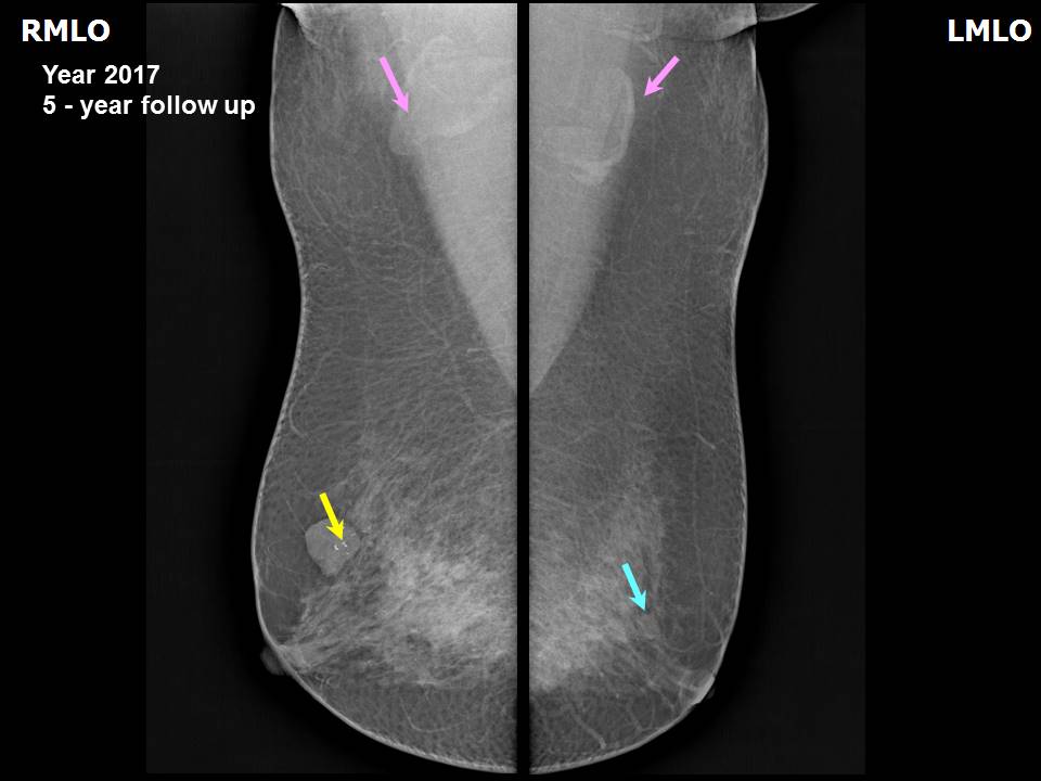
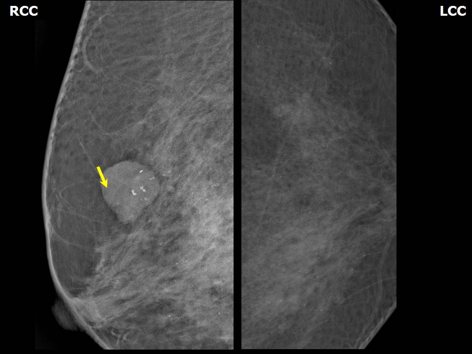
| ‣ Location of the lesion: | 2017: Right breast, upper outer quadrant at 11 oclock, anterior third |
| ‣ Mass: | |
| • Number: | 1 |
| • Size: | 1.9 × 1.5 cm |
| • Shape: | Oval |
| • Margins: | Circumscribed |
| • Density: | Equal |
| ‣ Calcifications: | |
| • Typically benign: | None |
| • Suspicious: | None |
| • Distribution: | None |
| ‣ Architectural distortion: | None |
| ‣ Asymmetry: | None |
| ‣ Intramammary node: | None |
| ‣ Skin lesion: | None |
| ‣ Solitary dilated duct: | None |
| ‣ Associated features: | None |
| Mammography features: | |
| ‣ Location of the lesion: | 2017: Left breast, upper inner quadrant at 11 oclock, anterior third |
| ‣ Mass: | |
| • Number: | 1 |
| • Size: | 1.0 cm in greatest dimension |
| • Shape: | Oval |
| • Margins: | Circumscribed |
| • Density: | Equal |
| ‣ Calcifications: | |
| • Typically benign: | None |
| • Suspicious: | None |
| • Distribution: | None |
| ‣ Architectural distortion: | None |
| ‣ Asymmetry: | None |
| ‣ Intramammary node: | None |
| ‣ Skin lesion: | None |
| ‣ Solitary dilated duct: | None |
| ‣ Associated features: | None |
Ultrasound:
 |  |
 |
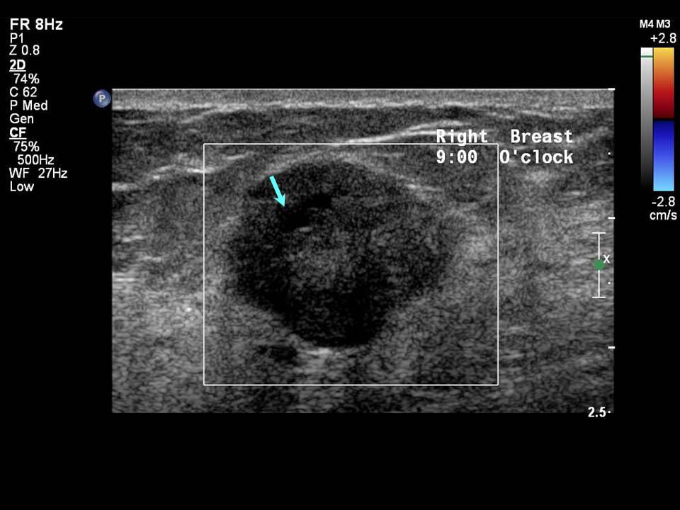
| Ultrasound features: 2011: Right breast, outer quadrants at 9 oclock | |
| ‣ Mass | |
| • Location: | 2011: Right breast, outer quadrants at 9 oclock |
| • Number: | 1 |
| • Size: | 1.7 × 1.5 cm |
| • Shape: | Oval |
| • Orientation: | Parallel |
| • Margins: | Circumscribed, 23 lobulations present |
| • Echo pattern: | Heterogeneous |
| • Posterior features: | No posterior features |
| ‣ Calcifications: | None |
| ‣ Associated features: | None |
| ‣ Special cases: | None |
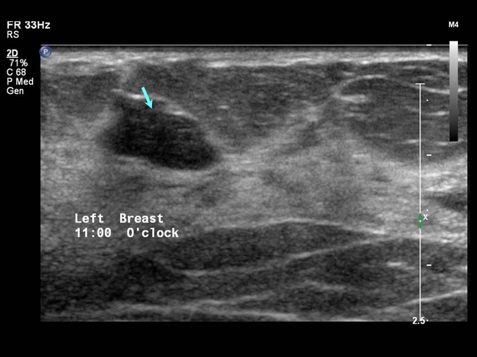
| Ultrasound features: 2011: Left breast, upper inner quadrant at 11 oclock | |
| ‣ Mass | |
| • Location: | 2011: Left breast, upper inner quadrant at 11 oclock |
| • Number: | 1 |
| • Size: | 1.0 × 10.5 cm |
| • Shape: | Irregular |
| • Orientation: | Parallel |
| • Margins: | Angular |
| • Echo pattern: | Hypoechoic |
| • Posterior features: | No posterior features |
| ‣ Calcifications: | None |
| ‣ Associated features: | None |
| ‣ Special cases: | None |
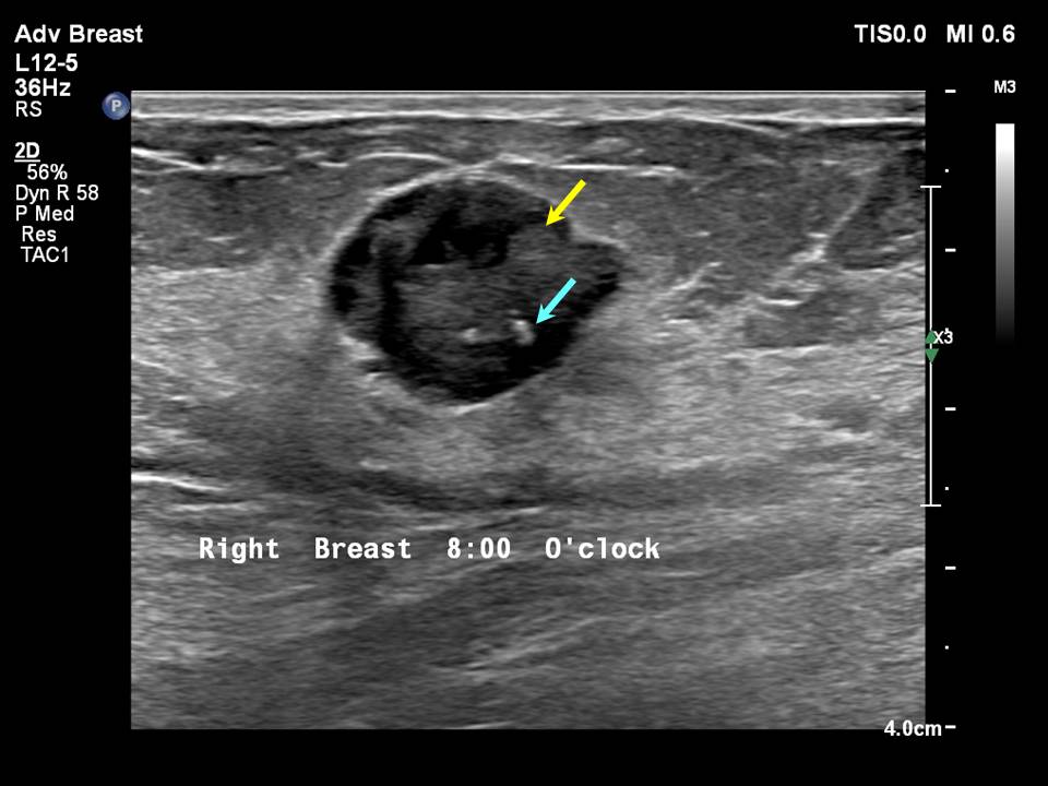 |  |
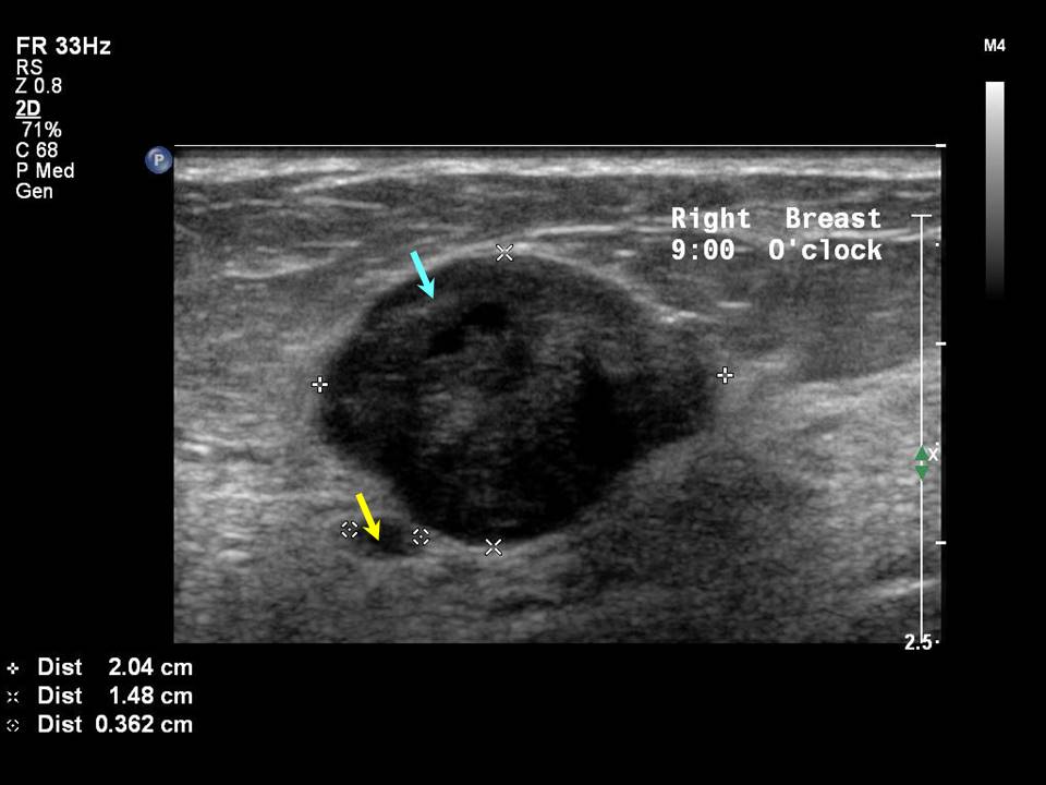
| Ultrasound features: 2017: Right breast, outer quadrants at 9 oclock | |
| ‣ Mass | |
| • Location: | 2017: Right breast, outer quadrants at 9 oclock |
| • Number: | 1 |
| • Size: | 1.7 × 1.5 cm |
| • Shape: | Oval |
| • Orientation: | Parallel |
| • Margins: | Circumscribed, 23 lobulations present |
| • Echo pattern: | Heterogeneous |
| • Posterior features: | No posterior features |
| ‣ Calcifications: | None |
| ‣ Associated features: | None |
| ‣ Special cases: | None |
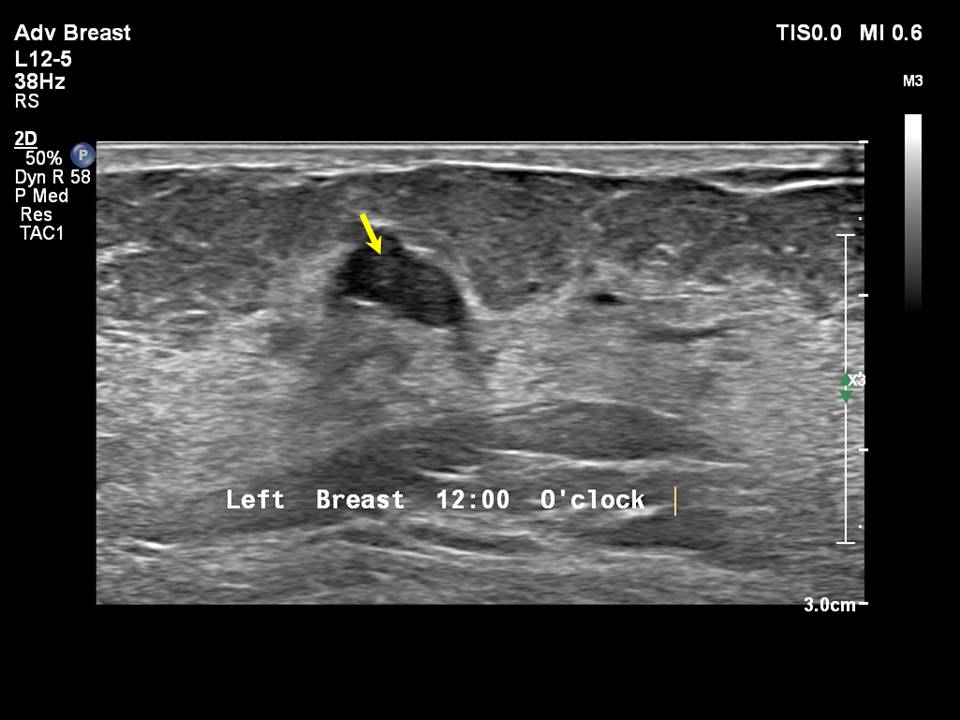
| Ultrasound features: 2017: Left breast, upper inner quadrant at 11 oclock | |
| ‣ Mass | |
| • Location: | 2017: Left breast, upper inner quadrant at 11 oclock |
| • Number: | 1 |
| • Size: | 1.0 × 10.5 cm |
| • Shape: | Irregular |
| • Orientation: | Parallel |
| • Margins: | Angular |
| • Echo pattern: | Hypoechoic |
| • Posterior features: | No posterior features |
| ‣ Calcifications: | None |
| ‣ Associated features: | None |
| ‣ Special cases: | None |
BI-RADS:
BI-RADS Category (2011): 3 (probably benign)BI-RADS Category (2012): 2 (benign)
BI-RADS Category (2017): 2 (benign)
MRI:
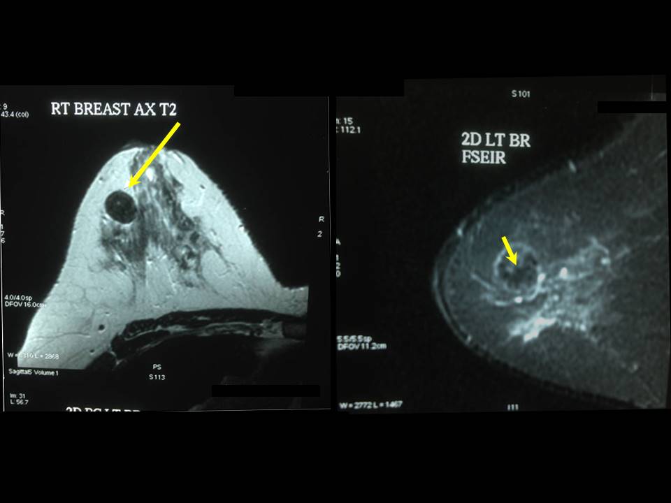 | 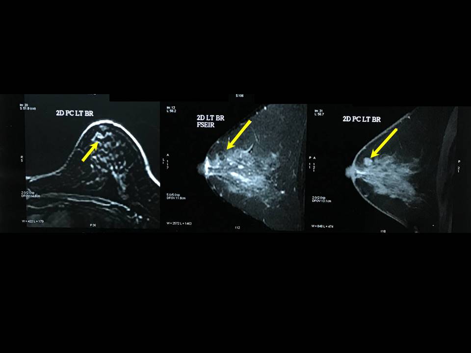 |
 |
| MRI features: | ||
| ‣ MRI features: | Amount of fibroglandular tissue: ACR category a (predominantly fatty with small proportion of glandular density). Background parenchymal enhancement: Mild (2550%), symmetrical | |
| ‣ Location: | Right breast at 11 oclock | |
| ‣ Focus: | No | |
| ‣ Mass: | ||
| • Shape: | Round | |
| • Margin: | Circumscribed | |
| • Internal enhancement: | Homogeneous | |
| • Kinetic curve: | Type I | |
| ‣ Non-mass enhancement: | ||
| • Distribution: | No | |
| • Internal enhancement: | No | |
| ‣ Non-enhancing findings: | T2 hypointense | |
| ‣ Associated features: | A few smaller lesions of similar morphology | |
| ‣ Axillary nodes: | None | |
Case summary:
| Premenopausal woman with increased risk of developing breast cancer presented with right breast lump diagnosed as involuting (partially calcified) fibroadenoma in right breast, BI-RADS category 2 on imaging. Left breast reveals probably benign lesion, BI-RADS category 3 on initial assessment. Regular annual follow-up done for 5 years and diagnosed as sclerotic fibroadenoma in left breast, BI-RADS category 2 on imaging. |
Learning points:
|