


Home / Training / Manuals / Atlas of breast cancer early detection / Cases
Atlas of breast cancer early detection
Filter by language: English / Русский
Go back to the list of case studies
.png) Click on the pictures to magnify and display the legends
Click on the pictures to magnify and display the legends
BI-RADS Category (at follow-up (2017)): 2 (benign)
BI-RADS Category (at follow-up (2018)): 2 (benign)
| Case number: | 161 |
| Age: | 44 |
| Clinical presentation: | Premenopausal woman with increased risk of breast carcinoma because of family history of breast cancer presented in 2012 for screening. CBE did not reveal any abnormality. |
Mammography:
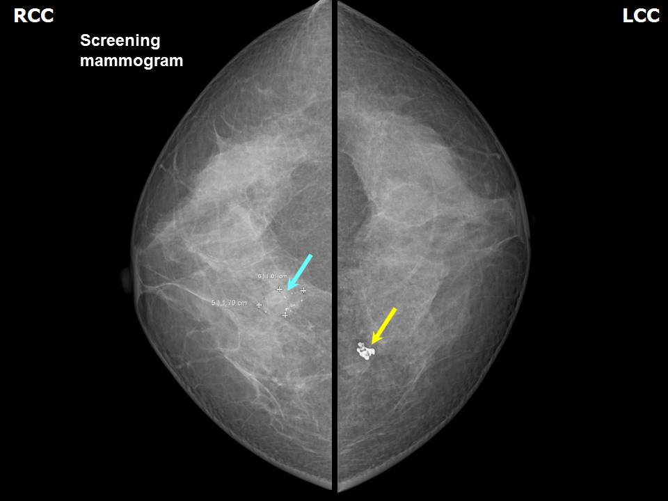 | 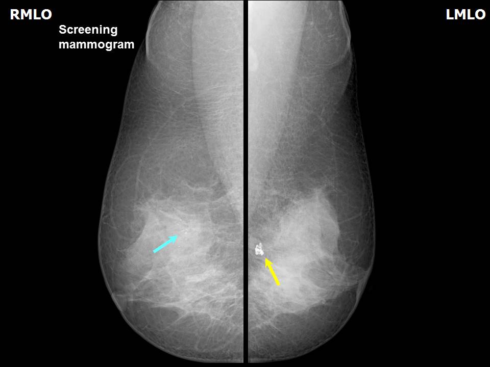 |
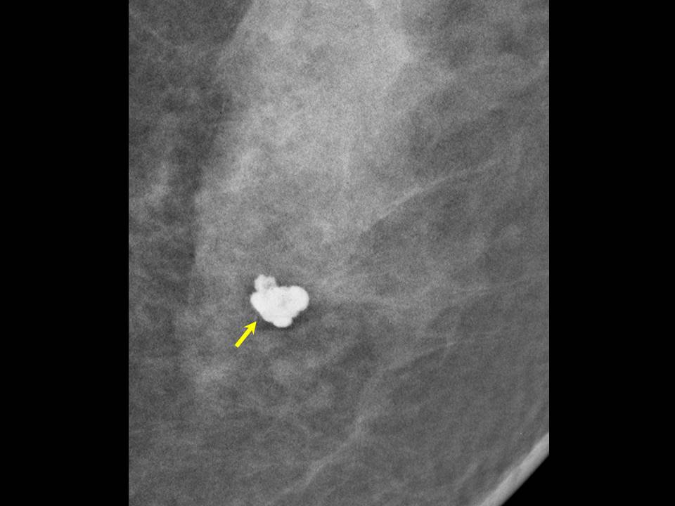
| Breast composition: | ACR category c (the breasts are heterogeneously dense, which may obscure small masses) | Mammography features: |
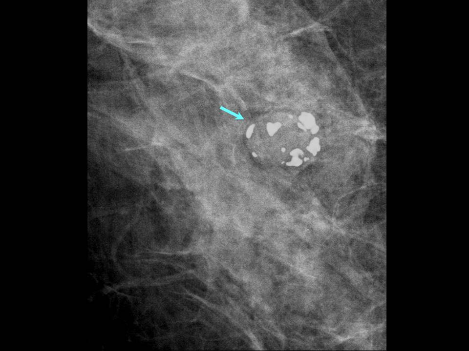
| ‣ Location of the lesion: | 2012: Right breast, upper inner quadrant at 1 oclock, middle third |
| ‣ Mass: | |
| • Number: | 1 |
| • Size: | 1.8 × 1.0 cm |
| • Shape: | Oval |
| • Margins: | Partly circumscribed and partly indistinct |
| • Density: | Equal |
| ‣ Calcifications: | |
| • Typically benign: | None |
| • Suspicious: | Suspicious calcifications |
| • Distribution: | Grouped, > 5 in number, in < 2.0 cm of breast tissue |
| ‣ Architectural distortion: | None |
| ‣ Asymmetry: | None |
| ‣ Intramammary node: | None |
| ‣ Skin lesion: | None |
| ‣ Solitary dilated duct: | None |
| ‣ Associated features: | None |
| Mammography features: | |
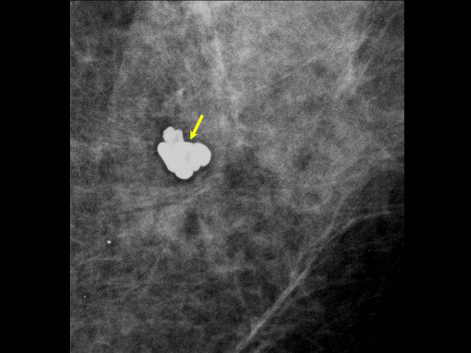
| ‣ Location of the lesion: | 2012: Left breast, upper inner quadrant at 10 oclock, posterior third |
| ‣ Mass: | |
| • Number: | 0 |
| • Size: | None |
| • Shape: | None |
| • Margins: | None |
| • Density: | None |
| ‣ Calcifications: | |
| • Typically benign: | None |
| • Suspicious: | None |
| • Distribution: | Single macrocalcification focus |
| ‣ Architectural distortion: | None |
| ‣ Asymmetry: | None |
| ‣ Intramammary node: | None |
| ‣ Skin lesion: | None |
| ‣ Solitary dilated duct: | None |
| ‣ Associated features: | None |
 |  |
 |  |
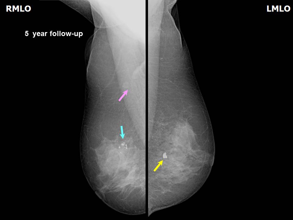
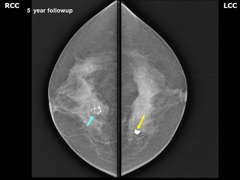
| Breast composition: | ACR category b (there are scattered areas of fibroglandular density) | Mammography features: |
| ‣ Location of the lesion: | 2017: Follow-up at year 5: Right breast, upper inner quadrant at 1 oclock, middle third |
| ‣ Mass: | |
| • Number: | 1 |
| • Size: | 1.7 × 1.0 cm |
| • Shape: | Oval |
| • Margins: | Partly circumscribed and partly indistinct |
| • Density: | Equal |
| ‣ Calcifications: | |
| • Typically benign: | Coarse macrocalcifications |
| • Suspicious: | None |
| • Distribution: | In mass |
| ‣ Architectural distortion: | None |
| ‣ Asymmetry: | None |
| ‣ Intramammary node: | None |
| ‣ Skin lesion: | None |
| ‣ Solitary dilated duct: | None |
| ‣ Associated features: | None |
| Breast composition: | ACR category b (there are scattered areas of fibroglandular density) | Mammography features: |
| ‣ Location of the lesion: | 2017: Follow-up at year 5: Left breast, upper inner quadrant at 10 oclock, posterior third |
| ‣ Mass: | |
| • Number: | 0 |
| • Size: | None |
| • Shape: | None |
| • Margins: | None |
| • Density: | None |
| ‣ Calcifications: | |
| • Typically benign: | None |
| • Suspicious: | None |
| • Distribution: | Single macrocalcification focus |
| ‣ Architectural distortion: | None |
| ‣ Asymmetry: | None |
| ‣ Intramammary node: | None |
| ‣ Skin lesion: | None |
| ‣ Solitary dilated duct: | None |
| ‣ Associated features: | None |
 |  |
 |  |
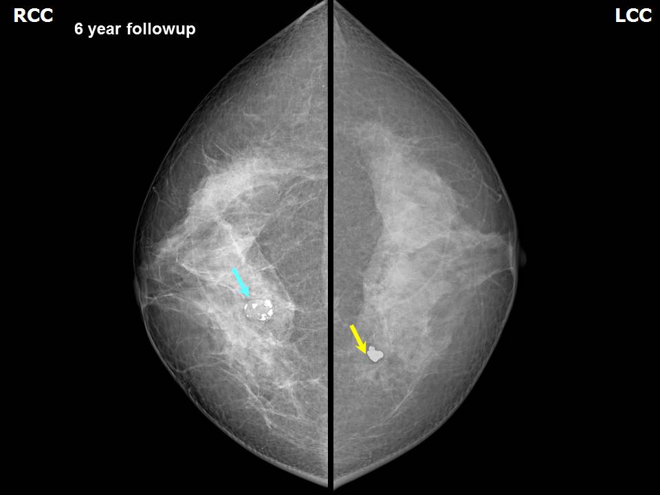
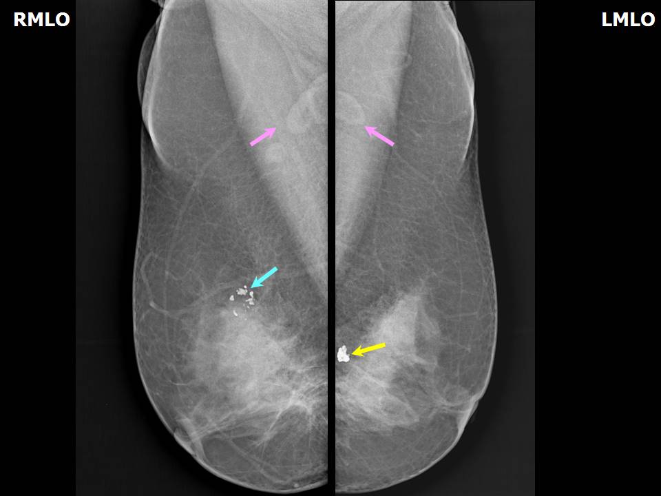
| Breast composition: | ACR category b (there are scattered areas of fibroglandular density) | Mammography features: |
| ‣ Location of the lesion: | 2018: Follow-up at year 6: Right breast, upper inner quadrant, 1 oclock, middle third |
| ‣ Mass: | |
| • Number: | 1 |
| • Size: | 1.7 × 1.0 cm |
| • Shape: | Oval |
| • Margins: | Partly circumscribed and partly indistinct |
| • Density: | Equal |
| ‣ Calcifications: | |
| • Typically benign: | Coarse macrocalcifications |
| • Suspicious: | None |
| • Distribution: | In mass |
| ‣ Architectural distortion: | None |
| ‣ Asymmetry: | None |
| ‣ Intramammary node: | None |
| ‣ Skin lesion: | None |
| ‣ Solitary dilated duct: | None |
| ‣ Associated features: | None |
| Breast composition: | ACR category b (there are scattered areas of fibroglandular density) | Mammography features: |
| ‣ Location of the lesion: | 2018: Follow-up at year 6: Left breast, upper inner quadrant at 10 oclock, posterior third |
| ‣ Mass: | |
| • Number: | 0 |
| • Size: | None |
| • Shape: | None |
| • Margins: | None |
| • Density: | None |
| ‣ Calcifications: | |
| • Typically benign: | None |
| • Suspicious: | None |
| • Distribution: | Single macrocalcification focus |
| ‣ Architectural distortion: | None |
| ‣ Asymmetry: | None |
| ‣ Intramammary node: | None |
| ‣ Skin lesion: | None |
| ‣ Solitary dilated duct: | None |
| ‣ Associated features: | None |
Ultrasound:
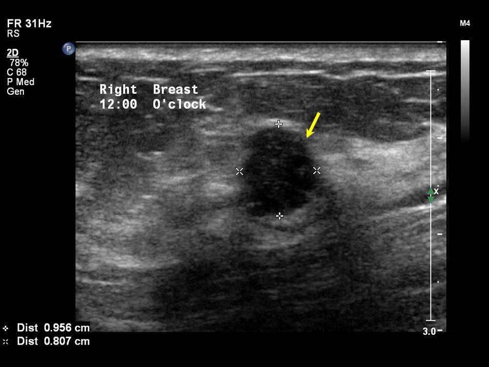 |
| Ultrasound features: 2012: Right breast, upper quadrants at 12 oclock | |
| ‣ Mass | |
| • Location: | 2012: Right breast, upper quadrants at 12 oclock |
| • Number: | 1 |
| • Size: | 1.0 × 0.8 cm |
| • Shape: | Oval |
| • Orientation: | Not parallel |
| • Margins: | Circumscribed, 23 lobulations present |
| • Echo pattern: | Hypoechoic |
| • Posterior features: | No posterior features |
| ‣ Calcifications: | None |
| ‣ Associated features: | None |
| ‣ Special cases: | None |
BI-RADS:
BI-RADS Category (2012): 3 (probably benign)BI-RADS Category (at follow-up (2017)): 2 (benign)
BI-RADS Category (at follow-up (2018)): 2 (benign)
Case summary:
| Premenopausal woman with increased risk of breast cancer because of family history of breast cancer in a first-degree relative presented for mammography screening. She attended for regular annual follow-up for 6 years. On initial assessment, diagnosed as grouped calcifications of suspicious morphology in right breast, BI-RADS 3 on imaging. At 6 year follow-up, diagnosed with involuting (partially calcified) fibroadenoma in right breast, BI-RADS 2 on imaging, and stable, typically benign calcifications in left breast, BI-RADS 2 on imaging. |
Learning points:
|